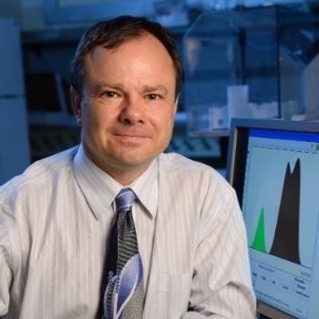
The 2019 Emerging Leader in Molecular Spectroscopy: Advancing Biomedical Raman Spectroscopy
By combining Raman spectroscopy, imaging, and chemometrics, Ishan Barman of Johns Hopkins University, the 2019 winner of the Emerging Leader in Spectroscopy award, develops novel approaches in which structural and molecular data converge to provide fresh insights into cancer, diabetes and an array of other diseases.
By combining Raman spectroscopy, imaging, and chemometrics, Ishan Barman of Johns Hopkins University, the 2019 winner of the Emerging Leader in Spectroscopy award, develops novel approaches in which structural and molecular data converge to provide fresh insights into cancer, diabetes and an array of other diseases.
When he was a student at the Massachusetts Institute of Technology (MIT), Barman worked on exploiting endogenous contrast in tissue for investigating metabolic disorders, and for developing Raman scattering-based pathophysiologic sensors. In his current role as a faculty member at Johns Hopkins, his laboratory has advanced plasmon-enhanced Raman scattering phenomenon to create highly specific reporters for in vitro and in vivo imaging, and tailored tags for ultrasensitive, multiplexed molecular analysis. The Barman Laboratory combines chemical imaging, nanoplasmonics, optical spectroscopy, and advanced chemometric tools for determining transformations affecting human health. This research is creating new tools for diagnosis, monitoring of treatment response, and improved understanding of disease-related mechanisms.
In recognition of this work, Barman is the 2019 winner of the Emerging Leader in Molecular Spectroscopy Award, which is presented by Spectroscopy magazine. The award will be presented to Barman at the SciX-FACSS Conference, October 13-18, 2019 in Palm Springs, California. This interview with Barman examines the latest developments in Raman spectroscopy for biomedical discovery.
Please tell us about some of your earliest research interests. How did you get started in science? What has kept you motivated? What is the most exciting part of your work day?
Curiosity. I was always curious to understand how and why things happen the way they do. The explosion of technological developments in the early 1990s further piqued this initial interest. I also had some amazing teachers who persuaded me to travel down the scientific path. There has not been much turning back.
Some of my earliest research interests were in exploring the interface between humans and robotics with a focus on haptic feedback. That changed during graduate school to finding new, cost-effective desalination methods before I entered the world of biomedical optics and spectroscopy.
The process of discovery is incredibly motivating. Make no mistake, it’s demanding and sometimes frustrating too. But then there’s that aha moment . . . and you feel all’s right with the world. If you want to be in an environment in which you can work on interesting and important problems in a rigorous way and with greater freedom than you may find elsewhere, pursuing a research career is the way to go.
What have been the most difficult aspects of your research to date? How have you worked to overcome these challenges?
Finding the right people to work with is, often, the most challenging aspect of undertaking biomedical research. How do you bring together people with complementary skills and create an environment where interdisciplinary efforts can thrive? In many ways, I’ve been very fortunate to attract the right mix of graduate students and postdoctoral scholars with distinct expertise to grow our program. Johns Hopkins’ intrinsic strengths in biomedical research has certainly been a huge plus, and has facilitated successful collaborations with medical scientists and physicians across various disciplines such as oncology, pathology, radiology, and hematology.
The integration of Raman spectroscopic measurement with biopsy needles is one of your recent design advances (1). This diagnostic tool provides a new device with the potential for minimally invasive real time cancer detection. Which medical diagnostic application of this device do you feel will have the greatest potential impact in medicine? What difficulties did you face in designing this device?
The miniature Raman probe can not only detect microcalcifications, a prominent mammographic marker of breast pathology, but also diagnose the specific breast lesions associated with the microcalcifications during core-needle biopsies. By providing real-time feedback to the radiologist, it has the potential to minimize patient anxiety by eliminating the currently unavoidable wait for pathology diagnosis. Crucially, it would also reduce the likelihood of a non-diagnostic biopsy that necessitates a repeat needle or surgical procedure. If successful, such an approach would significantly improve the quality of healthcare and lower associated costs for breast cancer patients.
In the future, integrating the rich molecular detail of Raman spectroscopy with spatially offset probes and tomography may even offer fully non-invasive molecular characterization and microstructure visualization- “optical biopsy”. Though our efforts have largely been focused in breast cancer, this approach would be advantageous to detection of malignancies in other tissues such as pancreas, colorectal, and prostate.
There are a number of challenges that needed to be overcome. A major challenge was to design and fabricate a miniature side-viewing probe that could be inserted into the narrow central channel of the biopsy needle for intermittent acquisition of spectra in vivo. Next, we needed to assess whether the spectroscopic information is capable of identifying the malignancy with sufficient statistical confidence. Often, the differences between the spectral signatures, particularly in the context of label-free detection, can be very subtle -which makes robust identification of the molecular changes associated with the pathology very difficult.
Raman spectroscopy and chemometric techniques were used to create a new tool for analysis of lung and primary breast tumor tissues for diagnosis of metastatic cancers very early in the metastatic cascade (2). How does one use Raman to observe morphological changes in tissues leading to an understanding of cancer etiology? What are the biological changes that can be seen using Raman?
Label-free molecular imaging approaches are particularly attractive, as they enable multiplexed measurements without needing to focus on a limited number of predefined biomolecules. Raman spectroscopy offers the ability to interrogate biomolecular changes in a label-free manner, and to visualize complex molecular heterogeneity directly from cells and tissues. It relies on the inelastic scattering of light, arising from its interactions with the biological specimen, to quantify the unique vibrational modes of molecules within its native context. We and others have leveraged its lack of sample preparation requirements and high molecular specificity for objective recognition of epithelial and stromal changes in cancers.
In this specific study, Raman spectroscopic measurements identified vibrational features of the collagenous stroma as well as proteoglycans in premetastatic mouse lungs, a common site for spontaneous dissemination of breast cancer. Importantly, the identified spectral markers of this distant site accurately classified the metastatic potential of the primary tumor by recapitulating the compositional changes -without requiring addition of dyes, molecular stains, or human interpretation.
Noninvasive glucose concentration measurements are a major medical challenge that many researchers have explored. You and your colleagues developed a dynamic concentration correction (DCC) scheme, using the mass transfer of glucose interstitial fluid (ISF) glucose and blood glucose (3). How successful were you in achieving a method for accurately measuring blood glucose using Raman spectroscopy? Do you think this technology will lead to a clinically viable tool for such measurements?
I sure hope so! It’s an area that is very close to my heart. There have been solid advances in developing optical and spectroscopic methods by examining the different issues that complicate noninvasive blood glucose sensing. The diffusional process that underlies the transport of glucose from the bloodstream to the interstitial fluid and the cells is one such issue, as the optically measured and clinically relevant variables are not identical. Addressing these aspects systematically is bringing us ever closer to the ultimate goal, and, given the recent pace of advances, a noninvasive sensor may be a reality in the not too distant future.
Of all your research papers so far, what are the most meaningful papers you would like to highlight for our readers?
Of our recent work, I would like to highlight our contributions to the development of surface-enhanced Raman spectroscopy (SERS)-based single cell analysis platforms (4,5) and the recognition of differential radiation sensitivity using Raman spectroscopy with the goal of guiding personalization and biological adaptation of radiotherapy (6).
What is the most difficult or challenging aspect of your current research? How do you manage a research group, while at the same time mentoring and directing students?
Certainly, one of the challenging aspects is striking the right balance between performing research, teaching, and providing quality mentorship to your advisees.
From the research perspective, I would say the biggest challenge resides in translating our advances from the laboratory to the clinic. Seeing one of our discoveries truly benefiting the patient population would be most gratifying.
Mentoring students is an absolute privilege. I thoroughly enjoy my role as an advisor. It’s a fantastic experience molding students and seeing them grow in the span of a few years. Some of my advisees are so smart, it’s amazing to think what they could achieve in the future!
How were you able to direct your research toward discovery of important medical problems? What made you choose the path of medical research versus industrial, chemical, or environmental problems?
The choice of medical research as our first and principal theme was a natural one. Inspired by the Star Trek tricorder, I have long wanted to design fully noninvasive sensors that could reveal the most complex pathologies. We are nowhere close to that yet but I would like to believe that the distance to realization of the magic wand has decreased!
As our laboratory has grown, we have been able to take some of these technological innovations and leverage them to address problems of significance in the industrial and environmental arena. For instance, we are currently developing a suite of optical sensors for the biopharmaceutical industry, and adapting our SERS assays for monitoring microcystin production during algal blooms.
What areas would you like to see this research expand into when looking toward the future? Do you plan to stay in Raman work or are there another analytical techniques that look promising?
As a scientist, I am always excited to ask fresh questions, use new tools, and, generally, poke around! Science is fun because it’s rarely predictable. While we will continue to build on our work in advancing Raman spectroscopy, exploring new areas will be a big part of our future. Our recent efforts provide a foundation for making forays in creating new plasmonic tools that can reveal the interplay between nanoscale deformations and macroscopic functions in live cells, in probing metastatic progression and organotropism, and in noninvasive monitoring of therapy response by simultaneous utilization of tumor and microevironment information. We are also intrigued by the recent developments in deep learning for bioimaging and in quantitative phase microscopy, and are eager to see how these could power our own work. Lots of excellent possibilities!
What would you tell those young people interested in science about how to prepare for a career in research?
Science is not linear. As you embark on a career in research, know that you are going to fail sometimes. You can’t take failure too seriously-indeed, it should encourage you to think outside the box.
Also, take the initiative and reach out to people. It will help you hone your people skills, gain a deeper appreciation of your research area, and expand your horizon.
References
(1) I. Barman, N.C. Dingari, A. Saha, S. McGee, L.H. Galindo, W. Liu, D. Plecha, N. Klein, R.R. Dasari, and M. Fitzmaurice, Application of Raman spectroscopy to identify microcalcifications and underlying breast lesions at stereotactic core needle biopsy. Cancer Res. 73(11), 3206–3215 (2013).
(2) S. K. Paidi, A. Rizwan, C. Zheng, M. Cheng, K. Glunde, I. Barman, “Label-free Raman spectroscopy detects stromal adaptations in pre-metastatic lungs primed by breast cancer”, Cancer Res. 77(2), 247-256 (2017).
(3) I. Barman, C.R. Kong, G.P. Singh, R.R. Dasari, and M.S. Feld, Accurate spectroscopic calibration for noninvasive glucose monitoring by modeling the physiological glucose dynamics. Anal. Chem.82(14) 6104–6114 (2010).
(4) Q. Jin, M. Li, B. Polat, S.K. Paidi, A. Dai, A. Zhang, J.V. Pagaduan, I. Barman, and D.H. Gracias, Mechanical Trap SurfaceâEnhanced Raman Spectroscopy for ThreeâDimensional Surface Molecular Imaging of Single Live Cells. Angew. Chem. Int. Ed. Engl.56(14), 3822–3826 (2017).
(5) W. Xu, S.K. Paidi, Z. Qin, Q. Huang, C.H. Yu, J.V. Pagaduan, M.J. Buehler, I. Barman, and D.H. Gracias, Self-folding hybrid graphene skin for 3D biosensing. Nano Lett.19(3), 1409–1417 (2018).
(6) S.K. Paidi, P.M. Diaz, S. Dadgar, S.V. Jenkins, C.M. Quick, R.J. Griffin, R.P. Dings, N. Rajaram, and I. Barman, Label-free Raman spectroscopy reveals signatures of radiation resistance in the tumor microenvironment. Cancer Res. 79(8), 2054–2064 (2019).
Newsletter
Get essential updates on the latest spectroscopy technologies, regulatory standards, and best practices—subscribe today to Spectroscopy.


