Analyzing Soil and Meat with Shifted Excitation Raman Difference Spectroscopy
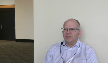
Kay Sowoidnich, PhD, is a research associate with Laser Sensors Lab at the Ferdinand-Braun-Institut, Leibniz-Institut für Höchstfrequenztechnik and one of the 2020 winners of the Society for Applied Spectroscopy William F. Meggers Award. His group have been able to demonstrate the potential of shifted-excitation Raman difference spectroscopy (SERDS) as an efficient tool for soil nutrient analysis.
Kay Sowoidnich, PhD, is a research associate with Laser Sensors Lab at the Ferdinand-Braun-Institut, Leibniz-Institut für Höchstfrequenztechnik and one of the 2020 winners of the Society for Applied Spectroscopy William F. Meggers Award. His group have been able to demonstrate the potential of shifted-excitation Raman difference spectroscopy (SERDS) as an efficient tool for soil nutrient analysis. Additionally, his previous research work that applied SERDS for the analysis of meat could enable rapid screening of large numbers of meat samples for authentication and quality monitoring purposes at different points along the distribution chain, from the slaughterhouse to supermarket. Spectroscopy spoke with Sowoidnich about his work applying SERDS, its impact and limitations, his work using spatially offset Raman spectroscopy (SORS) to diagnosis skeletal disorders, and more.
You’ve been applying SERDS for the analysis of soil, food, and other constituents for several years (1,2). How has this analytical technique proven to be advantageous for soil and food analysis? What type of impact could it have on precision agriculture and for food supplies?
Conventional Raman spectroscopy allows obtaining chemically specific information from numerous samples and is a powerful tool for qualitative and quantitative analysis. As Raman scattering is usually a weak effect, interfering contributions can easily mask the characteristic Raman signals and thus complicate or even prevent meaningful sample investigation. Prominent examples of such interferences are laser-induced fluorescence and, driven by improvements in the sensitivity of portable Raman instrumentation, various types of ambient light. Among other techniques, SERDS is a powerful instrumental approach to address this issue by separating Raman signals from unwanted light contributions using a dual-wavelength excitation light source. If two slightly shifted excitation wavelengths are used to record two Raman spectra, then the Raman signal positions will follow that shift in wavelength while fluorescence and other (quasi-)static contributions will remain virtually unchanged. Subtracting the two recorded Raman spectra can thus effectively retain the Raman spectroscopic information while removing the unwanted interferences.
Diode lasers are well-suited as excitation light source for SERDS, as they offer compact size, low-power consumption, and high efficiency. Such innovative and customized lasers are developed and realized at Ferdinand-Braun-Institut, Leibniz-Institut für Höchstfrequenztechnik (FBH). Heinz-Detlef Kronfeldt’s laser spectroscopy group at Technical University Berlin had established a fruitful collaboration with Dr. Bernd Sumpf and Dr. Martin Maiwald from FBH, and we were able to use unique diode lasers for our Raman and SERDS experiments. Within a collaborative research project, SERDS has successfully been applied for the non-invasive optical discrimination between meat of selected animal species, exemplarily demonstrated for beef, pork, chicken, and turkey. Furthermore, Raman investigations could demonstrate the non-invasive quantitative detection of meat spoilage with respect to legal limits. In the future, this could enable rapid screening of large numbers of meat samples for authentication and quality monitoring purposes at different points along the distribution chain, from the slaughterhouse to the supermarket. The non-destructive Raman technique could become a valuable asset for food inspectors, enabling analysis of large production batches rather than relying on random sampling and cumbersome laboratory analysis. The topic of Raman spectroscopy for food analysis has further been developed by my former colleague Dr. Heinar Schmidt at University of Bayreuth. His group conducted successful Raman trials for meat inspection in a slaughterhouse as well for on-site inspection of Australian lamb meat using a portable device at 671 nm excitation.
During my current role within the Laser Sensors Lab at FBH, our group was able to demonstrate the potential of SERDS as an efficient tool for soil nutrient analysis. Currently, soil investigations require sample collection followed by complex and time-consuming laboratory analysis. Optical methods could be very beneficial in this instance, as they enable rapid analysis without prior sample collection. Conventional Raman spectroscopy can provide information on the present molecular species but suffers from fluorescence from soil organic matter. Here, SERDS could enable in situ soil investigations at a large number of spots across the whole field, thus allowing mapping the distribution of relevant soil components, including nutrients. This could be a major advantage as the needs-based precision fertilization is already possible with state-of-the-art farming equipment. But to fully exploit this benefit it would require knowledge of spatially-resolved information about the actual soil status that is not yet available from current analytical methods.
What types of limitations does SERDS present when, for example, using it to investigate fluorescent heterogenous samples (3,4)? How do you overcome these limitations or challenges?
SERDS is based on the alternate and subsequent recording of Raman spectra excited at two slightly shifted wavelengths. Using a conventional charge-coupled device (CCD) detector, one SERDS cycle would then comprise the following steps: 1) exposure at first wavelength, 2) read-out of Raman spectrum recorded at first wavelength, 3) exposure at second wavelength, and 4) read-out of Raman spectrum recorded at second wavelength. If during that period of time unwanted background interferences are not constant, residual artefacts could remain after subtraction of the two spectra, and this would compromise qualitative and quantitative analysis. Potential examples are fluorescence quenching, sample or instrument movement in case of heterogeneous specimens, and varying ambient light intensities. Faster, ideally quasi-simultaneous, acquisition of SERDS spectra would therefore be highly beneficial to address this issue. On the excitation side, this will require a dual-wavelength light source with reproducible and fast-shifting capabilities between the two emission lines. Such lasers have readily been demonstrated, for example, by FBH with rapid modulation up to the kilohertz range. The limitation rather lies on the detection side, where one could reduce the individual exposure times. However, there is a fundamental physical limit to the time required to read out the charges from the CCD in conventional operation mode.
During my time at the United Kingdom’s Science and Technology Facilities Council (STFC), I was working in the Central Laser Facility (CLF) under the supervision of Prof. Pavel Matousek, who is a brilliant scientist full of exciting new ideas. He came up with a neat solution by combining the advantages of SERDS with the benefits of a charge-shifting approach. The basic concept of charge-shifting CCD operation was proposed in 1990, while a pilot study in 2008 by others has shown its suitability for conventional Raman spectroscopy. The major advantage here is that the read-out steps after each single exposure are omitted. Instead, the charges generated by Raman scattered light excited at the first excitation wavelength are electronically shifted towardsa non-illuminated area on the CCD before charges are generated by Raman scattered light excited at the second excitation wavelength. These charges, in turn, are then shifted to another unilluminated area on the CCD when additional charges are generated by Raman scattered light excited at the first excitation wavelength. This alternate shifting procedure is repeated until sufficient charge in the two distinct areas on the CCD, corresponding to charges generated by Raman scattered light at the two excitation wavelengths, is accumulated. Finally, only one read-out step at the very end is performed.
Through a prolific cooperation between STFC and FBH, which developed a custom 830-nm diode laser with tailored electro-optical properties, putting special emphasis on fast wavelength switching, the charge-shifting SERDS approach has been realized experimentally. Using a stock CCD that has been modified according to our requirements, we could successfully demonstrate alternate recording of Raman spectra at frequencies of 1,000 Hz, and that is about two orders of magnitude faster than speeds achievable in conventional CCD read-out mode. The reproducibility of SERDS spectra recorded on exemplarily chosen heterogeneous rock samples in charge-shifting mode was superior to spectra recorded in conventional CCD operation. This led to improved classification accuracy for the discrimination between rock species. We also applied the charge-shifting concept to suppress rapidly varying ambient light interference (5) and successfully translated it toward sub-surface analysis using spatially offset Raman spectroscopy (SORS).
Through your work using SORS to diagnosis skeletal disorders as well as other medical conditions, what did you learn about the technique (6)? How is it advantageous as a medical diagnostic technique? Is it currently used clinically or are there plans for clinical deployment?
The current gold standard for the diagnosis of bone conditions are X-ray-based methods. Despite being able to assess the mineral phase of the bone very well, this kind of technique is unfortunately insensitive to collagen, the second important component of the composite material bone. Raman spectroscopy, in contrast, is able to retrieve chemically specific information from both phases, for example, mineral and collagen, thus giving a more complete picture of the overall bone status. As conventional Raman spectroscopy is restricted to the investigation of surface-near layers, we applied SORS for efficient sub-surface analysis. This enables us to measure the bone composition, non-invasively, through the skin at selected anatomical locations, such as at lower legs and fingers.
SORS works in such a way that the point of laser illumination on the surface is set at a certain spatial distance from the point where the Raman-scattered photons are collected, thus probing sub-surface layers within the sample. For the development of SORS as medical diagnostic technique it is essential to know from what depth the detected Raman spectroscopic information is actually emerging for a chosen spatial offset. Our photon migration study was addressing this important point, using typical long bone tissue, as well as antler and bulla, to include two types of bone exhibiting among the lowest and highest mineralization levels found in nature. Results indicated that photons can more easily migrate inside less mineralized bone tissue and that porosity can play an important role as well. We were also able to estimate the approximate depth from which the major Raman signal contribution is collected for a given spatial offset within exposed bone during SORS investigations. Under safe laser illumination conditions compatible with in vivo applications even in highly mineralized bulla depths of up to 3.8 mm are still accessible using large spatial offsets in the range of 9–10 mm. The findings of our study significantly increased our understanding of SORS analysis through bones of different composition, thus providing vital information to assess the potential of SORS for medical diagnostics.
We are aiming to develop SORS as a valuable tool for the non-invasive in vivo diagnosis of bone disorders and diseases. In close collaboration with the Royal National Orthopaedic Hospital in London, we were running a pre-clinical SORS trial with a custom instrument to apply the technique in a clinical environment. More than 150 patients were recruited to-date, either having one of the diagnosed bone conditions-osteoporosis, osteoarthritis or osteogenesis imperfecta (also known as brittle bone disease)-or belonging to a matched healthy control group without any bone condition. Within the frame of this SORS trial, for the first time, the in vivo bone disease detection in case of osteogenesis imperfecta was demonstrated. For the other bone conditions, particularly for less severe cases, our results indicate a difference between healthy and diseased bone, but the differentiation is not yet statistically significant. We are, however, confident that improvements in instrument hardware and data analysis will pave the way for SORS to become a routine analytical technique for bone-quality assessment, complementing or even partially replacing existing methods.
What would you consider to be the most meaningful contributions of your work?
In my opinion, the charge-shifting SERDS approach has made a significant impact, as it has overcome existing fundamental limitations of conventional CCD operation. The potential applications are numerous, and the examples described in our corresponding publications only cover a fraction of what will actually be possible. The underlying technology has already been implemented into a novel CCD camera of one of the world’s leading companies for spectroscopic equipment. The future will show what exciting new research fields can be explored by the enabling capabilities of the charge-shifting technique. This is actually a hot topic, as in parallel to our study, similar research has been undertaken independently by Leibniz-Institut für Astrophysik Potsdam (Germany) and University of Tokyo (Japan).
Besides doing cutting-edge research, it is equally important to communicate the findings not only to scientific experts but also addressing the layman. Another important contribution is therefore related to public outreach activities. During my time at STFC’s Rutherford Appleton Laboratory, our group has developed a SORS demonstration setup within the frame of the International Year of Light 2015. The instrument was basically a simplified version of the commercial SORS devices used at airports to scan liquids inside containers. During numerous exhibitions, the SORS demo setup was one of the most popular and most reliable exhibits highlighting an important part of STFC’s science to the public. The instrument has been also presented to key stakeholders and decision-makers, including the UK science minister. Due to its tremendous success, a second device was realized that is now permanently installed in the CLF visitor’s centers at STFC’s Rutherford Appleton Laboratory.
What are your plans for the future, your next steps?
My immediate next steps are further research efforts to establish SERDS as a valuable tool for the analysis of soil, paving the way for efficient nutrient management in the frame of precision agriculture (the consortium Intelligence for Soil (I4S) in funding measure Soil as a Sustainable Resource for the Bioeconomy (BonaRes) by Federal Ministry of Education and Research). On a long-term scale, at FBH the Laser Sensors Lab provides excellent working conditions for me to make further significant contributions to application-oriented research aiming to address current as well as upcoming societal needs and global challenges. FBH’s big advantage is that the whole value chain is located in-house, with expertise reaching from the design and manufacture of novel diode lasers with unique properties to the application of these laser sources as part of customized portable SERDS instruments for in situ field experiments. As the area of photonic technologies in general and optical spectroscopy in particular is a very dynamic field, I am keen to see what opportunities and scientific challenges I will come across in the future.
References
- FBH research, April 16, 2020.
https://www.fbh-berlin.de/forschung/forschungsnews/detail/raman-spectroscopic-investigations-on-soil-using-a-785-nm-dual-wavelength-diode-laser .
- K. Sowoidnich and H.-D. Kronfeldt, Encyclopedia of Analytical Chemistry, Wiley Online Library, (2015).
https://doi.org/10.1002/9780470027318.a9510 .
- K. Sowoidnich, M. Towrie, M. Maiwald, B. Sumpf, and P. Matousek, Appl. Spectrosc.73, 1265–1276 (2019).
- K. Sowoidnich, M. Maiwald, B. Sumpf, M. Towrie, and P. Matousek, " Proc. SPIE11236, 112360K (2020).
- K. Sowoidnich, M. Towrie, and P. Matousek, J. Raman Spectrosc.50, 983–995 (2019).
- K. Sowoidnich, J.H. Churchwell, K. Buckley, et al., Analyst 142, 3219–3226 (2017).
Newsletter
Get essential updates on the latest spectroscopy technologies, regulatory standards, and best practices—subscribe today to Spectroscopy.


