Cell and Drug Interactions Studied Using Surface Plasmon Resonance and Fluorescence
Scientists from the Beijing University of Chemical Technology and the Chinese Academy of Sciences in Beijing, China recently tested a combination of surface plasmon resonance (SPR) and fluorescence analysis in studying cell and drug interactions. Their research was published in the journal Spectrochimica Acta Part A: Molecular and Biomolecular Spectroscopy (1).
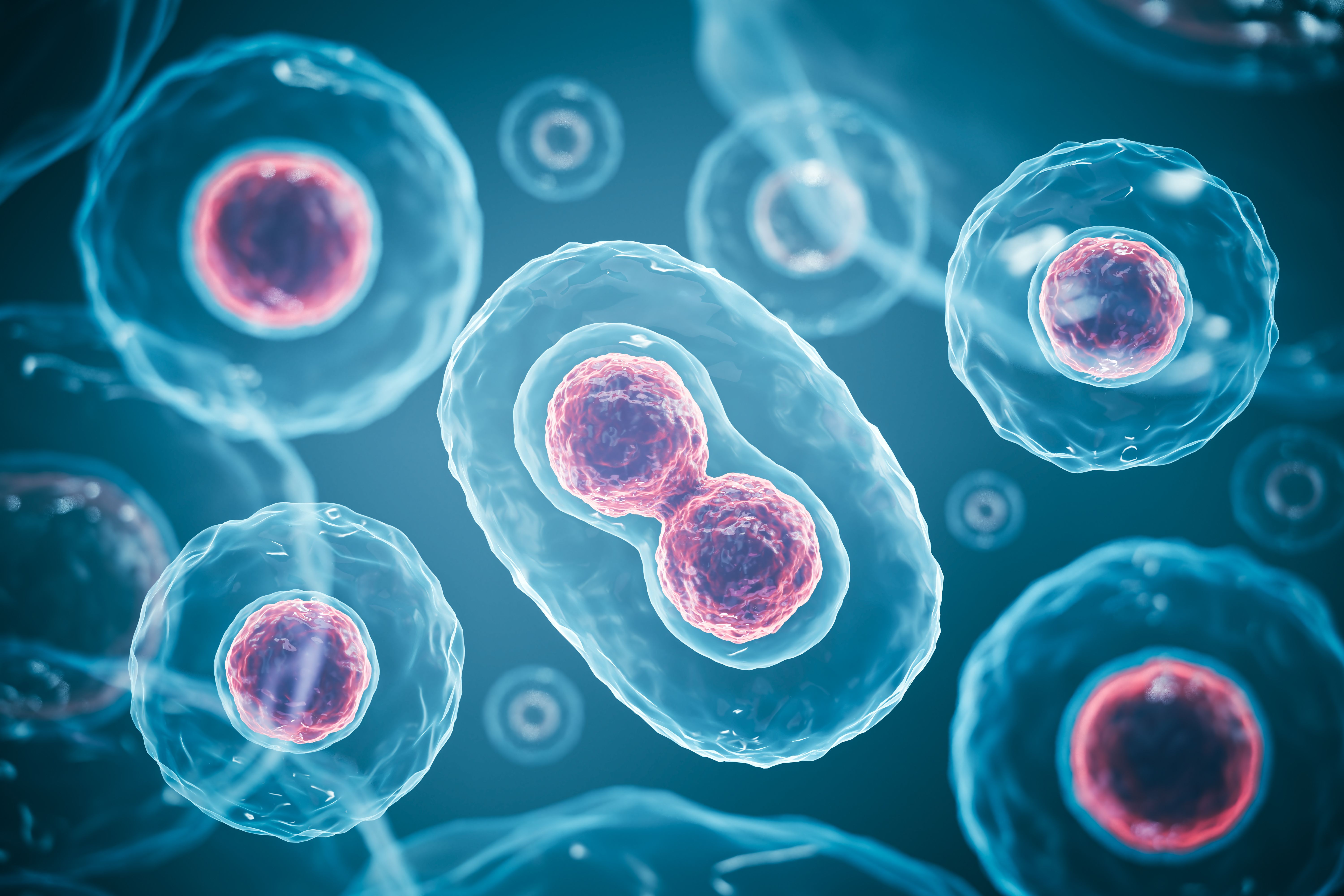
3d rendering of Human cell or Embryonic stem cell microscope background. | Image Credit: © Anusorn - stock.adobe.com

Cell-based measurements are becoming a popular tool in drug research, partly due to how they can assess the effect of drugs on fundamental biological processes and help extract intracellular information. The study of interactions between cells and drugs has been developing rapidly for decades and has become vital to modern pharmacological analyses. Traditional techniques for analyzing cell-drug interactions include scanning near-field optical microscopy (SNOM), high-throughput screening (HTS), and cell proliferation analysis; however, these methods have downsides, such as mainly being used for evaluating cell populations, possessing cumbersome processing, providing inadequate biological insights, and lacking precision in detecting drug impacts. This has led to a need for a rapid, high quantitative accuracy, and multidimensional method for analyzing cell-drug interactions.
For this study, the scientists used two types of methods for achieving cell localization, real-time cell image monitoring, and quantitative drug analysis: surface plasmon resonance (SPR) and fluorescence analysis. SPR is a detection technique based on physical optics, which detects changes in the refractive index of a medium on the surface of a metal film. It is typically used for quantitatively analyzing molecular recognition events, including the analysis of interactions between drugs and receptors on cell membranes. Meanwhile, fluorescence analysis methods are widely used for non-invasive and biomolecular tracking in the field of rapid analysis in biology and medicine. Compared to other bioluminescence methods, fluorescence analysis provides simpler image processing and allows for in vivo cellular imaging. Hence, this method provides many benefits in studying cellular growth and metabolic processes, in addition to analyzing the expressions of molecules within cells.
Using these two techniques, a dual-mode system was constructed to obtain more comprehensive, accurate, and real-time data, with a 4× magnification lens also being used. This enabled simultaneous analyses for an entire cell and specific regions of the cell membrane. An adaptive adjustment algorithm was used for distorted SPR images, which allowed for temporal and spatial matching of the dual-mode detection. Further, combining SPR and fluorescence complemented the qualitative and quantitative limitations of SPR or fluorescence alone. For system characterization, the SPR response signal increased with upticks of epidermal growth factor (EGF) within stimulated cells, showing the platform capable of quantitative detection of the cell membrane region. When adding EGF, a peak was seen in the SPR curve, with the corresponding cells in the SPR image turning whiter; this showed the platform capable of simultaneously monitoring SPR response signal and image changes.
The response time of fluorescence in EGF testing was earlier than SPR, showing that signal transduction first occurred in the whole cell before propagating to the cell membrane region. The detection limit of the method was shown to be 20 IU/mL for EGF. These findings highlight the benefits that come with SPR and fluorescence dual-mode techniques when it comes to analyzing cell-drug interactions. There is more research to be done, but this combination shows strong potential in drug screening.
Reference
(1) Zhang, L.; Liu, R.; Liu, L.; Xing, X.; Cai, H.; et al. Study of Cell and Drug Interactions Based on Dual-Mode Detection Using SPR and Fluorescence Imaging. Spectrochim. Acta Part A: Mol. Biomol. Spectrosc. 2024, 314, 124170. DOI: https://doi.org/10.1016/j.saa.2024.124170
New Study Reveals Insights into Phenol’s Behavior in Ice
April 16th 2025A new study published in Spectrochimica Acta Part A by Dominik Heger and colleagues at Masaryk University reveals that phenol's photophysical properties change significantly when frozen, potentially enabling its breakdown by sunlight in icy environments.
Tracking Molecular Transport in Chromatographic Particles with Single-Molecule Fluorescence Imaging
May 18th 2012An interview with Justin Cooper, winner of a 2011 FACSS Innovation Award. Part of a new podcast series presented in collaboration with the Federation of Analytical Chemistry and Spectroscopy Societies (FACSS), in connection with SciX 2012 ? the Great Scientific Exchange, the North American conference (39th Annual) of FACSS.
Advanced Raman Spectroscopy Method Boosts Precision in Drug Component Detection
April 7th 2025Researchers in China have developed a rapid, non-destructive Raman spectroscopy method that accurately detects active components in complex drug formulations by combining advanced algorithms to eliminate noise and fluorescence interference.