Cutting-Edge Biomedical Imaging Spotlighted at SciX 2024
At the 2024 SciX conference in Raleigh, North Carolina, scientists from around the world showcased cutting-edge spectroscopy technologies for diagnostic and biological applications. The session, organized by 2024 Charles Mann Award Winner Nicholas Stone, a professor of Biomedical Imaging and Biosensing at the University of Exeter, highlighted innovations in various areas of biomedical research.
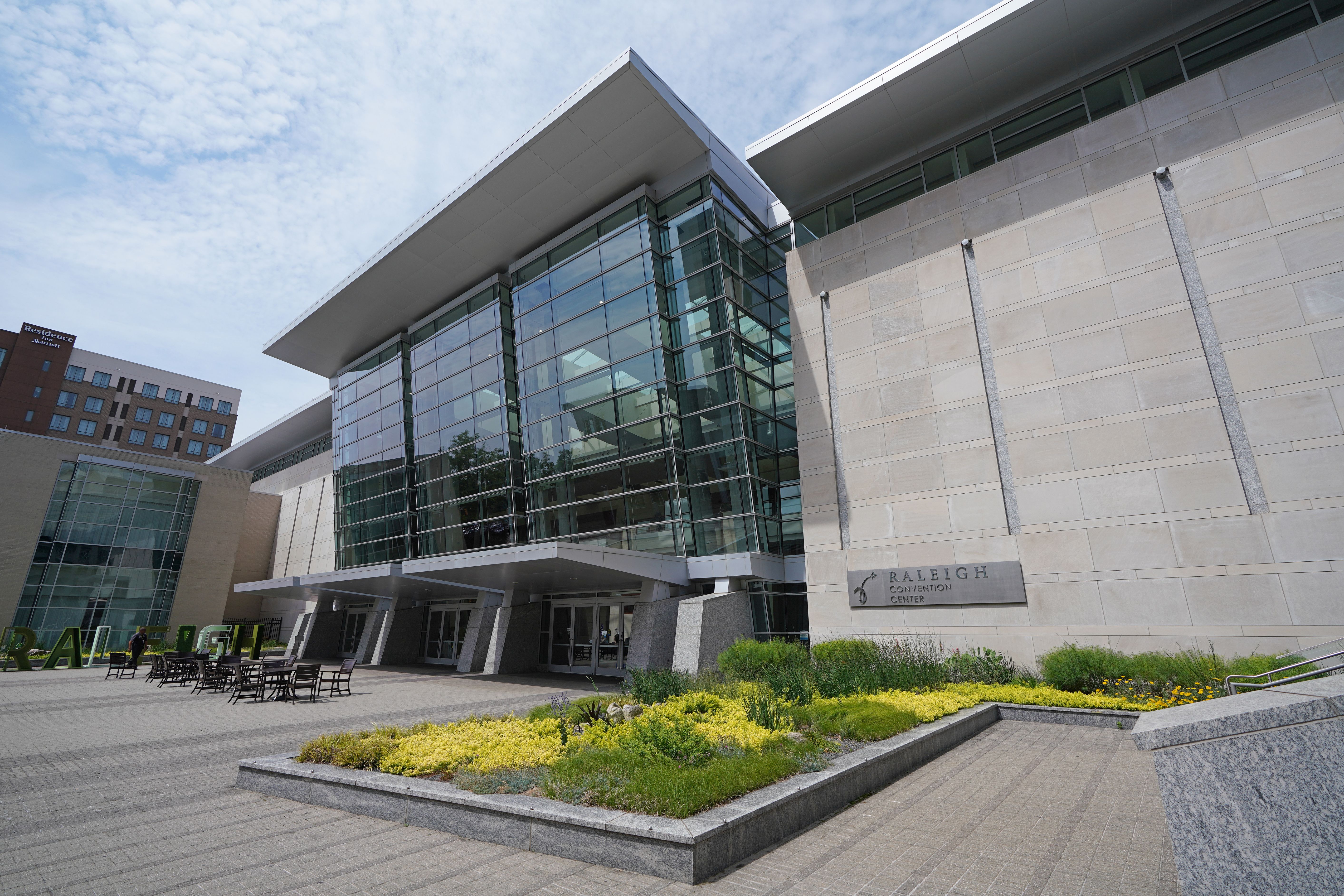
The 2024 SciX conference is taking place at the conference center in Raleigh, North Carolina © zimmytws - stock.adobe.com

Spectroscopy plays a crucial role in diagnostic and biomedical technology by providing non-invasive, real-time insights into the chemical composition and structural characteristics of biological tissues and fluids. By enhancing the sensitivity and specificity of medical assessments, spectroscopy improves the accuracy of cancer detection, monitoring of metabolic disorders, and evaluation of hydration levels, among other applications. Furthermore, its ability to analyze samples without extensive preparation makes it a valuable tool in clinical settings, where timely decision-making is essential.
To begin the session, Anita Mahadevan-Jansen from Vanderbilt University opened with her presentation on spatially offset high wavenumber Raman spectroscopy (SORS) for real-time physiological hydration assessment. She emphasized the limitations of current hydration monitoring methods, which are either invasive or provide delayed results, such as plasma osmolality and sweat-based tests. Her team developed a transfer function capable of detecting subtle hydration changes using high wavenumber Raman spectroscopy, showing its potential for non-invasive, real-time monitoring, which is crucial for preventing dehydration-related cognitive and physical impairments.
Next, Roy Goodacre, professor at the University of Liverpool, discussed his work on scaling metabolomics down to the single-cell level using optical photothermal infrared (O-PTIR) spectroscopy. Goodacre highlighted the challenge of analyzing rod-shaped bacteria (< 10 µm in size) at the cellular level, as most analytical systems require large biomass samples, limiting insights to population-level data. His team's innovative use of O-PTIR, in combination with Raman spectroscopy, allows for the identification of pathogens and exploration of phenotypic heterogeneity at the single-cell level, offering new insights into bacterial behavior and metabolism.
Laura Fabris, professor at Politecnico di Torino in Italy, followed with a presentation on using advanced colloidal chemistry and surface-enhanced Raman spectroscopy (SERS) for early cancer detection, patient stratification, and understanding viral evolution. Fabris stressed that these techniques hold promise for addressing global health challenges, potentially helping to predict and prevent future pandemics by enhancing diagnostic capabilities.
The session then shifted focus to cancer detection using spectroscopy. Ioan Notingher of the University of Nottingham presented his work on applying Raman microscopy during breast cancer surgery to improve the accuracy of surgical margin assessments and sentinel lymph node analysis. Notingher’s team combined Raman spectroscopy with auto-fluorescence (AF) imaging, enabling real-time detection of cancerous tissue during surgery. This approach reduces the need for additional surgeries by providing immediate feedback on the presence of cancerous cells in surgical margins and lymph nodes.
Peter Gardner from the University of Manchester concluded the session with a discussion on using quantum cascade laser (QCL) systems for full fingerprint hyperspectral imaging of prostate cancer tissues. Gardner addressed the challenge of long acquisition times in infrared spectroscopic imaging, which can take hours for high-resolution tissue scans. However, advancements in QCL technology now allow for rapid imaging of entire microscope slides in under 30 minutes, without compromising on data quality or frequency bands. His comparison of QCL and FT-IR spectrometers demonstrated that high-quality hyperspectral data can now be acquired quickly, making it feasible for use in clinical settings to detect aggressive cancer early and reduce overtreatment.
This session highlighted significant advancements in spectroscopy that could transform the fields of diagnostics and personalized medicine, offering more precise, real-time solutions for a range of medical applications.
The State of Forensic Science: Previewing an Upcoming AAFS Video Series
March 10th 2025Here, we provide a preview of our upcoming multi-day video series that will focus on recapping the American Academy of Forensic Sciences Conference, as well as documenting the current state of the forensic science industry.