Dominic Hare, the 2019 Emerging Leader in Atomic Spectroscopy, Applies LA-ICP-MS to the Fundamental Investigation of Neurogenerative Diseases
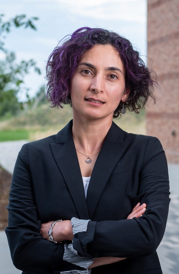
The challenge of tackling neurogenerative diseases like Parkinson’s and Alzheimer’s leads researchers to study the role of metals, for which atomic spectroscopy tools have become essential. Dominic Hare, the 2019 winner of the Emerging Leader in Atomic Spectroscopy award, presented by Spectroscopy, is a forerunner in the use of laser ablation-inductively coupled plasma-mass spectrometry (LA-ICP-MS) to image metals in biological tissue as part of the work to improve our fundamental understanding of neurodegenerative diseases. He recently spoke to us about this work. The award will be presented to Hare at The European Winter Conference on Plasma Spectrochemistry (EWCPS), in Pau, France, February 3-8, 2019.
The challenge of tackling neurogenerative diseases like Parkinson’s and Alzheimer’s leads researchers to study the role of metals, for which atomic spectroscopy tools have become essential. Dominic Hare, the 2019 winner of the Emerging Leader in Atomic Spectroscopy award, presented by Spectroscopy, is a forerunner in the use of laser ablation-inductively coupled plasma-mass spectrometry (LA-ICP-MS) to image metals in biological tissue as part of the work to improve our fundamental understanding of neurodegenerative diseases. He recently spoke to us about this work. The award will be presented to Hare at The European Winter Conference on Plasma Spectrochemistry (EWCPS), in Pau, France, February 3-8, 2019.
Atomic spectroscopy is being applied to the understanding of how redox-active metals, particularly iron, are involved in the pathology of diseases like Alzheimer’s and Parkinson’s disease. Dominic Hare, the head of the Atomic Pathology Laboratory in the Melbourne Dementia Research Centre at the Florey Institute of Neuroscience and Mental Health, in Melbourne, Australia, is one of the researchers studying the complex interactions between metal ions and the many proteins which rely on them for function. He and coworkers are exploring the direct application and understanding of metals to neurodegenerative disease research. The potential to develop chemical images relating to the atomic, functional protein, and other biomolecules in tissue provides a triad of tools that can be used to measure subtle chemical changes before, during, and after the appearance of structural changes caused by disease onset. This research may provide information on the inner workings of the cell and the fundamental basis of disease, leading to new therapies and earlier detection.
Please tell us about some of your earliest research interests. How did you get started in science? What has kept you motivated?
I started a degree in forensic science in 2002 at the University of Technology Sydney (UTS), though it wasn’t really science that took me there. In all honesty, I wanted a reason to move from a fairly boring part of Australia to the biggest and most exciting place I could think of, and my second choice was actually studying history. After my first year of undergraduate studies I picked up a summer job working at the Department of Forensic Medicine and State Coroner’s Court as the assistant to the clinical director, typing up his dictated post mortem reports. When university started again I landed a part time job in the histology laboratory and became more interested in the brain than forensic science-Australia isn’t exactly a country filled with criminal masterminds. Something about watching the neuropathologists examine and dissect human brains to understand how someone died seemed like a jigsaw puzzle, but in reverse. Sometimes it would take months to find a definitive cause of death, with pathologists poring over a microscope and ordering all kinds of tests until they had the answer they needed.
I suppose it started there. I lost a very close family member to Parkinson’s disease and seeing how difficult it could be to even diagnose a disease of the brain, even long after someone has died, made me want to change course and be part of the huge effort going into understanding these diseases while the patient is still alive. Rather than go back to square one and study medicine or biomedical sciences, my forensics degree was an applied chemistry course with a heavy analytical component and I was able to transfer what I had learned to developing new methods for medical research.
No one thing has kept me motivated all the time, and like any other career, mine has its ups and downs. Research funding is tight and there are more deserving scientists than there is money to go around. When some amazing young scientists joined my research team I gradually moved out of the lab-my clumsiness often makes me a bit of a hindrance rather than a help- and now I’m glued to a computer screen most of the day (and night). It’s actually what keeps me motivated now. There is nothing I enjoy more than designing experiments, analyzing data, and trying to work out how our results fit into the bigger, global picture that is medical research.
What have been the most difficult aspects of your research to date? How have you worked to overcome these challenges?
As a bit of a scientific outsider at the Florey, it was initially a bit of a challenge to get buy in from those who needed to understand what I do but might not know it yet. Laser ablation was originally intended for geologists, and it’s unrealistic to think neuroscientist or clinicians have any idea, let alone care, about what the technology can do for their own interests. Add to that the fact that cutting-edge analytical chemistry requires substantial investment in equipment long before you can even demonstrate its potential and you can see why my first 12 or so months at the Florey were certainly challenging. I spent a lot of time flying back and forth between Melbourne and Sydney, where my PhD supervisor (and very close friend) Prof. Philip Doble at UTS made sure I had access to all the equipment I needed.
It was worth it though. Thanks to a bunch of neuroscientists, too many to name them all here, who have traditionally been very out-of-the-box thinkers when it comes to neurodegenerative disease research, the technology I’d been working on since my PhD was starting to find a place in applied medical research. One person I have to name is Prof. Peter Crouch at the University of Melbourne, who believed mass spectrometry imaging was important enough to his work on motor neuron disease that he helped me raise the money I needed to purchase our own imaging system. Ever since, my fantastic team have gone from strength to strength, to a point now where we have more samples to analyze than there are hours in the day.
You did your doctoral research at the University of Technology Sydney from 2006 to 2009 with your thesis title: “Elemental bioâimaging: In situ analysis of trace elements in tissue by laser ablation–inductively coupled plasma–mass spectrometry,” with Prof. Philip Doble. What was the highlight of that work?
Handing in my thesis?
Seriously though, without a doubt it has been developing collaborations. Philip taught me the importance of working across disciplines and with industry to make sure a nifty new analytical method gets into the hands of those who will use it for a bigger purpose. To be honest, though, I think I only fully appreciate those lessons now that I’m nearly a decade older and supervising my own students. Philip and I still talk every few weeks, and often reminisce about my stubbornness and occasional near-insubordination during that time. Come to think of it, I can also say that the lifelong relationship I developed with Philip is just as big of a highlight.
That, and the time that first image of iron in a mouse brain I had made appeared on a computer screen in the lab in 2006. That was a pretty good day.
Of all your research papers so far, what are two of the most meaningful papers you would like to highlight for our readers?
My favorite paper has always been one of my slightly more obscure ones entitled, “Elemental bio-imaging of thorium, uranium and plutonium in tissues from occupationally exposed former nuclear workers,” in Analytical Chemistry (82, 3176-3182, 2010). I still think this paper tells the most interesting story from beginning to end. With collaborators from the United States Transuranium and Uranium Registry at Washington State University we were sent small pieces of tissue taken at autopsy from people who assembled nuclear weapons in the middle of the last century and had been involved in minor workplace accidents. Sure, they weren’t exactly Chernobyl-scale, but accidentally cutting a glove isn’t a small matter when plutonium is involved. The amazing thing was these workers survived, recovering completely and living well into old age. One case stuck out in particular, where a worker had breathed in plutonium oxide after a small fire broke out in 1965. For the rest of his life he carried particles of plutonium in the lymph nodes next to his windpipe, yet he passed away from unrelated heart failure in 2008 when he was over 90 years old!
As a card-carrying Fellow of the Royal Society of Chemistry I’m of course proud of all my papers in their fantastic transdisciplinary suite of journals, but right now I’m really pretty pleased with something one of my postdocs, Dr. Kai Kysenius, in collaboration with Peter Crouch, just published in Analytical and Bioanalytical Chemistry, in a themed topic collection on Elemental and Molecular Imaging by LA-ICP-MS, entitled, “A versatile quantitative microdroplet elemental imaging method optimized for integration in biochemical workflows for low-volume samples,” (doi:10.1007/s00216-018-1362-6, 2018). It’s a pretty typical method validation paper, but Kai is most definitely not an analytical chemist; he’s actually a neuroscientist from the University of Helsinki, here in Melbourne on a Sigrid Jusélius Fellowship. With a little advice here and there, mostly around the challenges writing for an analytical chemistry audience, Kai put together one of the most comprehensive method validation papers for quantifying elements in 1-microliter biological samples using LA-ICP-MS imaging I’ve had the pleasure of working on. It goes to show how far imaging technology has come, that someone with next-to-no experience in analytical chemistry can come into a method development lab, master what was once an incredibly complex process, and collect every shred of necessary data to show how LA-ICP-MS imaging can do something new and do it well. Every time I would call or email and suggest another experiment, he’d already have done it. Transferring not only knowledge, but a deep understanding of analytical chemistry to those with no background in the area is something I’m enormously proud of. It also helps that Kai is a pretty incredible young researcher too.
What is the most difficult or challenging aspect of your current research, group management, and mentoring positions?
Becoming a father and making the jump to leading a research team at the same time has been a challenge I’m still trying to work out two years later. There are not nearly enough hours in the day to work on everything that you find interesting or meaningful, or sometimes just plain fun, and it’s also taken some time to accept having to say no occasionally. That being said, I’m incredibly fortunate to have a team of people from a range of backgrounds, from nutrition to geosciences and everything in between who I trust and are all so eager and excited to take on new challenges. This year I was able to take on Jessica Billings, who actually provided me with all the mouse brain samples I used to develop the LA-ICP-MS imaging method I use now when I was completing my PhD and she was working on her master’s degree in neuroscience looking at iron as a possible causative factor in Parkinson’s disease. I was just using the tissue to work out how to make quantitative images, but I was fortunate to integrate this more directly with Jess’ biological findings after she completed her studies and went on to work for a company developing new Parkinson’s therapies. Ten years later, Jess is back working on the project we started together, and I couldn’t be happier.
There are always challenges, and you are only as good as the people you surround yourself with and the environment you create with them.
What is the most exciting part of your work day?
I’ve been travelling a lot recently, and unlike most researchers I’m not a huge fan of the international jet-setting life. Right now, the most exciting part of my work day is getting out of the taxi on the way back from Melbourne airport to see my family. That, and getting some sleep!
Jetlag and hotel rooms aside, what I’m really enjoying right now is spending time and talking with people working in clinical positions: pathologists, neurologists, and laboratory technicians in high-throughput labs where speed, accuracy, and precision sometimes literally mean life or death. My research is a long way from being that significant but learning how technology could make both their lives and those of their patients easier really does make the work that much more exciting.
Your research focuses on the use of LA-ICP-MS for imaging metals in biological tissue. Why did you decide to pursue this particular area of research?
I think the most fortunate thing about a PhD in analytical chemistry with Philip was the project was based around technology and he gave me freedom to seek out new people and use skills I learned from my time at the Coroner’s Court histology lab to find my own niche. He and I worked hard to develop relationships with my current colleagues at the Florey and I chose to work on Parkinson’s disease because of its known association with iron. It’s been nearly 100 years since the first paper suggesting something was amiss with iron metabolism was published, which was not only before the specific type of cells that die were identified, but also more than 30 years before dopamine, the chemical that is depleted in Parkinson’s disease, was even detected in the human brain! Also, having lost someone to this disease was a big driving factor; metals are involved in nearly half of all enzymatic reactions in the body and have a possible role in almost every single neurodegenerative disease.
In the last few years I’ve expanded my area of interest more broadly and would now say that my true interest is in pathology-all pathology. Every disease starts with a chemical reaction, and those reactions are driven by the elements of the periodic table. Our lab is working on everything from Parkinson’s disease, which will always be close to my heart and the subject of a lot of my work, to colorectal cancer (which is about as far from the brain as you can get!).
What new discoveries has your work revealed in the basic understanding of metals in neurodegenerative diseases?
LA-ICP-MS is a tool, and always will be. It will never solve these pressing challenges to human health on its own. I’ve had to complete what I consider a second PhD just to get up to speed on the complexities of neurodegenerative disease research. This wouldn’t be possible without input from experts in the neurosciences, and I have also been fortunate enough to have a decade-long mentor in Kay Double from the University of Sydney. Kay is a neuroscientist who has been working on Parkinson’s disease, mostly in either living patients or post mortem human tissue, and together we’ve been working on an evidence-based hypothesis of how reactions between iron and dopamine are a major disease-causing event in Parkinson’s disease that starts long before clinical symptoms appear. The significance of this has been twofold. Firstly, it’s given a direct biochemical mechanism that supports a current multi-site, European Union–supported clinical trial of an iron chelator in recently diagnosed patients being run by Prof. David Devos at the University of Lille in France. We’re eagerly awaiting the results of this trial and are looking to move this work further by testing whether it could actually prevent the disease if we can identify those at risk. That leads to the second point: It’s also given us a new needle in the haystack to look for that might help us single out those who are in the very early stages of the disease by measuring iron in their brains. This obviously can’t be done with LA-ICP-MS, but certain magnetic resonance imaging (MRI) techniques can. It’s a long journey still to be taken, but I’m excited about the future. For a disease that has no effective treatment beyond temporarily alleviating symptoms, new ideas are needed and I’m proud to be contributing to them.
Would you briefly describe your work of imaging metals in biological tissue? What are the unique challenges for method, instrumentation, and sample preparation when performing these analyses? What are the most surprising discoveries you have made to date?
The workflow is pretty simple now. Tissue sections are scanned by a laser and material that is ejected gets swept to an ICP-MS and detected as a function of time, allowing us to build two- and three- dimensional images depicting elemental content with pinpoint regional concentrations contained within pixels representing just a few square micrometers. It’s easily adaptable with routine methods used in medical research labs all around the world, where tissue sections are used to examine proteins and microstructure using histology and immunohistochemistry. Without the need for specialized targets or arduous preparation (in this case, less is more), any microscope slide will do.
That isn’t to say the technology and methods aren’t improving and we’re always keeping an eye on new developments. I work closely with both Agilent Technologies (Fred Fryer and Dr. David Bradley in particular) and Elemental Scientific (Drs. Ciaran O’Connor and Rob Hutchinson are also long time collaborators, and like Fred and David are now great friends too!) to access the latest developments in ICP-MS and laser ablation systems, respectively. Also, I’m always excited to read new work from groups led by people like Profs. Detlef Günther, Frank Vanhaecke, and Uwe Karst (and Philip, of course), who continue to push the technology to its absolute limits. I don’t want (and can’t) compete with the type of work they do, but it’s never hard to visualize the implications of new methods being developed in medical research by those groups and many others around the world.
The most surprising discovery is actually something I can’t quite talk about yet, but I hope to be able to sometime soon. I can say that it was so surprising we’ve had to go back and check it again and again and again, and now we’re doing it yet again with a much larger cohort of samples. Other than that, you’ll have to wait, but I hopefully not too much longer.
How were you able to direct your research toward discovery of important medical problems? What made you choose the path of medical research rather than industrial, chemical, or environmental problems?
There is a huge gap between the development of new technologies and their use in medical research, and it’s even bigger when it comes to application in a clinical setting. Part of this is the necessary validation that must be performed, and of course that takes time, but it’s also very much a result of a general disconnect between those developing methods and those who can get the most from them. For me, the opportunity to set up a method development and “blue sky” discovery lab in one of the world’s finest neuroscience research institutes has been amazing. To have those people I want to know about this technology in the same lab, on the same floor, or even in the same office (shout out to my fantastic officemate Sarah Gordon!) is something few analytical chemists get to experience. It also helps to work with other inspirational chemists who are tackling the same problem. Two of my emerging researcher colleagues Prof. Elizabeth New of the University of Sydney and Dr. Amy Heffernan, formerly at the Florey and now working in an applications specialist role for Sciex, are both incredibly talented chemists by training who have shown how applications of technology can be accelerated through collaborations and a bigger, world-view picture of how analytical chemistry fits into medical research.
Why medical research? We’re fast approaching a cliff few people want to talk about. The world’s population is living longer and longer, yet we still have no way of slowing the two biggest neurodegenerative diseases- Alzheimer’s and Parkinson’s disease. That’s where I feel I can make the biggest contribution.
How do you go about putting together a skilled and experienced research team for this specialized work?
By understanding that duplicating skills in a research team will actually slow progress. I can teach new team members how to generate images in a few days. What I would rather have is new team members with backgrounds that are diverse. That way, we’re all learning from one another, and all these skillsets contribute to everyone’s career goals. I hope that each person who spends time in my group leaves with skills in analytical chemistry they will use for the rest of their research lives.
What areas would you like to see this research expand into when looking toward the future?
Speaking of Parkinson’s disease in particular, while we may be approaching a global health crisis (really, we’re already in it), we have the tools at our disposal to make real differences, and soon. The problem is very much about awareness and, of course, funding. It’s my opinion that this disease, which affects millions around the world and numbers are only going up, is horrendously underfunded compared to Alzheimer’s disease or cancer. Years of research targeting specific disease pathways have come to naught, so now is the time that government funding agencies start investing in new ideas.
Also, technology is advancing faster than we can keep up. Modern technology, and not just LA-ICP-MS, can collect more information than we can dream of using at the moment. The answers we seek might already be there in data collected, so I’m a firm believer in the principles of open science. Who knows what other people might find in the data someone else has produced looking for something entirely different?
What is your most optimistic view of the fundamental understanding of the action of metals related to neurodegenerative diseases that you would hope to achieve over your lifetime? Do you think that the integration of immunohistochemistry and chemical imaging will lead to some of the most important new discoveries in understanding certain disease pathogenesis?
Absolutely, and they already are. Prof. Devos’ work using iron chelation is a new concept using an old drug, and if it proves to have a disease-modifying effect it will usher in a whole new approach to understanding and treating the disease. Our work with immunohistochemistry and chemical imaging has helped us refine and improve our understanding of the mechanism this class of drug targets, and if we can find a way to stop disease-causing reactions happening before cells begin to die it’s not entirely unreasonable to think that diseases like Parkinson’s can be managed, like how insulin revolutionized the treatment of diabetes.
Newsletter
Get essential updates on the latest spectroscopy technologies, regulatory standards, and best practices—subscribe today to Spectroscopy.


