Fabrication of Electrophoretic Microdevices for Fluorescence Detection: An Interview with AES Lifetime Achievement Award Winner James P. Landers
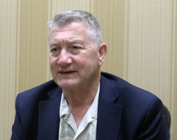
The AES Lifetime Achievement Award is given for exceptional career contributions to the fields of electrophoresis, electrokinetics, and related areas. This year’s recipient, James Landers of the University of Virginia, recently published (along with his colleagues) a paper illustrating a technique for fabricating electrophoretic microdevices for fluorescence detection.
As part of an ongoing series of interviews conducted by Spectroscopy with the winners of awards that will be presented at the annual SciX conference, which will be held this year from October 8 through October 13, in Sparks, Nevada, Landers spoke to us about this paper and his thoughts on receiving this award.
In a recent paper (1), you discuss the fabrication of electrophoretic microdevices for fluorescence detection. What are the benefits and challenges of creating such a device?
For decades, capillary electrophoresis (CE) has been a popular and sensitive methodology for polynucleic acid, protein, and small molecule analysis, and arguably the analytical workhorse for molecular biology-driven advances. Microfluidic chips offered an alternative to CE in a more compact form factor and with faster analysis times. While foundries offered generic and custom-designed electrophoretic ‘microchips’ in hard polymers (COC, PC, and so forth), these were not cost-effective for research labs. We had developed a fabrication method, termed “print, cut and laminate (PCL),” for the simple fabrication of microchips from common office materials and equipment (2). Not only is this fabrication approach cost-effective, but it also yielded microdevices that allowed for execution of many of the complex “unit operations” utilized on microchips, especially when centrifugal force was exploited for mobilizing fluids. Having shown that reagent/sample mixing, heating, cooling, and so forth, could be accomplished on centrifugal discs and, in fact, just about any reaction-based chemistry (including enzymatic), we questioned whether electrophoresis might be possible.
The benefits of creating a channel for ‘microchip’ electrophoresis were numerous, including the ease for creating short effective length of the capillary (Leff), thus, generating rapid analysis times. The challenges involved creating separation channels that were small enough, ideally, cross-sectional dimensions of 50 µm (w) x 50 µm (d).
What are some of the limitations with other analytical devices currently being used?
While CE has seen widespread adoption over decades for providing a robust and sensitive separation technology, only certain manufacturers/models provide the flexibility to change buffers, injection parameters, cleaning protocols and/or change the separation length (Leff rarely <10 cm). In contrast, microchip electrophoresis provides carte blanche with respect to buffers, injection, but allows (if not demands) short Leff in the 1 cm (or less) range – Harrison and associates showed this three decades ago (3). The microscale nature of microfluidics requires custom-built fluorescence detection systems, but control over design and Leff is vast.
Please summarize the findings of your research and describe what is most surprising or exciting about it.
The laser print, cut, and laminate (PCL) approach we defined for microfluidic device fabrication was ideal for inexpensive prototyping owing to the use of overhead transparencies (polyethylene terephthalate; PeT), printer toner as glue and an office laminator for assembly and bonding. While do Lago was the first to show the use of overhead transparencies for creating microfluidic devices (4), then using the toner layer itself to creates channels (where toner was absent), we saw immense value in creating the microfluidic architecture by cutting through the thickness of an internal layer. The ‘high tech’ approach for this was a trophy etcher (CO2 laser ablation), but simpler, more cost-effective alternatives exist in fabric cutting tools (for example., Cricut) from craft shops, but with resolution limitations.
We showed that electrophoretic microchips could, indeed, be crafted with this approach but, importantly, that the high background fluorescence of PeT could be circumvented by sheets of the favored hard polymer for microfluidics—cyclic olefin copolymer (COC)—and that this could be toner-coated for exploiting the PCL technique. Using a separation channel with an Leff of 4-6 cm: 1) centrifugal force could effectively load sieving polymer into the separation architecture less than 3 min, and 2) separation of DNA out to 400-bases could be achieved with a resolution of 3-4 bases. This provides a simple, rapid and cost-effective means for generating prototype electrophoretic architecture for application-specific testing.
Were there any limitations or challenges you encountered in your work?
First, scientists might be concerned, and rightfully so, that having the ‘gunk’ that is commercial printer toner as part of the microfluidic architecture used for an analytical process, particularly, when executing bioassays. It turns out that, for bioprocesses are prone to inhibition (enzymatic), the multiple (mostly proprietary) components that make up toner, have not been problematic for the most part. In our experience, this includes the polymerase chain reaction (PCR), where ‘inhibitors’ can adversely affect amplification efficiency. For electrophoretic separations, we have not seen any evidence that toner indirectly affects performance. Second, the method used for ‘cutting’ channels into the architecture of the middle layer(s) (laser ablation, in this case) determines the channel width, and this is limited by the laser cutter optics. With this approach, we can reproducibly ablate channels that are 100 µm x 100 µm, but for high resolution separations aiming for single base resolution, this is inadequate.
Ten authors from three distinct departments of the University of Virginia are credited on this paper, and you yourself are a member of all three departments. What skills and experiences from each department proved to be beneficial in this work?
Microfluidics, perhaps more so that the development of any other analytical technique, is a multidisciplinary field. It brings together the need for understanding aspects of organic chemistry, clinical chemistry, biochemistry, polymer chemistry, materials science and engineering, electrical engineering, physics, and mechatronics. For two decades, our work (and that of many others) have exploited the expertise in other departments as a means of expediting developments and, importantly, avoiding “reinventing the wheel.”
Can you please summarize the feedback that you have received from others regarding your efforts?
The positive feedback we receive on the developments from my group are of two threads and, without question, are the result of bright graduate students provided with an environment that allows them create and innovate. The first often refers to the simplicity of what we develop. The “keep it simple” principle has always served us well, from the development of infrared (slide projector bulb) PCR through to the use of craft-store gold leaf paper for electrodes. The second, is the highly integrated nature of the devices we create. Going back as far as my post-doc at the Mayo Clinic, it was clear that clinical assays where not monolithic, but an orchestrated series of processes where sample preparation needed to be seamlessly integrated with the analytical step. If microfluidics were to advance assay developments, integration was essential.
What are the next steps in this research?
The next steps are to fuse the ability to carry out electrophoretic separations on a centrifugal microdevice with highly integrated sample preparation. We have done this (and have a Science Advances paper in preparation) for DNA analysis applied to human identification. The electrophoresis, the process that provides the critical readout of genotype, is facilitated by sample prep from buccal swab on CD-size microfluidic disc that trades off all fluidic pumping hardware for the simplicity of centrifugal force.
What does your being named the recipient of the AES Lifetime Award mean to you professionally? Personally?
From both perspectives, it is clearly flattering. First, to join the cadre previous recipients who have all be pioneers in their own right is humbling.With a massive amount of brilliant innovation appearing in the literature at an ever-increasing rate from my colleagues in the field (which includes those speaking in this session), to be selected from a landscape of extremely innovative peers, by my peers, leaves not only flattered, but grateful.
What advice can you offer those starting out in their post-doctorate career or even those younger scientists looking for guidance?
As odd as it might sound, the key to success in the world of scientific and engineering innovation is failure. The scientific method is defined as “a method of procedure that has characterized natural science since the 17th century, consisting in systematic observation, measurement, and experiment, and the formulation, testing, and modification of hypotheses.” We modify hypotheses because we test and, often, fail. Hence, failure is the intangible variable in the equation that is the scientific method, and it is important—no, essential— that young scientists understand that it’s not only OK to fail, but that it’s a foundational tenet.
References
(1) Nelson, D. A.; Thompson, B. L.; Scott, A.-C.; Nouwairi, R.; Birch, C.; DuVall, J. A.; Le Roux, D.; Jingyi Li, J.; Root, B. E.; Landers, J. P. Rapid, Inexpensive Fabrication of Electrophoretic Microdevices for Fluorescence Detection. Electrophoresis 2022, 1–9. DOI:
(2) Thompson, B., Ouyang, Y., Duarte, G. et al. Inexpensive, Rapid Prototyping of Microfluidic Devices Using Overhead Transparencies and a Laser Print, Cut and Laminate Fabrication Method. Nat. Protoc. 2015, 10, 875–886. DOI:
(3) Harrison, D. J.; Fluri, K.; Seiler, K.; Fan, Z.; Effenhauser, C. S.; Manz, A. Micromachining a Miniaturized Capillary Electrophoresis-Based Chemical Analysis System on a Chip. Science 1993,261 (5123), 895. DOI:
(4) do Lago, C. L.; Neves, C. A.; D. Pereira de Jesus; da Silva, H. D. T.; Brito-Neto, J. G. A.; Fracassi da Silva, J. A. Microfluidic devices obtained by thermal toner transferring on glass substrate. Electrophoresis 2004, 25, 3825–3831 DOI:
About the Interviewee
Newsletter
Get essential updates on the latest spectroscopy technologies, regulatory standards, and best practices—subscribe today to Spectroscopy.


