Inside the Laboratory: The Calhoun Laboratory at the University of Tennessee
Inside the Laboratory is a joint series with LCGC and Spectroscopy, profiling analytical scientists and their research groups at universities all over the world. This series will spotlight the current chromatographic and spectroscopic research their group is conducting, and the importance of their research in analytical chemistry and specific industries. In this edition of “Inside the Laboratory,” Tessa Calhoun, PhD, an associate professor of biochemistry & cellular and molecular biology and chemistry at the University of Tennessee – Knoxville, discusses her group’s most recent work employing the optical technique, second harmonic scattering (SHS), to probe how living bacterial membranes uptake and transport small molecules, including antibiotics.
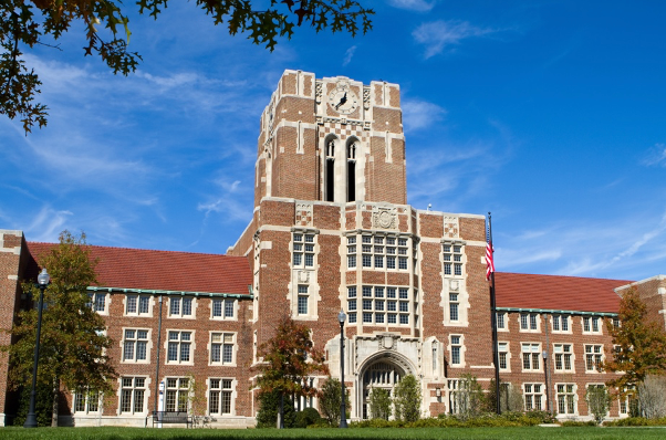
The Calhoun Laboratory is an interdisciplinary research group with appointments in the University of Tennessee’s Departments of Biochemistry & Cellular and Molecular Biology and Chemistry, in Knoxville, Tennessee. The Calhoun Laboratory specializes in the application and advancement of nonlinear spectroscopy and microscopy techniques for the study of a variety of systems. The main objective of the Calhoun Laboratory is to understand the impact of environmental effects on dynamics at complex interfaces.


University of Tennessee | Image Credit: © sframe – stock.adobe.com
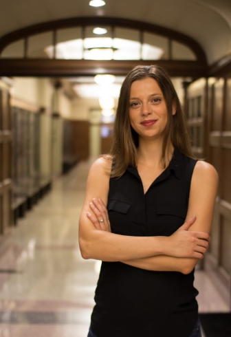
Tessa Calhoun, PhD, is the principal investigator of the Calhoun Laboratory. She received her BS in Chemistry from Iowa State University, and her PhD in Chemistry from the University of California, Berkeley. She completed her postdoc at Princeton University in their Lewis-Sigler Institute for Integrative Genomics before establishing her research group at the University of Tennessee.
We sat down with Calhoun to discuss her group’s most recent work connecting trends in small molecule structure with their transport dynamics in living bacterial membranes using a surface-specific optical approach.


Tessa Calhoun, PhD, Associate Professor in the Departments of Biochemistry & Cellular and Molecular Biology and Chemistry at the University of Tennessee – Knoxville. Photo Credit: © Tessa Calhoun
Can you talk about the spectroscopic techniques that your group used in your most recent research project?
We primarily used the nonlinear optical technique, second harmonic scattering (SHS). In SHS, two photons are combined to generate a single photon with twice the energy. This process has a few important properties that make it an ideal tool to study biological membranes. First, due to the symmetry requirements for the technique, signal will only come from the membrane in our samples, reducing significant background from the bulk solution. Given the complex media that living cells prefer, this is especially beneficial. Second, signal from molecules on one side of the membrane will cancel signal arising from molecules on the other side of the membrane. This allows us to directly track a population of small molecules as they move across the membrane. As the membrane is only 3–5 nm thick, there are few ways to directly monitor how molecules move across this space. Finally, the direction the signal scatters is dependent on the size of the particles, or in our case, cells. This provides another way to isolate our signal in complex, living samples.
Can you explain the importance of your research within the broader field of spectroscopy or in a specific industry/application?
To my knowledge, we are currently only one of three groups in the world applying this technique to bacterial cells. By embracing the complexity of a living sample, we aim to better understand the primary factors that impact how bacteria, and specifically their membranes, interact with small molecules. This can have a wide range of applications, from designing or improving antibiotics to optimizing biofuel extraction.
How do you stay updated with advancements in spectroscopy techniques and technologies? Can you discuss a recent innovation or development that you find particularly impactful or exciting?
Staying updated with advancements really requires time spent with recent literature and participation in conference opportunities. Overall, it is a hard challenge as it feels that there is never enough time to keep up with the seemingly furious pace of new work.
When probing with light, work on living cells is predominately focused on imaging techniques due to the system heterogeneity. The diffraction limit of light becomes a severe barrier when interrogating bacterial cells due to their already small size. As such, I’m always interested in new innovations that find clever ways to circumvent or avoid this limitation (like SHS!). I’m even more drawn to interdisciplinary and/or collaborative approaches that cross fields. At this point, I’m most excited about the pile of papers I’ve set aside to read about new mathematical and experimental approaches to push capabilities for super-resolution dynamic information.


 delivering his Wallace H. Coulter keynote lecture at Pittcon in Boston, Massachusetts, on March 2nd, 2025. Photo Credit: © LCGC International.")
Pittcon 2025: Keynote Coulter Lecture Highlights Work in Regenerative Engineering
March 3rd 2025Yesterday, at 5:00 pm in Ballroom East, the Wallace H. Coulter Lecture took place, and it was delivered by Cato T. Laurencin, MD, PhD, who is well-known as a scientist and entrepreneur with an extensive career in regenerative engineering. His lecture highlighted the work he and his team has done in this space.
 is an Assistant Professor at Sam Houston State University. Christopher Zall (center) is an Associate Professor at Sam Houston State University. Jared Estevanes is a Forensic Scientist at Microtrace LLC. Photo Credits: Geraldine Monjardez, Christopher Zall, and Jared Estevanes.")
Investigating ANFO Lattice Vibrations After Detonation with Raman and XRD
February 28th 2025Spectroscopy recently sat down with Dr. Geraldine Monjardez and two of her coauthors, Dr. Christopher Zall and Dr. Jared Estevanes, to discuss their most recent study, which examined the crystal structure of ammonium nitrate (AN) following exposure to explosive events.

Advancing Zebrafish Research: FT-IR Imaging Sheds Light on Tissue Preservation in Zebrafish
February 5th 2025Researchers at the University of Lublin and the Medical University of Lublin have demonstrated the first application of FT-IR imaging in zebrafish larvae, revealing that frozen samples better preserve tissue structure than chemical fixation.

Distinguishing Horsetails Using NIR and Predictive Modeling
February 3rd 2025Spectroscopy sat down with Knut Baumann of the University of Technology Braunschweig to discuss his latest research examining the classification of two closely related horsetail species, Equisetum arvense (field horsetail) and Equisetum palustre (marsh horsetail), using near-infrared spectroscopy (NIR).

