Laser-Induced Plasmas and Atomic Spectroscopy
Laser-induced plasmas are formed by the application of a laser pulse to a target surface, which instantly excites, ionizes, and vaporizes the material into a very hot vapor plume. One of the main uses of these plasmas is in laser-induced breakdown spectroscopy, a rapidly evolving and exciting field of study. Alessandro De Giacomo is a professor in the Department of Chemistry at the University of Bari in Italy and an associated researcher at CNR-NANOTEC, and he and his group are involved with the study of laser-induced plasmas and the use of nanoparticles (NPs) in laser-induced breakdown spectroscopy to enhance signal. We recently spoke with him about this research.

Laser-induced plasmas are formed by the application of a laser pulse to a target surface, which instantly excites, ionizes, and vaporizes the material into a very hot vapor plume. One of the main uses of these plasmas is in laser-induced breakdown spectroscopy, a rapidly evolving and exciting field of study. Alessandro De Giacomo is a professor in the Department of Chemistry at the University of Bari in Italy and an associated researcher at CNR-NANOTEC, and he and his group are involved with the study of laser-induced plasmas and the use of nanoparticles (NPs) in laser-induced breakdown spectroscopy to enhance signal. We recently spoke with him about this research.
A review article on laser-induced plasma emission you coauthored with Jörg Hermann of Aix-Marseille LP3 CNRS presented a discussion of the temporal evolution of laser-induced plasmas that describes species changes from ions to atoms to molecules (1). The main spectroscopic techniques associated with laser-induced plasmas are elemental analysis approaches such as laser-induced breakdown spectroscopy (LIBS). What types of molecular analysis can be done using laser-induced plasmas?
Indeed laser-induced plasmas are used for elemental analysis in analytical chemistry, and also the detection of molecular bands is used to support the determination of elemental composition. As a matter of fact, the molecules detected in a laser-induced plasma are those formed with the recombination of atoms in the plasma phase and between the plasma and the background environment, so they are not linked to the original chemistry of the sample. However, sometimes, detecting the molecular band can be easier than detecting the element, like in the case of halogen species, or the isotopic detection proposed by the Russo group, namely laser-ablation molecular isotopic spectrometry (LAMIS), where molecular bands are used because the shift of the molecular band of an isotopic biatomic molecule is greater than the line shift of the individual isotopic atom. Molecular bands also have been used as fingerprints of organic compounds, plastics, and explosives. But ultimately, as you said, laser-induced plasmas are associated with elemental analysis.
How has the development of femtosecond laser plasma techniques affected the field of study? What are the primary differences between the use of nanosecond lasers and femtosecond lasers?
Femtosecond lasers became a big fashion in the LIBS community some years ago, mainly because they enable a completely different and “cleaner” ablation process compared to nanosecond laser pulses. Like in the case of architecture and clothing, fashion does not always coincide with practical use, however, and nanosecond lasers allow a better LIBS analysis, in terms of sensitivity and equilibrium stability of the plasma for emission spectroscopy. Although only a little portion of the laser pulse really interacts with the sample, the remaining part of the pulse is what gives energy to the plasma, heating the electrons and further ionizing the ablated material. In this view I would be more interested in long-pulse LIBS than ultrashort-pulse LIBS. Femtosecond-laser pulses have been crucial for the comprehension of laser–matter interaction and are extremely useful for material processing and for some specific kinds of LIBS experiments where very high irradiance is required, such as filament LIBS, or in other techniques where further excitation is given by supplementary sources (double-pulse LIBS or laser-ablation inductively coupled plasma–mass spectrometry [LA-ICP-MS]), but, in general, I would not suggest using a femtosecond laser pulse for LIBS. For example, if you want to transport a lot of people would you prefer to have a fast Ferrari for doing several trips or a bus, which is slower but roomier? Considering the costs and effectiveness I would have no doubt.
What challenges remain to be investigated in the field of laser-induced plasma research?
Well, talking in general about laser-induced plasmas, there are plenty of things to understand and exploit. Generally, the laser-induced plasma approach is a method for interacting with matter by separating the matter itself into ions and atoms and in this context there are unlimited applications to be explored. Also, from a fundamental point of view, beyond the state of the art and the accepted vision of the plasma, there are many concepts that should be investigated further and with an open mind. It may seem a paradox, but the growing expectations in real-world applications, somehow, are quenching the interest in looking at new questions or for approaches that are different from the commonly accepted ones. Concerning laser-induced plasmas for LIBS, I think that every time we find a limit we find a new challenge, and most of the efforts are currently devoted to improving the analytical performance of the technique. Researchers are doing good work to improve the spatial resolution of micro-LIBS, to apply lasers with energy levels as low as possible for LIBS, and to improve the accuracy of calibration-free analysis. The extension of LIBS to analyzing fresh biological tissue may be an important issue for exploiting LIBS for rapid screening during surgery or for fast diagnosis in specific cases.
What I personally think is that the increased use of LIBS for laboratory analysis requires a significant improvement in the limit of detection of the technique to justify its use in place of conventional analytical techniques. Much has been done to optimize the signal detection, but there is still a lot to do to improve the ablation efficiency-when I refer to ablation efficiency I don't mean the amount of ejected material from the sample, but the amount of the ejected material that contributes to the analytical signal, which is a completely different issue. This consideration pushed us toward investigating the use of nanotechnology for laser ablation–based analytical techniques.
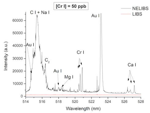
Your group has applied nanoparticle-enhanced laser-induced breakdown spectroscopy (NELIBS) to the analysis of sub-part-per-million analyte concentrations in microdrops of solutions (2). How does the use of noble metal nanoparticles (NPs) enhance the LIBS signal? What are the main advantages of this approach?
NELIBS is an amazing story. In 2013 we were producing NPs for a national contract by pulsed laser ablation in liquid, and we developed a fast way to estimate NP size and concentration by LIBS. During these experiments we noted that the emission signal of the substrate, where NPs were collected, was hugely enhanced. Initially it looked like magic, because nobody had reported this effect with LIBS and the most pertinent studies were done with very different experimental conditions. After 5 years I began to have a clear understanding of what happens and it seems like magic. In metallic NPs, such as Au, Ag, Cu/CuO, Pt, and so forth, upon laser irradiation, surface electrons oscillate collectively and induce a dipole on the particle. This electron density displacement from the equilibrium position in the lattice, namely the plasmon, allows the modulation of the incident laser pulse energy distribution on the sample in different ways, depending on the interparticle distances and excitation wavelength of the incident laser. When a high-energy laser pulse is used, the coupling with the local field induced on the zone around the NPs results in an enhancement of the incident electromagnetic field of orders of magnitude and consequently in an extremely efficient ionization and excitation of the ablated sample.
In the case of liquid solution drops deposited on a nanoparticle layer, the local field enhancement caused by the coupling of the laser pulse with the NP system and the strong affinity of solutes for the NP surface induces, during ablation, a complete transfer of the analyte to the active plasma phase, which allows the detection of sub-picogram elemental compositions by single-shot LIBS.
The amplification of the electromagnetic field of the incident laser pulse by NP plasmon activity distributes several peaks of energy on the sample surface and in turn strongly improves the ablation efficiency, in the sense I mentioned above, and so enhances the emission signal by one to two orders of magnitude. Clearly, specific approaches are required for different types of samples, but year after year, operating with NPs becomes easier and easier. We have learned a great deal about this technique, which some years ago, as I noted earlier, was obscure and just looked like magic.


Figure 1: NELIBS experiment on microdrop solution.
In a review article you coauthored about NELIBS (3), sample preparation is mentioned as an important factor in the technique. Can you please describe the basic process of preparing a target surface for NELIBS analysis? What are the important considerations when choosing the correct nanoparticles to use in a particular analysis?
Sample preparation is the main factor for the success of a NELIBS experiment. The first good practice to keep in mind is to use pure metallic NPs to avoid interference from contaminants with the analyte signal. This is why the use of commercial NPs, generally obtained by chemical reduction of metal salts, should be approached with caution, because they can contain a lot of chemical species besides the NP element. NPs produced by pulsed laser ablation in liquid work very well-they can be produced with very high purity-so they are my first choice. After you have found the right NPs you have to deposit them on the sample. In a simple case, depositing a few microliters of a colloidal solution containing the NPs is sufficient for observing the signal enhancement. However, to obtain good reproducibility the NPs should be deposited homogeneously with an optimal distance between them.
Unfortunately, working with NPs is a very delicate process. It requires some expertise, because of their high surface reactivity and high susceptibility to every kind of perturbation. For this reason we are working, in collaboration with other groups in the field of material science, to prepare standard solutions of NPs with various organic molecules that can act as spacers between the NPs when they are dried on the sample. The use of organic molecules, such as polymers, stabilizers, and proteins, indeed improves the sample preparation operation, but it gives lower enhancement, because these molecules affect the permittivity of the NP system and introduce high ionization energy species in the plasma. We are working to find an optimal compromise to accelerate the implementation of NELIBS in those laboratories in which operators do not have great expertise with nanotechnologies.

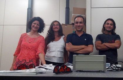
Figure 2: The group that started NELIBS in 2013 (from left to right): Dr. Rosalba Gaudiuso, post-doc (now at University of Massachusetts Lowell); Dr. Marcella Dell’Aglio senior researcher at CNR-NANOTEC; Prof. Alessandro De Giacomo, head of the laser-plasma lab at University of Bari and CNR-NANOTEC; and Dr. Can Koral (then a PhD student, now postdoc at Federico II University of Naples).
What are the next steps in your research with LIBS and laser-induced plasmas?
I have a long list of things that I would like to investigate, like the early stages of laser-induced plasmas, the formation and vaporization of particles in the plasma, the distribution of energy and the consequent evolution in the plasma, the use of specific properties of some NPs still unexplored in laser ablation, the use of laser-induced plasmas in medicine, and so on. Clearly the list of things to do is longer than the list of things I can do in one life, and it continues to grow. Every time I understand something that I learn and observe in the lab, I always feel that there are many more things to understand and to learn compared to what I do catch. This awareness is frustrating but also very inspiring. I used to tell my coworkers that I am a professor just because I am too old to be a student, but learning is still the main task for me and-believe me-it is not just an excuse for avoiding paperwork!
References
1. A. De Giacomo and J. Hermann, J. Phys. D.: Appl. Phys.50, 183002 (2017). doi: https://doi.org/10.1088/1361-6463/aa6585
2. A. De Giacomo, C. Koral, G. Valenza, R. Gaudiuso, and M. Dell’Aglio, Anal. Chem.88, 5251–5257 (2016). doi: 10.1021/acs.analchem.6b00324
3. M. Dell’Aglio, R. Alrifai, and A. De Giacomo, Spectrochimica Acta Part B148, 105–112 (2018). doi: https://doi.org/10.1016/j.sab.2018.06.008
Investigating ANFO Lattice Vibrations After Detonation with Raman and XRD
February 28th 2025Spectroscopy recently sat down with Dr. Geraldine Monjardez and two of her coauthors, Dr. Christopher Zall and Dr. Jared Estevanes, to discuss their most recent study, which examined the crystal structure of ammonium nitrate (AN) following exposure to explosive events.
Distinguishing Horsetails Using NIR and Predictive Modeling
February 3rd 2025Spectroscopy sat down with Knut Baumann of the University of Technology Braunschweig to discuss his latest research examining the classification of two closely related horsetail species, Equisetum arvense (field horsetail) and Equisetum palustre (marsh horsetail), using near-infrared spectroscopy (NIR).
An Inside Look at the Fundamentals and Principles of Two-Dimensional Correlation Spectroscopy
January 17th 2025Spectroscopy recently sat down with Isao Noda of the University of Delaware and Young Mee Jung of Kangwon National University to talk about the principles of two-dimensional correlation spectroscopy (2D-COS) and its key applications.
Measuring Microplastics in Remote and Pristine Environments
December 12th 2024Aleksandra "Sasha" Karapetrova and Win Cowger discuss their research using µ-FTIR spectroscopy and Open Specy software to investigate microplastic deposits in remote snow areas, shedding light on the long-range transport of microplastics.
The Fundamental Role of Advanced Hyphenated Techniques in Lithium-Ion Battery Research
December 4th 2024Spectroscopy spoke with Uwe Karst, a full professor at the University of Münster in the Institute of Inorganic and Analytical Chemistry, to discuss his research on hyphenated analytical techniques in battery research.