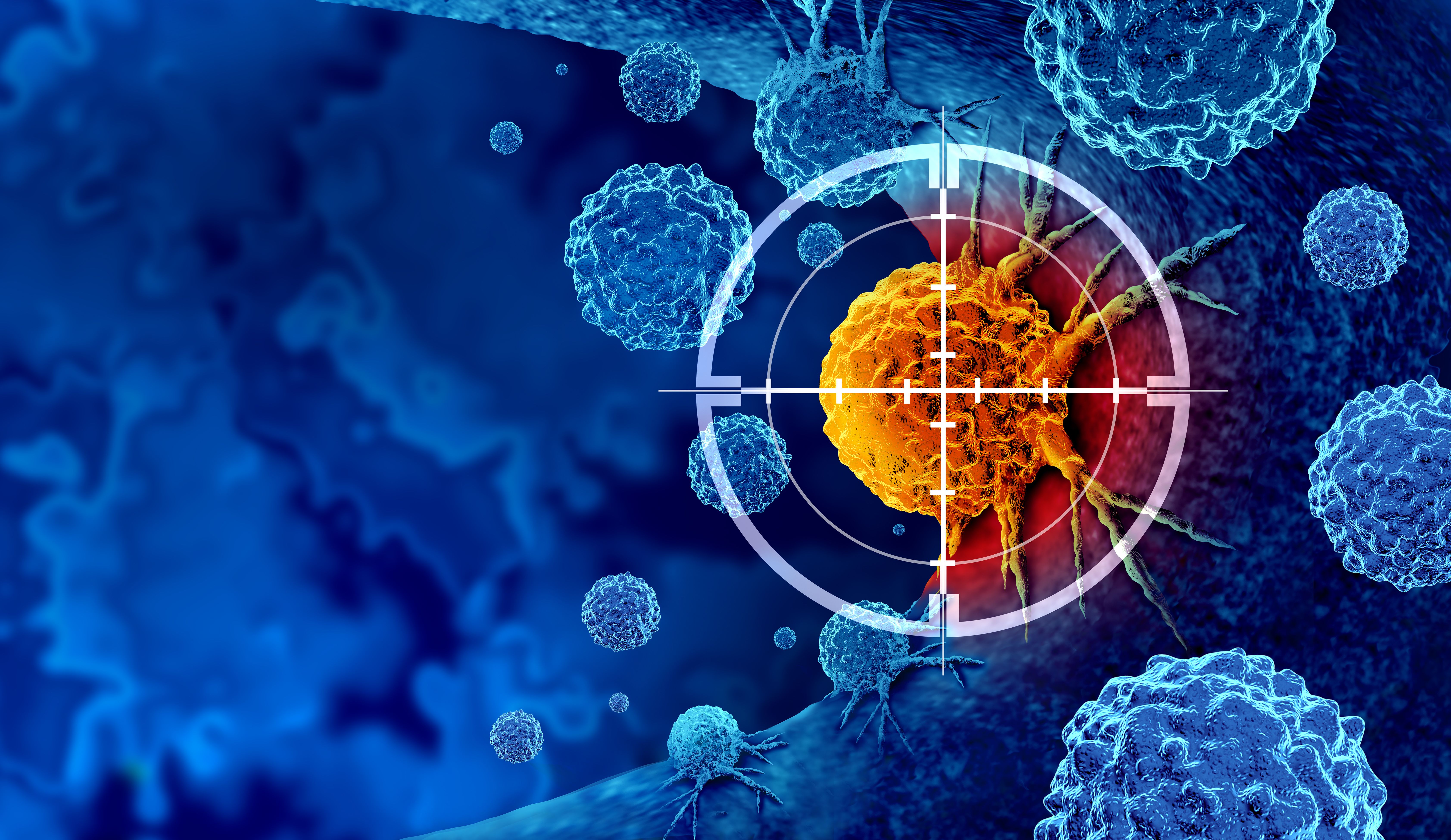
Machine Learning and Synchrotron Imaging: Advancing Early Cancer Diagnostics
Machine learning and synchrotron radiation-based micro X-ray fluorescence imaging show promise for early cancer diagnostics by identifying trace biometals as potential cancer biomarkers. The research demonstrates the feasibility of using machine learning algorithms to analyze the spatial distribution of biometals and classify cancer pathogenesis stages, offering potential advancements in non-invasive cancer detection.
Trace biometals in biological samples have shown potential as cancer biomarkers, but their spatial analysis for early cancer diagnostics remains challenging. In a recent study lead by J. J. Okonda, and conducted at the Department of Physics at the University of Nairobi, researchers have explored the feasibility of utilizing machine learning and synchrotron radiation-based micro X-ray fluorescence imaging to detect trace biometals and identify their spatial distribution as cancer biomarkers. This research was published in the journal Spectrochimica Acta Part B: Atomic Spectroscopy (1).


Cancer detection and screening as a treatment for malignant cells with a biopsy or testing caused by carcinogens and genetics with a cancerous cell as an immunotherapy symbol | Image Credit: © freshidea - stock.adobe.com
The team employed principal component analysis (PCA)-enabled artificial neural networks (ANN) to simultaneously determine the spatial profiles of biometals, including manganese (Mn), iron (Fe), copper (Cu), and selenium (Se), in model human cell cultures (DU145 and Vero). By culturing the cell lines on silicon nitride membranes and performing micro X-ray fluorescence (micro XRF) analysis at the TwinMic beamline, Elletra synchrotron source, the researchers obtained high-resolution 2D maps of the trace biometals.
To analyze the complex multivariate relationships between the trace biometals spatial distribution and cancer pathogenesis stages, PCA was employed to reduce the data dimensions. The ANN model, trained using the pixel spectral profiles of the biometals, successfully classified the cell cultures into cancerous and healthy groups. Moreover, the selected spectral profiles enabled the ANN to classify cancerous cells into early, intermediate, and advanced stages based on the fluorescence lines of Fe and Cu, as well as Compton scatter.
The study demonstrated promising results, with high spatial correlations observed between Cu and Fe in cancerous cell cultures compared to normal ones. These findings pave the way for the early diagnosis of cancer by utilizing the alterations in the spatial distribution and multivariate properties of trace biometals as cancer biomarkers.
The integration of machine learning and synchrotron radiation-based micro X-ray fluorescence imaging offers a powerful tool for early cancer detection, providing valuable insights into the intricate relationships between trace biometals and cancer pathogenesis stages. This research contributes to the development of non-invasive and precise diagnostic techniques, bringing us closer to improved cancer management and patient outcomes.
Reference
(1) Okonda, J. J.; Angeyo, H. K.; Dehayem-Kamadjeu, A.; Rogena, A. E. Feasibility for early cancer diagnostics by machine learning enabled synchrotron radiation based micro X-ray fluorescence imaging of trace biometals as cancer biomarkers. Spectrochimica Acta Part B: At. Spectrosc. 2023, 204, 106671. DOI:10.1016/j.sab.2023.106671


 is an Assistant Professor at Sam Houston State University. Christopher Zall (center) is an Associate Professor at Sam Houston State University. Jared Estevanes is a Forensic Scientist at Microtrace LLC. Photo Credits: Geraldine Monjardez, Christopher Zall, and Jared Estevanes.")
Investigating ANFO Lattice Vibrations After Detonation with Raman and XRD
February 28th 2025Spectroscopy recently sat down with Dr. Geraldine Monjardez and two of her coauthors, Dr. Christopher Zall and Dr. Jared Estevanes, to discuss their most recent study, which examined the crystal structure of ammonium nitrate (AN) following exposure to explosive events.

Applications of Micro X-Ray Fluorescence Spectroscopy in Food and Agricultural Products
January 25th 2025In recent years, advances in X-ray optics and detectors have enabled the commercialization of laboratory μXRF spectrometers with spot sizes of ~3 to 30 μm that are suitable for routine imaging of element localization, which was previously only available with scanning electron microscopy (SEM-EDS). This new technique opens a variety of new μXRF applications in the food and agricultural sciences, which have the potential to provide researchers with valuable data that can enhance food safety, improve product consistency, and refine our understanding of the mechanisms of elemental uptake and homeostasis in agricultural crops. This month’s column takes a more detailed look at some of those application areas.

