Organic Nitrogen Compounds, Part I: Introduction
Spectroscopy
So far, we have restricted our discussion to organic functional groups that contain carbon, hydrogen, and oxygen, with past columns addressing the theory of infrared spectral interpretation of C-H bonds, C-O bonds, and the C=O functional group. We now turn our attention to interpretation involving organic nitrogen compounds.
What a long, interesting, and hopefully informative for my readers trip it has been. By my reckoning, this is the 23rd installment of this column, and it is about to go into its fifth year. There has, however, been a method to my madness. We have covered, in order, the theory of infrared spectral interpretation, C-H bonds, C-O bonds, and, in the last column, we finished up our study of the infrared spectroscopy of the carbonyl, or C=O, functional group. So far, we have restricted our discussion to organic functional groups that contain carbon, hydrogen, and oxygen. Traditionally, the chemical element nitrogen is considered part of organic chemistry. In this column, and the several following it, we will turn our attention, then, to organic nitrogen compounds.
The nitrogen atom has an atomic number of 7 and five outer shell electrons. The drive of chemical elements, of course, is to get their outer shells filled. In most organic compounds, nitrogen tries to fill its outer shell by forming three chemical bonds to other atoms, be they hydrogens, carbons, or whatever. Carbon and nitrogen can form single, double, or triple bonds, as seen in Figure 1.

Figure 1: Examples of nitrogen-carbon bonds. From left to right: a single bond, double bond, and triple bond.
Note that, for the C-N bond seen at left in Figure 1, the bond angles are approximately 120 °, and that there are three atoms bonded to the nitrogen. For the C=N bond seen in the middle of Figure 1, the bond angles are approximately 90 ° (1), and there are two atoms attached to the nitrogen. Lastly, for the C≡N bond seen at right in Figure 1, the bond angles are approximately 180 ° and there is one atom, the carbon, attached to the nitrogen.
Nitrogen has an electronegativity of 3, which means it tends to hog the electrons in any bonds it forms. Carbon and hydrogen have electronegativities of 2.5 and 2.1, respectively (1), so carbon-nitrogen and N-H bonds are decidedly covalent. A question we would like to answer is whether there is a way of using infrared spectroscopy to determine the presence of an organic nitrogen functional group in a sample.
You might think that, since C-N bonds are common, they might serve our purpose. However, this is not the case as illustrated in Figure 2.
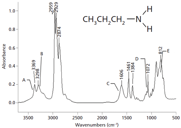
Figure 2: The infrared spectrum of propylamine. Note the weak C-N stretching peak at 1072 cm-1 labeled D, and the somewhat broadened N-H stretches at 3369 cm-1 and 3298 cm-1, labeled A and B.
Recall (2) that bond dipole moments are calculated using equation 1.

where µ = dipole moment, q = charge, and r = bond length.
The dipole moment is a measure of charge distribution asymmetry. Chemical bonds with large electronegativity differences between the atoms will have large positive and negative charges at either end of the bond, giving a large dipole. We have already seen this with carbonyl groups, for example (3). Thus, ionic and polar bonds have large dipole moments. If the electronegativity difference between the two atoms in a chemical bond is small, or zero, there will be a small charge at either end of the bond, and the dipole moment will be small. For example, C-H and C-C bonds typically have small dipole moments (1).
We have seen (2) that one thing that determines peak intensity in infrared spectra is the change in dipole moment with respect to bond length during a vibration. This is summarized in equation 2.

where A = absorbance, dµ = change in dipole moment during a vibration, and dx = change in bond length during a vibration.
Vibrations where dµ/dx is large, such as C=O stretches, will have big peaks (3). Vibrations where dµ/dx is small will have small peaks, such as C-C stretches. One of the reasons we have not discussed C-C stretching peaks much in these columns is that they are weak and hard to see.
The electronegativity difference between carbon and nitrogen is 0.5, which means these bonds are non-polar, and have a small dipole moment. Stretching a normal C-N bond, then, means there is a small value of dµ/dx, and the resultant peak is medium to weak in intensity and not always easy to see. Additionally, C-N stretches fall between 1400 and 1000 cm-1, right in the middle of the fingerprint region, the busiest area in most spectra. Thus, your typical C-N stretch is a small, non-distinct peak in a busy area, making it difficult to see. An example is the C-N stretch of propylamine seen at 1072 cm-1 in Figure 2 and labeled D. This is the tenth most intense peak in the spectrum, and the only reason we can see it here is that propylamine is a small molecule with a relatively simple fingerprint region. Such a small, nondescript, difficult to find peak is not an example of a good group wavenumber, and hence cannot be used to determine the presence of nitrogen in an organic molecule.
So, if the C-N bond is not useful, what about the C=N bond? Although these bonds exist, they are unstable (1), and thus not useful for our purposes. What about the carbon nitrogen triple bond, C≡N? These bonds are stable (1), and have a wonderful C≡N stretching vibration that is strong, narrow, intense, and shows up at a unique wavenumber (4). However, C≡N bonds are not that common, and hence are not a useful indicator for the presence of nitrogen in a sample.
So, we have been stymied so far in our search for an infrared feature that will disclose the presence of a nitrogen atom in an organic compound. As it turns out, the best infrared signature for nitrogen are the peaks from nitrogen-hydrogen bonds stretching vibrations. Two of these are seen in Figure 2 at 3369 and 3298 cm-1, labeled A and B. Note that these peaks are broadened at their base; this is due to hydrogen bonding (more below), but they come to a relatively sharp point, and that they are of medium intensity. In general, N-H stretching peaks fall from 3500 to 3100 cm-1. Recall that the O-H stretching peaks of functional groups like alcohols and carboxylic acids also fall in this range (5,6), and that these peaks are broad and strong, as illustrated by the infrared spectrum of ethyl alcohol seen in Figure 3.
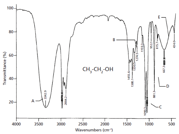
Figure 3: The infrared spectrum of ethyl alcohol. Note the large and broad O-H stretch labeled A at 3342 cm-1.
The peak labeled A in Figure 1 at 3342 cm-1 is the O-H stretch. Note how broad and strong it is. Compare this peak to the peaks in Figure 2 at 3369 and 3298 cm-1, labeled A and B, respectively. Note how these peaks are somewhat broadened and medium in intensity. This is typical of N-H stretches; they are of medium intensity and width compared to O-H stretches. Although O-H and N-H stretches show up in the same wavenumber region, if you remember that O-H stretches are broad and strong, and the N-H stretches are weaker and narrower, you will not get them confused. This is illustrated in Figure 4, which shows the N-H stretching peaks of propylamine to the left, and the O-H stretching peak of ethyl alcohol to the right (the left hand spectrum is in absorbance so the peaks point up, the right hand spectrum is in percent transmittance so the peak points down).
The reason that N-H stretching peaks are narrower and smaller than O-H stretching peaks goes back to electronegativity differences. We stated above that nitrogen has an electronegativity of 3.0. Hydrogen has an electronegativity of 2.1, and oxygen of 3.5. Since oxygen is more electronegative than nitrogen, O-H bonds are more polar than N-H bonds. This means that O-H bonds form stronger hydrogen bonds than N-H bonds. Recall (2) that one of the things that determines peak widths in infrared spectra is the strength of intermolecular interactions. Hydrogen bonds are a strong type of intermolecular interaction. The hydrogen bonding scheme for alcohols is seen in Figure 5. In the figure, the R- groups are substituents attached to the oxygen. The δ+ and δ- symbols represent partial positive and negative charges, respectively. The oxygen of one alcohol molecule with a partial positive charge on it coordinates with the hydrogen of a neighboring molecule with a partial positive charge on it. The resultant hydrogen bond is denoted by a dotted line. It is the strength of this bonding that causes the O-H stretches of alcohols to have such broad infrared peaks, as seen in the spectrum of ethyl alcohol in Figures 3 and 4. The N-H stretches in Figures 2 and 4 are narrower because the sizes of the partial charges in the N-H bond are lower than in O-H's because nitrogen is less electronegative than oxygen. To summarize, then:
O-H > N-H
Polarity
Dipole moment
Peak intensity and width
As we discuss the infrared spectroscopy of organic nitrogen compounds in the next several columns, it will be the stretching and bending vibrations of the N-H bond that will be our focus. However, there are organic functional groups that contain nitrogen but no N-H bonds, such as the nitrile or -C≡N functional group. In these cases, we will find it may sometimes be difficult to determine the presence of organic nitrogen in a sample based on the infrared spectrum by itself.
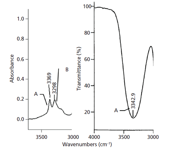
Figure 4: Left: the N-H stretching peaks of propylamine plotted in absorbance (the peaks point up). Right: The O-H stretching peak of ethyl alcohol plotted in percent (%) transmittance (the peak points down).
Conclusion
Nitrogen is one of the atoms that makes up the science of organic chemistry. Nitrogen forms single, double, and triple bonds with carbon. Although it would be nice if peaks from one or more of these functional groups would be diagnostic for the presence of organic nitrogen in a sample, this is, unfortunately, not the case. Instead, N-H stretching peaks are the best indicator for the presence of nitrogen in a sample. N-H stretches appear in the same wavenumber region as O-H stretches, but are narrower and weaker, because N-H bonds engage in weaker hydrogen bonding than O-H bonds. Nitrogen containing functional groups that do not have N-H bonds may be difficult to detect by infrared spectroscopy.
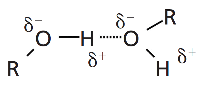
Figure 5: The hydrogen bonding scheme for alcohols.
Infrared Spectral Interpretation Workshop
The workshop is taking time off for the holidays. It will return in the next issue.
References
(1) A. Stretiweiser and C. Heathcock, Introduction to Organic Chemistry (Macmillan, New York, 1976).
(2) B.C. Smith, Spectroscopy, 30(1), 16–23 (2015).
(3) B.C. Smith, Spectroscopy, 32(9), 31–36 (2017).
(4) B.C. Smith, Infrared Spectral Interpretation: A Systematic Approach (CRC Press, Boca Raton, 1999).
(5) B.C. Smith, Spectroscopy, 32(1), 14–21 (2017).
(6) B.C. Smith, Spectroscopy, 33(1), 14–20 (2018).
>Brian C. Smith, PhD

>Brian C. Smith, PhD, is a research chemist at the Center for Food Safety and Applied Nutrition at the U.S. Food and Drug Administration, in College Park, Maryland. Direct correspondence about this article to: Jon.Wong@fda.hhs.gov.

The Big Review V: The C-O Bond
March 20th 2025In the fifth installment of “The Big Review” of infrared (IR) spectral interpretation, we review the spectroscopy of functional groups containing C-O bonds, discuss alcohols and phenols, and see how to use IR spectroscopy to distinguish these alcohols from each other. We then discuss ethers and see how to use IR spectroscopy to distinguish the three different types from each other.
The Big Review IV: Hydrocarbons
January 25th 2025In the fourth installment of our review of infrared spectral interpretation, we will discuss the spectroscopy of hydrocarbons. We will look at the stretching and bending vibrations of methyl (CH3) and methylene (CH2) groups, how to distinguish them, and how to know whether one or both of these functional groups are present in a sample. We will also discuss aromatic hydrocarbons, specifically the C-H stretching and bending peaks of mono- and disubstituted benzene rings, and how to distinguish them.
The Big Review III: Molecular Vibration Theory
January 2nd 2025It has occurred to me that, in the 10+ years I have been writing about molecular vibrations, I have never introduced my readers to its basic theory! I will rectify that now. Some of this is new material, and some will be review. Either way, it is important that all this material be covered in one place.
The Big Review II: The Physical Mechanism of Infrared Absorbance and Peak Types
October 10th 2024In the second installment of “The Big Review,” we discuss the physical mechanism behind how molecules absorb infrared (IR) radiation. Because light can be thought of as a wave or a particle, we have two equivalent pictures of IR absorbance. We also discuss the quantum mechanics behind IR absorbance, and how this leads to the different peak types observed in IR spectrum.