Spectroscopy for Analysis of Botanicals: Raman-Based Agriculture and Detection of Important Chemical Constituents in Plant Materials
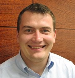
Dmitry Kurouski of the Department of Biochemistry and Biophysics at Texas A&M University in College Station, Texas, is applying Raman spectroscopy to important applications in agriculture for identification and classification of various plant species, for quantification of essential plant constituents, for disease diagnosis in plants, and for Raman-based digital and precision farming. He and his colleagues are at the forefront of using the advantages of Raman based measurements for non-invasive and non-destructive analyses for important field and remote measurement applications.
You have said that “Raman spectroscopy (RS) is an emerging analytical technique that can be used to develop and deploy precision agriculture” (1). Why is precision agriculture an important concept?
Continuing population growth makes global food security a critical issue for maintaining a healthy civilization. Currently, over a billion people suffer from malnutrition due to a lack of food. This problem can be solved by expansion of agricultural territories or by development of what is being called digital farming. While the first approach is destructive and inefficient, the second strategy is focused on the enhancement of farming efficiency. By other means, digital farming, or precision agriculture, aims to maximize crop yield with minimal environmental impact. This can be achieved by timely detection and identification of biotic (plant diseases) and abiotic (drought, nutrient deficiency) stresses. This is very important because plant diseases caused by fungi and viruses can reduce crop yields on an average of 40%, whereas losses associated with lack of nutrients, water or hyper salinity may reach 70%. Thus, solving the problem of timely diagnostics of biotic and abiotic stresses, we aim to solve the problem of food security that our civilization is going to face in the near future.
The question is, What can Raman do to solve those problems? Our group discovered that Raman spectroscopy could be used for highly accurate, nondestructive and label-free diagnostics of fungal, viral, and bacterial diseases on a large variety of plants. Moreover, we showed that Raman spectroscopy can assist in plant breeding enabling digital selection of plants. Our experimental evidence shows that nutrient content of fruits and seeds can be probed using this non-invasive and non-destructive approach. These pieces of evidence demonstrate that this technique can be used to solve the problem of timely disease diagnostics, which currently cannot be solved by any molecular (polymerase chain reaction [PCR], enzyme-linked immunosorbent assay [ELISA]) or imaging (satellite and drone) techniques. Thus, Raman spectroscopy can transform digital farming in the United States and overseas.
You have reported that Raman spectroscopy can be used for Huanglongbing (HLB) diagnostics on both orange and grapefruit trees, as well as detection and identification of various fungal and viral diseases in plants. What has led you to pursue this research, and when did you discover that there was merit in the idea of applying Raman spectroscopy to perform analyses where previously quantitative PCR (qPCR) had been used as the “gold standard” for pathogen diagnostics in plants?
HLB is a devastating disease that obliterates citrus trees in the United States. The disease is caused by bacteria that are vectored by psyllids. Once they appear in the plant, bacteria migrate into the phloem, thus clotting this very important transportation system of the tree. Such trees exhibit premature fruit drop, yellowness of leaves, and ultimately the death of the entire plant. Currently, all citrus plants in Florida are infected, just like the trees in three counties in the South of Texas. The disease spreads so quickly that it is highly likely that all citrus trees in Texas and California will be infected soon. Proliferation of HLB can be stopped by timely diagnostics of bacteria. However, pathogen detection is extremely challenging to do by qPCR or ELISA due to the high cost of such tests. Also, PCR and ELISA are often not sensitive enough to detect the pathogen on the early stages of disease proliferation.
Our group was privileged to collaborate with Prof. Kranthi Mandadi from Texas A&M Research and Extension Center located in Weslaco, Texas, to investigate the potential of Raman spectroscopy for timely diagnostics of HLB. Our findings exceeded our expectations. We demonstrated that this technique could be used to detect HLB on early and late stages, as well as to identify plants that exhibit nutrient deficiencies. These findings show that Raman spectroscopy is not only an excellent tool for detection and identification of bacterial diseases, but it also has a far reaching implications in diagnostics of abiotic stresses caused by nutrient deficiencies.
We also questioned whether Raman spectroscopy is more accurate that qPCR, the “gold standard” of plant disease diagnostics. To answer this question, we selected a group of plants that was monitored by both qPCR and Raman spectroscopy every 3 months. Our results showed that Raman spectroscopy was more sensitive than qPCR, as it could predict the infectious status of orange trees three months before this conclusion could be made by qPCR. It should be noted that in hot summer months, bacteria migrate from leaves into roots of trees. Thus, if leaves of such a tree are sampled for PCR in June–August, the results will be false negative. Our data conclusively show that Raman cannot be misled by the lack of bacteria in the leaves and will diagnose the tree as infected if the pathogen is located in the roots.
Using Raman spectroscopy, you have reported the development of a label-free, non-invasive and non-destructive approach for remote and field detection and identification of poison ivy (Toxicodendron radicans) and for other plant constituents (2,3). You have applied a miniature Raman spectrometer for identification of this species in one second using roaming robotic mounted Raman spectroscopy or unmanned aerial vehicle (UAV) mounted Raman spectroscopy. How successful is this application for surveying a large area for poison ivy? Is this method applicable for identifying many other plant species in a designated area?
This work showed that different plant species have unique Raman spectra that can be used for their confirmatory identification. In fact, our recent findings show that such sensitivity can be reached for different plant varieties or cultivars within a species. Thus, Raman spectroscopy can be used to scan an agricultural or forest area to identify plant species that grow there. This finding will likely transform botanical approaches in identification of species and potentially revolutionize plant taxonomy. Currently, we have designed a robot that will be able to survey a large agricultural area identifying plant species that grow in such ecosystems.
This study also broughtgreat public recognition of our work in general and of Raman-based plant identification in particular. I received numerous calls from excited people appreciating the development of a fully non-invasive and non-destructive approach that can be used for identification of poison ivy, a very dangerous plant species that is present in all U.S. states except Alaska and Hawaii. Considering current technological development that enabled miniaturization of Raman spectrometers to the size of a cell phone, our discovery can be used for inspection of nearly any territory ranging from a personal backyard to a state park.
Further investigations have led you to apply Raman spectroscopy for nondestructive discrimination between hemp and cannabis (4). You determined whether this technique can be used to detect cannabidiol (CBD) and cannabigerol (CBG) in hemp, and to differentiation between hemp, cannabis, and CBD-rich hemp. What have you learned from this work?
This was by far the most fascinating and crazy project that we had in our lab. At that time, neither cannabis nor hemp could be grown in Texas legally. So, we jumped on the plane and went to Denver, Colorado, where we were collecting spectra from industrial hemp, CBD-rich hemp, and cannabis.
The biggest difference between industrial hemp, CBD-rich hemp, and cannabis is the presence of cannabinoids. In hemp, tetrahydrocannabinolic acid (THCA), the psychologically active compound, is present in a very low quantity, below 0.3%. In cannabis, the percentage of THCA varies from 4 to 12% or even 15% sometimes. CBD-rich hemp has below 0.3% of THCA, but contains cannabidiol (CBD) in high quantities. CBD has unique pharmacological applications. Therefore, there is a growing interest in farming of CBD-rich hemp nation-wide. However, it is important for farmers to prove that their plants have below 0.3% of THCA or total THC.
Raman spectroscopy can detect the presence of THCA, CBD, and other cannabinoids by non-invasive spectroscopic analysis of plants. Moreover, we can measure intensities of bands that originate from those cannabinoids to predict quantities of those cannabinoids in the plant material. Altogether, this discovery opened up a possibility to use Raman to probe the cannabinoid content of plants, as well as identify different varieties of CBD-rich hemp and cannabis.
You have also investigated the application of Raman spectroscopyfor the fast and accurate identification of peanut genotypes, and for the prediction of nematode resistance and oleic-linoleic oil (O/L) ratio in peanuts (5). How accurate is this analysis and are you able to determine other constituents with high confidence using Raman spectroscopy?
This analysis is highly accurate. In addition to the O/L ratio, we were able to probe the concentration of proteins, carbohydrates, oils, fiber, esters, and unsaturated fatty acids.
Have you had to develop any specialized chemometric techniques for your various applications of Raman spectroscopy? What has been your greatest data analysis challenge in performing your research?
We typically use supervised chemometric algorithms such as partial least squares–discriminant analysis (PLS-DA). Data analysis for plants is a challenging task. Primarily because we work on live field- and greenhouse-grown plants that exhibit substantial variability associated with small differences in biology of these subjects. Therefore, we have to collect hundreds and often thousands of spectra to make a reasonable conclusion about the feasibility of Raman-based diagnostics for a certain disease. The burden of data analysis is handled by Charlie Farber, the fourth-year graduate student in my lab. So, the success of our group is to a large extend dependent upon his professionalism and persistence in data analysis.
In a more general sense, what were some of the major challenges you have encountered during your career of laboratory research? How important is sound experimental design in your work?
Launching a lab is a challenge. Launching a physics–chemistry lab with a ton of instrumentation is a challenge to the fourth power. In addition to this, a large portion of our work is in the field. Several times, together with my team, we have continued working until 2:00 AM doing measurements in a corn field, which is somewhat spooky. Nevertheless, a great team of young researchers is what has allowed us to make such dramatic progress (we now have more than 40 papers published) in only three years.
Experimental design and planning is the key for everyone who works with living (life) systems. We do very careful planning of our experiments often months ahead. After that, strong commitment and professionalism of all group members is what determines our success.
What would you consider to be the most meaningful contributions of your research work? What is most exciting to you about your work?
We see Raman spectroscopy transforming agriculture in the United States. Our work shows that Raman has high sensitivity and specificity in diagnostics of both plant diseases and nutrient deficiencies. Raman spectroscopy also provides a chemical-free diagnostics approach, which essentially reduces the cost of analysis to zero. Lastly, measurements can be done directly in the field. We expect that once our approach is fully automated, the customer will receive a text message with the type of problem and the location of this problem or danger area within their field. This precise diagnostic can be used to apply pesticides of fertilizers only to the area where they are needed, saving a large amount of money to farmers, while simultaneously minimizing pollution risks to the environment.
Since the world’s population is continuously increasing, the problem of food security will only get more important in the future. We understand that poor digitalization of current agriculture causes approximately 50% crop loss each year worldwide. This makes more than a million people suffer from all types of malnutrition. Therefore, one can envision that Raman spectroscopy will help to feed the planet in the future.
Would you share with our readers to describe your work ethic philosophy, and how you plan your daily or weekly work schedule?
On my opinion, the key component of any company is people. I am privileged to lead a great team of young enthusiasts who work very hard to make all of us succeed. I also try to gather a team of top experts to solve problems that we work on. We are professionals in spectroscopy, but we understand that this is not enough to succeed in the world of plants or agriculture. The success comes from our working together with professionals in plant pathology, plant breeding, and plant biology. We also interact closely with industrial partners. Such collaborations make our work sound and interesting not only to the spectroscopic community, but also to a large audience of farmers and biologists.
What words of wisdom do you have for any young people interested in a scientific research career?
Don’t be afraid to explore uncharted territories. Three years ago, no one would believe that there is any practicality of Raman in agriculture. We were the first who stuck the flagpole in the ground. Today, our experimental findings show that Raman can be used to enable digital farming. Its use in the field will reduce the cost of farming and help feed the world.
One of the best markers of usefulness of any academic research is the interest from companies. Currently, our group works with several companies expanding applicability of our fundamental discoveries. We are positive that industrial interest in our work will only increase in the future.
References
(1) Sanchez, L., Pant, S., Mandadi, K., Kurouski, D. (2020) Raman Spectroscopy vs Quantitative Polymerase Chain Reaction In Early Stage Huanglongbing Diagnostics. Nature Sci. Rep. 10, 10101.
(2) Farber, C., Sanchez, L., Kurouski, D. (2020) Confirmatory Non-Invasive and Non-Destructive Identification of Poison Ivy Using A Hand-Held Raman Spectrometer. RSC Advances 10, 21530–21534.
(3) Sanchez, L., Ermolenkov, A., Tang, X.-T., Tamborindeguy, C., Kurouski, D. (2020) Non-Invasive Diagnostics of Liberibacter Disease on Tomatoes Using a Hand-held Raman Spectrometer. Planta 251, 64
(4) Sanchez, L., Baltensperger, D. and Kurouski, D. (2020) Raman-Based Differentiation of Hemp, Canabdiol-Rich Hemp and Cannabis. Anal. Chem. 92, 7733–7737.
(5) Farber, C., Sanchez, L., Rizevsky, S., Ermolenkov, A., McCutchen, B., Cason, J., Simpson, C., Burow, M., Kurouski, D. (2020) Raman Spectroscopy Enables Non-Invasive Identification of Peanut Genotypes and Value-Added Traits. Nature Sci. Rep. 10, 7730.
Dmitry Kurouski

Dmitry Kurouski earned his M. S. in Biochemistry from Belarusian State University, Belarus and Ph.D. (Distinguished Dissertation) in Analytical Chemistry from SUNY Albany, NY, USA. After a Postdoc in the laboratory of Professor Richard P. Van Duyne at Northwestern University, Dr. Kurouski joined Boehringer-Ingelheim Pharmaceuticals, where he worked as Senior Research Scientist. In 2017, Dr. Kurouski joined the Biochemistry and Biophysics Department of Texas A&M University as an Assistant Professor. His research interests are focused on nanoscale characterization of biological and photocatalytic systems using tip enhanced Raman spectroscopy (TERS), and atomic force microscope infrared-spectroscopy (AFM-IR).
Investigating ANFO Lattice Vibrations After Detonation with Raman and XRD
February 28th 2025Spectroscopy recently sat down with Dr. Geraldine Monjardez and two of her coauthors, Dr. Christopher Zall and Dr. Jared Estevanes, to discuss their most recent study, which examined the crystal structure of ammonium nitrate (AN) following exposure to explosive events.
Distinguishing Horsetails Using NIR and Predictive Modeling
February 3rd 2025Spectroscopy sat down with Knut Baumann of the University of Technology Braunschweig to discuss his latest research examining the classification of two closely related horsetail species, Equisetum arvense (field horsetail) and Equisetum palustre (marsh horsetail), using near-infrared spectroscopy (NIR).
An Inside Look at the Fundamentals and Principles of Two-Dimensional Correlation Spectroscopy
January 17th 2025Spectroscopy recently sat down with Isao Noda of the University of Delaware and Young Mee Jung of Kangwon National University to talk about the principles of two-dimensional correlation spectroscopy (2D-COS) and its key applications.
Measuring Microplastics in Remote and Pristine Environments
December 12th 2024Aleksandra "Sasha" Karapetrova and Win Cowger discuss their research using µ-FTIR spectroscopy and Open Specy software to investigate microplastic deposits in remote snow areas, shedding light on the long-range transport of microplastics.
The Fundamental Role of Advanced Hyphenated Techniques in Lithium-Ion Battery Research
December 4th 2024Spectroscopy spoke with Uwe Karst, a full professor at the University of Münster in the Institute of Inorganic and Analytical Chemistry, to discuss his research on hyphenated analytical techniques in battery research.