Using NMR to Understand Protein Unfolding
A recent study from the University of Guelph used nuclear magnetic resonance (NMR) spectroscopy to learn more about protein unfolding.
By using nuclear magnetic resonance (NMR) spectroscopy, researchers can uncover an atomic-level picture of the thermally induced unfolding of a membrane-embedded α-helical protein, human aquaporin 1 (hAQP1). The findings of this study were published in Science Advances (1).
Membrane proteins are present in the cell membrane and play crucial roles in numerous biological processes (1,2). Membrane proteins perform essential functions in the human body, including several dynamic cellular processes (2). These processes include ionic and molecular transport, enzymatic reactions, and electron transport, to name a few (2). Although scientists know quite a bit about membrane proteins and their functions, there is still little information about how their amino acid sequences dictate their structure and stability.
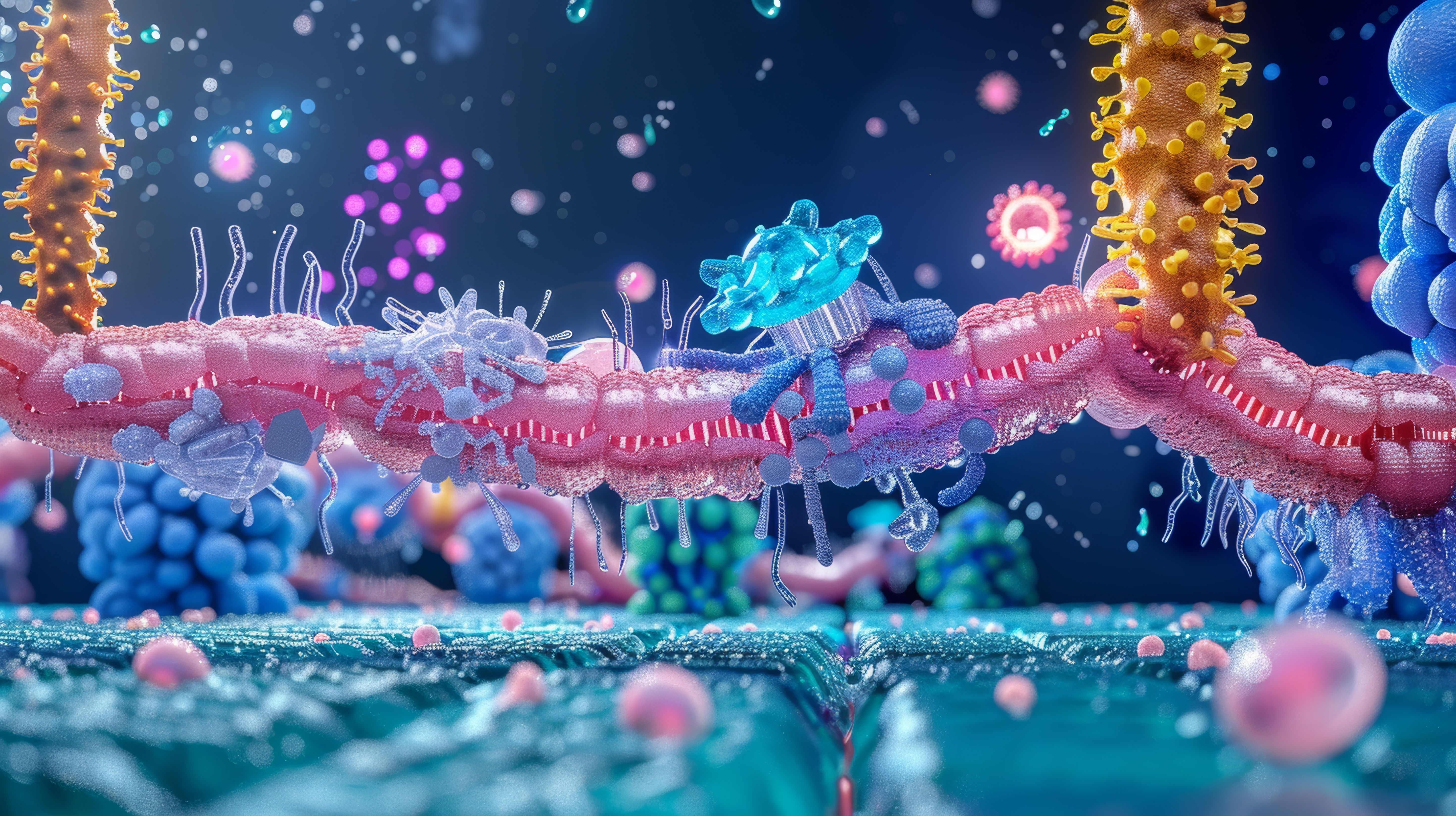
3D illustration of a cell membrane with various proteins and molecules, including a receptor protein, a transmembrane protein, and a lipid molecule, all rendered in vibrant colors and intricate detail. Generated with AI. | Image Credit: © Ponchita - stock.adobe.com

Researchers from the University of Guelph, led by Leonid S. Brown and Vladimir Ladizhansky, explored this topic. In their study, the research team examined the thermally induced unfolding of a membrane-embedded α-helical protein, hAQP1, using NMR spectroscopy (1). Doing so allowed them to obtain an atomic-level picture of the protein unfolding, which allowed them to gain more insights into this biological process.
The study demonstrated that the unfolding of hAQP1 begins with the extracellular loop C, a structured loop that plays a pivotal role in maintaining the stability of the entire protein (1). This loop unfolds first, driven by the breaking of strong hydrogen bonds and the solvent exposure of hydrophobic side chains, resulting in a high activation barrier of at least 300 kJ/mol (1). This initial step is followed by the unraveling of the transmembrane (TM) domain as a single unit, leading to a heterogeneous misfolded state characterized by high helical content but nonnative helical packing (1).
By employing site-specific measurements of HDX rates, the researchers were able to identify unfolding domains within hAQP1 that exchange in an internally correlated manner (1). This advanced methodology allowed them to follow the temperature-dependent kinetics of unfolding at the level of individual amino acid residues, offering a detailed view of the unfolding process (1).
The researchers also used Fourier-transform infrared (FT-IR) spectroscopy to learn more about the sequencing of unfolding intermediate and misfolded states (1). For the FT-IR measurements, a sample was prepared separately and incubated in H2O at increasing temperatures (25°, 40°, 50°, 55°, 60°, and 65 °C) (1). After each incubation, the sample was cooled to room temperature, and the FT-IR spectra were recorded (1). This method aimed to avoid isotopic shifts from exchange and focused on identifying spectroscopic signatures of secondary structures in trapped intermediate (I) and misfolded (M) states (1).
For the HDX-ssNMR measurements, the process involved the sample was incubated in D2O at increasing temperatures (20°, 55°, 60°, 62°, and 64 °C), cooled to 5 °C, and analyzed using ssNMR after each incubation (1). The high-temperature incubation created partially unfolded states, which, upon cooling, could either return to the folded state (reversible unfolding) or remain in intermediate or misfolded states (1).
Membrane proteins are difficult to study because of their complex environment within the lipid bilayer. By using solid-state NMR spectroscopy, the researchers were able to overcome a lot of the challenges that previous studies encountered when studying membrane proteins.
References
(1) Xiao, P.; Drewniak, P.; Dingwell, D. A.; et al. Probing the Energy Barriers and Stages of Membrane Protein Unfolding Using Solid-State NMR Spectroscopy. Sci. Adv. 2024, 10 (20), eadm7907. DOI: 10.1126/sciadv.adm7907
(2) Jelokhani-Niaraki, M. Membrane Proteins: Structure, Function and Motion. Int. J. Mol. Sci. 2023, 24 (1), 468. DOI: 10.3390/ijms24010468