Using Raman Spectroscopy for Characterization of Defects and Disorder in Two-Dimensional Materials
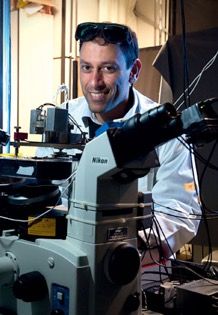
Raman spectroscopy has been demonstrated as an analytical technique for characterizing disorder in two-dimensional (2D) crystalline material structures caused by the presence of defects (1). This disorder in 2D crystalline structures may be described from a dimensionality point of view, zero-dimensional (0D), or one-dimensional (1D) defects, expressed as points or lines, respectively. For characterization of the quantity of 0D and 1D defects respectively, two Raman measurement parameters are required as defect-induced activation of forbidden Raman modes, and defect-induced confinement of phonons. Professor Ado Jorio, of the Department of Physics at the Universidade Federal de Minas Gerais in Brazil, recently talked to us about his research in this field.
Raman spectroscopy has been demonstrated as an analytical technique for characterizing disorder in two-dimensional (2D) crystalline material structures caused by the presence of defects (1). This disorder in 2D crystalline structures may be described from a dimensionality point of view, zero-dimensional (0D), or one-dimensional (1D) defects, expressed as points or lines, respectively. For characterization of the quantity of 0D and 1D defects respectively, two Raman measurement parameters are required as defect-induced activation of forbidden Raman modes, and defect-induced confinement of phonons. Professor Ado Jorio, of the Department of Physics at the Universidade Federal de Minas Gerais in Brazil, recently talked to us about his research in this field.
You have recently described advances in defect determination in crystalline materials using Raman spectroscopy. What prompted you to investigate this problem? What is unique about your approach?
This subject has been under research investigation since 1970, when graphitic structures began to be studied, for example graphite intercalated compounds, important today in the batteries for cars and cell phones. The development of related materials, for example the isolation of single-layered graphene, made it possible to develop a detailed understanding of the relationship between defects and Raman spectroscopy signatures. We were able to use two standardized materials with a well-controlled evolution of the quantity and type of defects to unravel the spectroscopic signature of defect dimensionality. We were able to develop a much more accurate model to relate Raman spectroscopy to the number of defects, and we demonstrated, for the first time, that Raman spectroscopy can be used to distinguish between the two most fundamental classes of defects in 2D materials, which are the zero-dimensional and the one-dimensional defect types. Zero-dimensional defects are vacancies, substitutional atoms, and isolated functional groups. One-dimensional defects are grain boundaries and dislocations. These two classes of defects have strikingly different consequences in the properties of two-dimensional materials.
Would you briefly explain the theory behind how Raman measurements of the defect-induced activation of forbidden Raman modes, and the defect-induced confinement of phonons allows you to characterize specific information regarding disorder in crystalline structures? How would you describe these defects in simple terms?
Raman spectroscopy is defined as the inelastic scattering of light. You can imagine the photon as a particle that collides with the material and is scattered with different energy and momentum (this is why it is considered inelastic). Therefore, with Raman spectroscopy there is a conservation of energy and momentum. We note that a very beautiful property of nature is the relation between symmetry and the conservation laws. The conservation of momentum is a direct consequence of the translation symmetry. When you have a defect in a crystal, you break the translation symmetry in the ordered structure, and the typical rule of momentum conservation does not apply. Without the momentum conservation restriction, the light can be scattered in ways that were forbidden for the perfect crystalline structure, and this gives rises to new peaks in the Raman scattering spectra. We then identify and characterize these peaks to infer defects within the crystalline structures.
You have published research regarding stimulated Raman scattering (SRS) and coherent anti-stokes spectroscopy (CARS) for characterizing two dimensional materials (2). How does this work differ from your research described in reference 1 above?
This specific work is not related to defect identification; it is actually related to the difference in CARS when comparing graphene with hexagonal boron-nitrite (hBN). Graphene is a semimetal and hBN is an insulator. The difference in electronic structure generates differences in the CARS response. We were able to characterize these differences within this study.
What do you believe is the potential for Raman to be used routinely for characterizing two-dimensional materials for detailed physical or chemical properties?
The potential is huge, and this is not only my belief; it has been proven by many scientific studies. I believe the important breakthrough here is that when you enter the nanoworld, the characterization techniques have to be more gentle, less invasive, for you to access the properties of the materials without modifying the materials with the probing technique. In this sense, light is a massless and chargeless noninvasive probe that, in the visible range of the spectrum, causes no effects on the studied material. Furthermore, Raman spectroscopy has the extra advantage, within the optics techniques world, of being very specific and spectrally accurate. The spectral accuracy in Raman spectroscopy is in the order of 0.1 meV, much more accurate that what you can achieve with other optical, electronic, or physical transport measurement techniques.
For those having greater interest in this topic, what reference papers or books would you recommend they obtain to read or study?
For studying the basics, I recommend the book edited by A. Jorio, M. Dresselhaus, R. Saito, and G.F. Dresselhaus, entitled, Raman Spectroscopy in Graphene Related Systems (John Wiley and Sons, 2011) (3). For the state-of-the-art developments I recommend the papers you cited here and references that may be obtained from these research papers.
What recent advances in Raman instrumentation, software, or sampling methods have you been most active in over the recent past?
I am highly involved in the development of tip-enhanced Raman spectroscopy (TERS), a technique that will allow us to characterize materials using Raman spectroscopy with nanometer resolution. A microscope usually can focus the light onto a 1-micrometer squared area. Graphene, in a one- micrometer square has about 39 million atoms. To push nanotechnology even further, we have to be able to achieve near nanometer resolution in materials characterization. This is what we are doing.
What have been your greatest challenges in scientific discovery over your career? What is your general approach to problem solving in your scientific work?
The great challenge, overall, is to be able to surpass the limits of other previous work. In materials science, this is strongly related to being able to break the limits of your measurements, in energy or in resolution. When we move into nanoscience and nanotechnology, the most important limit is resolution. My team and I made a very important breakthrough in the past showing how to do single nanotube Raman spectroscopy, and demonstrating all the wonders you might achieve when you are able to do this. In your questions you have highlighted the strong advance in the ability to characterize defects. And here I have highlighted the importance of going beyond the diffraction limit to perform optical measurements with nanometer resolution. All these achievements are related to my general approach for solving problems in the scientific world that is based on “try it first, check if it is possible or not later”. It might sound absurd, but I believe ignorance is very important in the world of scientific discoveries. When you can calculate or estimate whether something is possible or not, it means you are not dealing with something truly new.
What are some major gaps in knowledge for Raman technology that you would like to see more research and development time devoted to?
Now that we are entering the world of nano-optics in depth, it is clear that required improvements of resolution come along with a price. The light–matter interaction phenomena at the nanometer scale are different from those at the micrometer scale and higher. This research is a completely new and open avenue for the further development of Raman technology as applied to nanomaterials and molecular structures. In addition, we have recently discovered that two photons can interact inside a transparent material in a way that is similar to the interaction observed between electrons in superconductivity Cooper pairs. To study this new phenomena again we need novel instrumentation to merge Raman spectroscopy with quantum optics techniques. In short, Raman spectroscopy is rapidly moving into the nanoworld and into the quantum optics world.
What do you anticipate is your next major area of research or application in your field?
Unfortunately, I had a serious laboratory accident in the past, and I lost 80% of the vision in one eye, burned by an infrared laser. As a consequence, I decided to build an intraocular spectrometer. Of course there is a lot of research on this field, which is all about ophthalmology. But to my knowledge, not much using Raman spectroscopy. We know Raman spectroscopy has been very powerful to characterize materials, and we know the eye is the window to the brain. So, I want to build a machine that will allow us to study the optical nerve using Raman spectroscopy.
References
- A. Jorio, and L.G. Cançado, in Raman Spectroscopy of Two-Dimensional Materials (Springer, Singapore, 2019), pp. 99–110.
- L.M. Malard, L. Lafeta, A. Cadore, T. Grasiano, K. Watanabe, T. Taniguchi, L. Campos, and A. Jório, Bull. Amer. Phys. Soc. (Mar. 7, 2018).
- A. Jorio, M. Dresselhaus, R. Saito, and G.F. Dresselhaus, Eeds., Raman Spectroscopy in Graphene Related Systems (John Wiley and Sons, Hoboken, NJ, 2011).

Professor in the Department of Physics at the Federal University of Minas Gerais, Ado Jorio works in research and development of scientific instrumentation in optics for the study of nanostructures, with applications to novel materials and biomedicine. He received his PhD in physics at the same institution in 1999, working with phase transitions in incommensurate systems, followed by a postdoctoral study at the Massachusetts Institute of Technology (MIT), Cambridge, USA (2000-2001), working with optical properties of nanomaterials, with a specialization on Raman spectroscopy and optical properties of carbon nanomaterials. Jorio received the Somiya Award, from the International Union of Materials Research Societies (2009), the Scopus Brazil award from Elseviar & CAPES (2009), the ICTP Prize (2012), the Georg Forster Research Award from the Humboldt Foundation (2015), and the Inconfidence Medal from the Minas Gerais State (2016). He is a member of the Brazilian Physical Society, the Brazilian Academy of Sciences, the National Order of Scientific Merit, and the American Chemical Society. He held the positions of Coordinator for Strategic Studies and Information at INMETRO (2008-2009) and, at UFMG, of Director of the Coordination of Technological Innovation and Transfer (2010-2012), Head of the Physics Department (2015-2016), and Dean of Research (2016-2018).
Investigating ANFO Lattice Vibrations After Detonation with Raman and XRD
February 28th 2025Spectroscopy recently sat down with Dr. Geraldine Monjardez and two of her coauthors, Dr. Christopher Zall and Dr. Jared Estevanes, to discuss their most recent study, which examined the crystal structure of ammonium nitrate (AN) following exposure to explosive events.
Distinguishing Horsetails Using NIR and Predictive Modeling
February 3rd 2025Spectroscopy sat down with Knut Baumann of the University of Technology Braunschweig to discuss his latest research examining the classification of two closely related horsetail species, Equisetum arvense (field horsetail) and Equisetum palustre (marsh horsetail), using near-infrared spectroscopy (NIR).
An Inside Look at the Fundamentals and Principles of Two-Dimensional Correlation Spectroscopy
January 17th 2025Spectroscopy recently sat down with Isao Noda of the University of Delaware and Young Mee Jung of Kangwon National University to talk about the principles of two-dimensional correlation spectroscopy (2D-COS) and its key applications.
Measuring Microplastics in Remote and Pristine Environments
December 12th 2024Aleksandra "Sasha" Karapetrova and Win Cowger discuss their research using µ-FTIR spectroscopy and Open Specy software to investigate microplastic deposits in remote snow areas, shedding light on the long-range transport of microplastics.
The Fundamental Role of Advanced Hyphenated Techniques in Lithium-Ion Battery Research
December 4th 2024Spectroscopy spoke with Uwe Karst, a full professor at the University of Münster in the Institute of Inorganic and Analytical Chemistry, to discuss his research on hyphenated analytical techniques in battery research.