The Investigation of Laser-Induced Breakdown Spectroscopy for Detection of Biological Contaminants on Surfaces
The potential utility of laser-induced breakdown spectroscopy (LIBS) as a means to detect biological contaminants on painted surfaces is investigated.

Laser-induced breakdown spectroscopy (LIBS) offers an in-situ capability for instantaneous qualitative analysis of the elemental composition of materials. This technique differs from other forms of emission spectroscopy in that the sample is irradiated and evaluated in one step with little or no sample preparation: a laser pulse is focused on the sample to a sufficient energy density, volatizing a small amount of material into a microplasma or laser spark; light from the laser spark is spectrally resolved to identify the emitting species present in the plasma; compositional information about the target material is then deduced from the resulting spectrum. Since the phenomenon's discovery in 1962 (1), LIBS has found its way into such diverse applications as screening for toxic metals (2,3), industrial process control for analysis of ores in the mining industry and sorting of waste materials (4), inspection of art objects to detect fraudulent copies, and many others. A number of review articles (5,6) and, just recently, two books on LIBS have been published. The new book by Creamers and Radziemski (7) traces the historical development of LIBS, explains the underlying physical processes involved, describes the components of a LIBS apparatus and issues that arise in their assembly, and addresses various practical issues in the use of LIBS for qualitative and even quantitative analysis. The book concludes by summarizing current LIBS-related research and application activity and then projects the trajectory of the field into the near future. Those same authors treat LIBS history and fundamentals more succinctly in the first chapter of the new book edited by Miziolek, Palleschi, and Schechter (8), subsequent chapters of which feature a succession of prominent LIBS researchers discussing the nature of the LIBS plasma, experimental measurement and data analysis issues, and a wide variety of LIBS applications.
Considered as a means to detect contaminants, LIBS is best suited for circumstances in which the sought-for contaminant contains one or more elements that are not proper (or desired) constituents of the test sample. The problem of detecting lead and other toxic metals in paint provides an excellent example. Lead-based paint, which once was preferred for use on building exteriors due to its durability, is now prohibited by environmental regulations in the U.S. and elsewhere because of the hazard it presents to anyone exposed to it, especially small children. Nevertheless, lead paint is still found on many older residences and other structures as well as in waste disposal sites holding their remains, and detection of old lead paint is obviously a necessary precursor to the remediation of this environmental hazard. Figure 1 shows a LIBS spectrum of an "unknown" paint that clearly contains lead. A qualitative analysis such as this can be performed instantaneously using LIBS. A more traditional laboratory analysis would be required to obtain a quantitative measure of the lead concentration in the paint; nevertheless, LIBS might eventually find application as a means to survey suspected sites for the presence of lead or other elemental contaminants. A thorough investigation into the potential of LIBS to detect environmental lead contamination was reported recently by Wainner and colleagues (2).
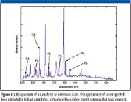
Figure 1
Successfully detecting and discriminating biological materials using LIBS is a far more challenging problem because the same elements occur in all biological organisms; obviously, any success to be had will have to rely on analysis of the relative proportions of the biological elements present rather than their simple presence or absence. Indeed, the extensive LIBS literature includes a limited number of papers that specifically address biological analysis. They typically have a well-defined focus and scope and utilize a specific approach, with the intent to determine the feasibility of using LIBS for some form of biological analysis. The focus varies from biologically important inorganics (9–11), to pollens (12–15), molds (12,13), and proteins (13), to bacteria (12,13,15,16); sample types vary from solids (9,10), to aerosol particles (11,14,17,18), to dense aerosol clouds (12), to concentrations of organisms compressed into pellets (15), to thin films of biological material deposited onto porous silver (13). Some of these studies tried to distinguish between different types of biologicals (12,13,15), in one case even to the level of specific bacterial species (16). The present study is extending that line of inquiry to the problem of identifying biological contaminants on exterior, painted surfaces, and classifying them to the level of major categories like mold, pollen, and bacterial spores — with the latter constituting our primary objective.
Hybl and colleagues (12) provide a thorough description of a study on the use of LIBS to analyze biological aerosols. They utilized both a broadband, multiparticle apparatus and a narrowband, single-particle apparatus to take LIBS spectra of an assortment of biological aerosols. Peak/base ratios were calculated for the atomic spectral responses, and principal components analysis (PCA) was used to discover distinguishing patterns. Their PCA results clearly distinguish between fungal spores, pollen, and the spore form of Bacillus globigii (a nonpathogenic sporifying bacterium that is frequently used as a simulant for anthrax); other biologicals are distinguishable as well. But the authors caution that a mixture of compounds could provide a spectral signature similar to a biological warfare agent, which could then be interpreted as a (false) positive.
Wormhoudt and colleagues (19) suggest that poor precision, difficulties in quantitation, and the inadequate detection limits that typically plague LIBS analysis might be less critical when testing biologicals because their detection may rely less on absolute quantification and more on a qualitative comparison to a known, finite set of "spectral fingerprints." Indeed, in all of the biological studies we reviewed, data analysis relied on some form of pattern recognition — attempting to identify distinguishing spectral patterns between different classes of organisms. The biological signatures or specific elemental lines for each organism were manipulated by several different techniques in attempts to identify indicative patterns. The elemental signatures of calcium, sodium, magnesium, manganese, silicon, potassium, phosphorus, iron, carbon, oxygen, and nitrogen, plus the transient recombinant molecular species cyanide and phosphate — all derivable from components of biological systems or their breakdown — were employed in the various biological studies.
Of particular interest to us was a study by Samuels and colleagues (13), which utilized LIBS to measure pure preparations of bacterial spores, molds, pollens, and proteins deposited as thin films on porous silver substrates. Except for those attributable to the silver substrate, all of the lines in their spectra arise from the biological layer. They utilized PCA and demonstrated that the technique can distinguish between different specimen types, but suggested that a more robust modeling scheme would be needed to distinguish between individual species of bacteria. Of all the works we reviewed, this one bore the greatest similarity to our problem.
More recently, DeLucia and colleagues (20) reported on a study in which LIBS was used to detect hazardous contaminants of several kinds, to include bacterial spores. Using data measured against the pure biological contaminants as well as the same contaminants in soil, they obtained excellent automated discrimination performance using an algorithm they devised that employed linear correlation analysis against a spectral library.
The work presented here expands upon those previous studies by treating the case in which the sought-for biological contaminant exists as a thin layer over a complex substrate — paint — which will necessarily impart its own spectral signature as a background against which the signature of the contaminant layer must be processed to discern the presence of a target biological threat. This article presents the design of our apparatus and the nature of the first set of data acquired under our current effort to investigate whether biological materials on a surface painted "Navy gray" can be classified based upon LIBS spectra taken on the surface layer in situ, despite interference from the paint that is taken up in the process. Our initial intention was to collect data that could be analyzed to give an indication of whether any reasonable level of discrimination of biologicals is possible based upon the LIBS spectra of thin coatings over such a complex substrate as paint. So we chose to assemble our apparatus from components that were on hand in our laboratory for this first phase of our study in which basic feasibility needed to be established, even though the characteristics of our spectrometer and its associated collection optics limited the available spectral range to about a third of what we desired. Nevertheless, we enjoyed a level of classification performance well in excess of what we hoped to achieve, on the basis of which we have undertaken to upgrade the apparatus to support continued progress on this problem.
Experimental
Figure 2 illustrates the apparatus we designed and assembled based upon our literature review, the highlights of which are summarized above. We utilized our laboratory's New Wave Research (Fremont, California) ACL-1 Q-switched Nd:YAG laser as the excitation source because its characteristics fall within the scope of the lasers typically used for biological-related LIBS studies: it generates a 35-mJ pulse at the Nd:YAG fundamental wavelength (1064 nm), and it can be used to deliver a single pulse or to operate at a pulse repetition frequency of 1 Hz. Several optical components were included beyond the basic requirements for LIBS to facilitate careful control of the excitation of the laser spark and the collection of the resulting LIBS emission for spectral analysis. The beam was expanded through the microscope objective to fill the clear aperture of the collimating lens and was then reflected by the mirror, which could be adjusted to direct the collimated beam downward toward the sample at any desired angle of incidence. The sample was mounted with its top surface horizontal to accommodate the possibility that the contaminant is not adsorbed strongly onto the substrate — a precaution that turned out to be unnecessary. The laser-focusing lens between the mirror and the sample provided the means to vary the spot size, hence the energy density, of the incident pulse. We included the collection lens between the sample and the coupling lens to permit the region from which light is collected for analysis by the spectrometer to be limited virtually to a pinpoint. The desired adjustability was achieved by mounting this pair of lenses on a rotational xyz stage configured to permit extended motion along the axis of the fiber-optic cable, which in turn transmitted the emission collected from the laser spark to the spectrometer. The coupling lens and the fiber assembly (Ocean Optics [Dunedin, Florida] 74-ACR and P600-2-VIS-NIR, respectively) were intended for the visible and near-infrared regions of the spectrum, so attenuation in the UV region increased progressively starting about 380 nm and the assembly approached opacity near 300 nm.
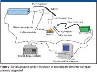
Figure 2
We performed a study that catalogued the principal relevant elemental emission lines and noted their separation in wavelength, based upon which we identified the spectral range of interest as ~200–1100 nm at a resolution of about 0.5 nm; this is comparable to several of the previous biologically oriented LIBS studies and includes the great majority of elemental emission lines we can reasonably expect to observe. For the experiment reported here, however, we had available only a single Ocean Optics HR 2000 spectrometer configured for ~200–650 nm. So in view of the aforementioned absorption characteristics of the collection optics, the spectral information on which our results are based is confined to the range ~300–650 nm.
Ocean Optics OOIBase software running on a Microsoft Windows-based computer controlled the apparatus. The computer was interfaced to the spectrometer through its USB port. A data collection event was triggered by activating the laser controller, which fired the flashlamp and simultaneously signaled our Signal Recovery (AMETEK, Wokingham, UK) model 9650A digital delay generator. After the programmed delay, the delay generator triggered the spectrometer via its control port. The software automatically collected data sets from the spectrometer and stored them along with the values of all settings and a date-time stamp.
A great advantage of LIBS — one of its characteristics that motivates this study — is the fact that the technique can be used to analyze samples in situ without the special preparation procedures that most analytical methods require. Even so, we have found that careful preparation of test samples strongly facilitates the effective use of the experimental control functionality that has been incorporated into the apparatus to obtain reproducible results. A sample substrate is fabricated by mounting a piece of pure sheet copper in phenolic resin, which is then polished to a ~1-μm finish using standard metallurgical methods. Subsequently, these substrates were coated with our "Navy gray" paint and then set aside in ambient air or a drying chamber for at least two weeks, until the paint had thoroughly hardened. This paint — a silicone alkyd anti-stain finish by International Marine Coatings, Inc. (Akzo Nobel, Arnhem, The Netherlands) called Interlac 1 — is used commonly on the weather surfaces of U.S. Navy ships. Before use, hardness was verified by pressing a hard metal probe onto the coatings, which were judged to be cured fully if no observable indentation resulted.
Our means of applying the contaminant to the substrate is motivated by the way pollen is typically observed to adhere to an automobile windshield through the agency of morning dew: wind will not remove the pollen layer after the dew evaporates; one must rub or wash it off. We place a drop of water (to include some alcohol if required, which was the case for our mold specimens) on the substrate and then mix in a small quantity of the contaminant to form a thin slurry, which is then left to dry in the ambient air or in a drying chamber — typically for two days or until all of the liquid has evaporated and the remaining biological material adheres firmly to the substrate. Finished samples are stored in a desiccator to ensure that the coating does not absorb water from the ambient air. Figure 3 shows the key stages in their preparation. The density of the resulting contaminant layer varies over the substrate, but is everywhere sufficiently thin that each LIBS shot will ablate a portion of paint as well as the overlying contaminant. No attempt was made to obtain a layer of uniform thickness at this first stage of our project, because our technique is meant to respond to the relative intensities of emission from the sample's elemental constituents rather than absolute measures of intensity. In the future, we will be performing sensitivity studies for which precise quantitation of the contaminant layer will be required; here, however, our intention was to replicate, as closely as possible, the manner in which a biological contaminant would adhere to an exterior painted surface in the outdoor environment.

Figure 3
In the early months of our study, we applied various contaminants directly to the unpainted polished-copper substrates, then LIBS spectra were taken with an earlier version of our apparatus that consisted of the laser, spectrometer, digital delay generator, and computer previously described, but with single-lens optics for laser focusing and data collection. A few examples are shown in Figure 4 to illustrate their elemental content and the resolution of our spectra. The copper lines obviously come from the substrate, while all others must be attributed to the contaminant. Excellent signal-to-background ratios clearly are apparent in these spectra, so it is not hard to convince oneself that a classification strategy based upon relative line intensities should have some chance of success.

Figure 4
In an experimental run performed on February 6, 2006, 341 single-shot LIBS spectra were taken against 11 separatepainted substrates: one uncontaminated, the other 10 each coated with one of nine different biological materials. The sample was rotated slightly between shots to present a fresh target spot each time. For these data, which are listed in Table I, the gating delay was set to 183 μs — a figure we had arrived at previously by taking repeated measurements against a bare copper substrate to find the optimal signal-to-background ratio. This delay, far longer than one would typically find in the LIBS literature, includes the time interval between the triggering of the laser flash lamp and the subsequent opening of the Q-switch — which we cannot determine precisely from available documentation or data. The spectrometer's integration time was set to 3 ms (the minimum possible for that instrument), which far exceeds the duration of the laser spark. Each spectrum was processed by the operating software to correct for electronic noise and then stored for subsequent analysis. This entire set of spectra was processed using the analysis methodology described below, without screening for quality, content, or any other criterion.

Table I: Specimens for LIBS analysis of biological contaminant coatings over paint
We developed an automated software package in MATLAB (The Mathworks, Natick, Massachusetts) that first preprocesses the spectra, then builds a classification tool based upon a training set consisting of the major fraction of the spectra, and finally applies that classification tool to the remaining fraction — the test set. Each spectrum is treated as an ordered set of 2048 intensities. Except where indicated in the following discussion, the procedures described were implemented based upon the book by Beebe and colleagues (21). Each individual spectrum is preprocessed by first taking derivatives with respect to index using the running simple difference method and then integrating the result to correct for baseline drift; after that, autoscaling consisting of variance scaling and mean centering is applied (in that order) to remove the effect of weightings imposed by the scale of intensities. Then, using an automated procedure that randomly permutes the spectra into the mutually exclusive training and test sets, 70% of the spectra from each sample (rounding down if necessary to get an integer) are assigned to the former and the remainder to the latter; thus, the data set was partitioned into a 232-member training set and a 109-member test set. A PCA was performed on the preprocessed spectra in the training set using an algorithm developed originally for facial recognition applications (22). Subsequently, a linear discriminant analysis (LDA) was performed to sharpen the achievable classification performance. Although PCA is standard fare, LDA has been introduced only recently (23) as a procedure for chemometric applications and is not discussed in our primary chemometric reference (21). To perform LDA, one selects enough PCA coordinates to represent the spectral features that appear above the noise floor and then computes the linear combination that simultaneously minimizes the intraclass separations and maximizes the interclass separations. Through repeated trials against an earlier data set, we settled on using the first 11 PCA coordinates as the basis set for the LDA transformation, so that strategy is currently hard-coded into our analysis package. Hierarchical cluster analysis (HCA) was then applied against the training set, as represented in the 11-dimensional PCA or LDA space, to derive an objective classification tool, which was then used to classify the members of the test set in order to assess its performance against an independent set of observations.

Figure 5
Results
Figure 5 shows three examples from the February data — spectra of Bacillus subtilis spores, oak pollen, and cladosporium mold over paint. The paint contributes so many lines to the background that the baseline is masked completely, and it might appear rather optimistic to expect to develop a classification tool capable of distinguishing different types of contaminant layers based on spectra such as these. Nevertheless, we were able to distinguish the bacterial spores from other biological and nonbiological contaminants with a high degree of confidence.
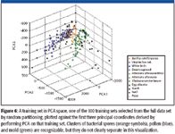
Figure 6
A total of 100 instances of the procedure described earlier were performed, each time with a different random partitioning of the same 341 spectra to form the training and test sets. Figure 6 shows a typical training set plotted against the first three principal coordinates. In Figure 7, the complementary test set is plotted against those same three coordinates. The fundamental output of a PCA is a set of so-called loading vectors, each of which is (in our case) a 2048-element vector; one takes the inner product between a loading vector and a spectrum to obtain the value of that spectrum's principal component along the coordinate corresponding to that loading vector. Loading vectors such as these can be regarded as spectra in their own right; by examining them, one can recognize features attributable to the major biological elements in addition to the titanium — a nonbiological element that is a constituent of the paint. Thus, each point in these figures represents the "position" of a certain spectrum in the first three dimensions of the PCA space; however, the HCA is performed over the first 11 coordinates. The circles that appear around some of the symbols in Figure 7 denote spectra that were assigned erroneously by the HCA classifier; where a circle has the same color as the symbol, the spectrum was assigned to a different contaminant in the same class — cladosporium classified as alternaria, for example.
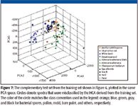
Figure 7
Figure 8 clearly illustrates the enhanced separation that develops when the same training set of Figure 6 is plotted against the first three so-called canonical coordinates of the LDA transformation. Figure 9 shows that same test set of Figure 7 plotted in the LDA space of Figure 8, with circles representing erroneous HCA classifications as before.
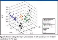
Figure 8
Table II summarizes the performance measures derived from the 100 different partitionings of the data set between the training and test sets, both when PCA alone is used prior to the HCA derivation and when the LDA is included. With respect to the summary performance of the classifier against the test sets, performance measures are provided according to the stringent criterion, whereby the specific contaminant specimen must be correctly assigned for the classification to be declared successful, and the class criterion, whereby success is declared if the classifier assigns the spectrum to the correct class of contaminant. A breakdown of classification performance by class is also included, in which — for each class — the success rate according to the class criterion is given, followed by the false positive and false negative rates. The false negative rate is the percentage of spectra belonging to the given class (pollen, for example) that failed to be classified as such; clearly, the success rate and the false negative rate must sum to 100%. The false positive rate is the percentage of spectra that were classified erroneously as belonging to the class.
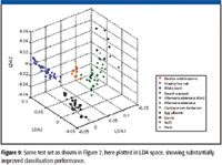
Figure 9
Discussion
The primary significance of our results is the strong indication they provide that LIBS spectra of contaminant layers over a complex substrate contain the information on the basis of which their biological content can be discerned. For our purpose, what is most noteworthy is the fact that the B. subtilis spores were classified with false-positive and false-negative performances of 1% and 3%, respectively. This far exceeded the modest indication of potential success that we were seeking as a justification to continue this line of applied research. The low false-positive rate is particularly gratifying.

Table II: HCA classification performance against February 2006 Bio-LIBS data
At this early stage, it would be well to acknowledge the preliminary nature of these results. While we do believe that our sample preparation methodology well imitates the manner in which contamination naturally occurs in the outdoor environment, there is a need to expand the number and variety of test specimens to adequately stress our developmental analysis methodology. Especially, we need to obtain several different species of bacterial spores and to obtain specimens of the same species that have been grown or prepared in different ways. We need to take data against contaminants formed from mixtures of bacterial spores with other biologicals in order to assess the robustness of our methodology and to develop experimental procedures to bring out the spore signature in the presence of interferents. The apparatus also should be improved to accommodate the greater part of the spectral region in which LIBS data can be measured. There is also the need to control for the x-scale drift that inevitably afflicts raw spectrometric data; obviously, we will need to include a calibration procedure in our data acquisition methodology along with a preprocessing step that uses the calibration data to compensate for drift. And despite the success of our chemometric methodology, which attempts to classify each individual specimen uniquely, it would be well to explore how classification performance could be improved further by employing procedures designed to recognize bacterial spores (or some other single class of contaminant) only. Finally, the need to include automated logic for outlier detection has been identified.
Conclusion
Thus, we see that LIBS spectra, measured against thin contaminant layers drawn from a limited set of representative specimens and applied over a representative exterior marine paint, contain the information on the basis of which the contaminants can be classified, with considerable confidence, into major categories such as bacterial spores, pollen, mold, and others. This summary result was taken as justification to proceed with this line of applied research. As this is being written, we are nearing the completion of a major upgrade of our apparatus, to include a more powerful and flexible laser that is capable of generating pulses at the Nd:YAG fourth harmonic (266 nm) having a higher pulse energy than we get at the fundamental from the laser described herein — which remains available to facilitate experiments involving dual-pulse excitation. We have moved to using Ocean Optics' new, more capable SpectraSuite software to control our apparatus. We have expanded the set of biological materials for our experiments to include spores of other bacterial species, spores of the same species that have undergone different preparation procedures, and a number of nonbacterial biologicals. Finally, we have initiated a thorough review of our existing chemometric processing scheme — which was designed to determine whether our spectra actually had the required information content — with the objective of developing a robust prototype analysis and classification tool oriented to the operational field-detection paradigm.
We are thus poised to execute the program of experimentation we are planning, which is intended to systematically explore the potential of LIBS as a means of spectrally analyzing contaminants on painted surfaces, and other complex substrates, to indicate the presence of biologicals that may, themselves, pose a threat or indicate that a covert biological attack has occurred. We plan to report the results of these studies as they become available.
Acknowledgment
The authors wish to thank the Joint Science and Technology Office for Chemical and Biological Defense (JSTO–CBD) of the Defense Threat Reduction Agency (DTRA) for supporting this work. The portion that was performed at The Pennsylvania State University was executed for the Naval Surface Warfare Center–Panama City under Delivery Order 0308 of Contract No. N00024-02-D-6604 with the Naval Sea Systems Command.
D.W. Merdes, J.M. Suhan, J.M. Keay, and D.M. Hadka are with the Applied Research Laboratory, The Pennsylvania State University, State College, Pennsylvania.
W.R. Bradley is with the Naval Surface Warfare Center – Panama City, Florida.
References
(1) F. Brech, Appl. Spectrosc. 16, 59 (1962).
(2) R.T. Wainner, R.S. Harmon, A.W. Miziolek, K.L. McNesby, and P.D. French, Spectrochim. Acta, Part B 56, 777–793 (2001).
(3) B.T. Fisher, H.J. Johnsen, S.G. Buckley, and D.W. Hahn, Appl. Spectrosc. 55, 1312–1319 (2001).
(4) D. Body and B.L. Chadwick, Spectrochim. Acta, Part B 56, 725–736 (2001).
(5) D.A. Rusak, B.C. Castle, B.W. Smith, and J.D. Winefordner, Trends Anal. Chem. 17, 453–461 (1998).
(6) L.J. Radziemski, Microchem. J. 50, 218–234 (1994).
(7) D.A. Creamers and L.J. Radziemski, Handbook of Laser-Induced Breakdown Spectroscopy (John Wiley & Sons, Ltd., New York, 2006).
(8) A.W. Miziolek, V. Palleschi, and I. Schechter, Eds., Laser-Induced Breakdown Spectroscopy (LIBS) Fundamentals and Applications (Cambridge University Press, UK, 2006).
(9) A. Assion et al., Appl. Phys. B-Lasers and Optics 77, 391–397 (2003).
(10) O. Samek et al., Spectrochim. Acta, Part B 56, 865–875 (2001).
(11) D.W. Hahn and M.M. Lunden, Aerosol Sci. Technol. 33, 30–48 (2000).
(12) J.D. Hybl, G.A. Lithgow, and S.G. Buckley, Appl. Spectrosc. 57, 1207–1215 (2003).
(13) A.C. Samuels, F.C. DeLucia, K.L. McNesby, and A.W. Miziolek, Appl. Opt. 42, 6205–6299 (2003).
(14) A.R. Boyain-Goitia, D.C.S. Beddows, B.C. Griffiths, and H.H. Telle, Appl. Opt. 42, 6119–6132 (2003).
(15) S. Morel, N. Leone, P. Adam, and J. Amouroux, Appl. Opt. 42, 6184–6191 (2003).
(16) T. Kim, Z.G. Specht, P.S. Vary, and C.T. Lin, J. Phys. Chem. B 108, 5477–5482 (2004).
(17) J.E. Carranza, B.T. Fisher, G.D. Yoder, and D.W. Hahn, Spectrochim. Acta, Part B 56, 851–864 (2001).
(18) D.W. Hahn, Appl. Phys. Lett. 72, 2960–2962 (1998).
(19) J. Wormhoudt, A. Freedman, D.K. Lewis, B.W. Smith, and D.W. Hahn, "Portable Laser Induced Breakdown Spectroscopy (LIBS) Sensor for Detection of Biological Agents, Final Report to U.S. Army Research Office," Research Triangle Park, North Carolina, NTIS Accession No. ADA415814 (2003).
(20) F.C. DeLucia et al., IEEE Sensors Journal 5, 681–689 (2005).
(21) K.R, Beebe, R.J. Pell, and M.B. Seasholtz, Chemometrics: A Practical Guide (John Wiley & Sons, Inc., New York, 1998).
(22) M. Turk and A. Pentland, "Face Recognition Using Eigenfaces," Proc. IEEE Conf on Computer Vision and Pattern Recognition (1991).
(23) E.M. Enlow, et al., Appl. Spectrosc. 59, 986–992 (2005).
LIBS System Built on Microjoule High PRF Laser Identifies Aluminum Alloys for Recycling Potential
January 2nd 2024Differing grades of aluminum alloys have large differences in their composition, especially when it comes to trace elements, emphasizing the need for them to be evaluated for means of production, use, and recycling.