Measuring the Crystallinity of PHBHx with Varying Amounts of Sidechains on a Benchtop Instrument
Current benchtop instruments with edge filters providing Raman spectra down to 50 cm-1 can be used to study the crystallization of PHBHx (polyhydroxybutyrate-hexanoate) without the complexity of larger instruments. By collecting the averaged signal from spherulites, the effects of orientation can be averaged out, enabling the use of multivariate techniques to compare samples. These polymers are being commercialized because they are created by fermentation and are biodegradable, making them an ideal replacement for petroleum-derived polymers that do not biodegrade. In addition, it is understood how to control the polymer’s physical properties by controlling the molecular weight and the percent of sidechains which, in turn, controls the crystallinity. To compare spectra of different samples, it is necessary to remove the effects of polymer chain orientation, which averaging the spectrum of a spherulite will enable.
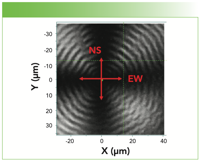
Although a polymer physicist is well aware of the formation of spherulites in semicrystalline polymers, it is an amazing experience to see them for the first time in an optical microscope with crossed-polar illumination. Figure 1 shows a spherulite in a film of PHBHx polyhydroxybutyrate-13% hexanoate that had been annealed for 36 h at 70 oC. Our understanding of the structure that accounts for what is observed is that the crystallites are nucleated at the center of these features and grow radially along the axis of maximum crystal growth. The sizes of the spherulites will depend on a variety of factors (including the number of nucleation sites, the structure of the molecules, and the cooling rate or annealing temperature), and can range between micrometers to centimeters. One might assume that the radial direction corresponds to the chain axes, but, in fact, it is often a different axis. In PHBHx, it is the a-axis that is the axis of optimum growth (1), and a is also the axis of the unusual interaction between one of the methyl protons on one chain in the unit cell and the carbonyl in the second chain in the unit cell (2).
FIGURE 1: Image of a spherulite of PHBHx (13% hexanoate) recorded with 100x magnification between crossed polars. The directions are labeled so that the polarization properties can be discussed.
The spherulites are actually a three-dimensional (3D) spherical structure comprised of lamellae that align along the radial axes. Lamellae are polymer chains that fold back and forth on each other in a direction normal to the growth direction. Lamellae define the crystalline regions, with the disordered chains between the lamellae being amorphous. For those readers who are unfamiliar with these concepts, I recommend looking at the Wikipedia article on polymer spherulites. In the case of PHBHx, the lamellae actually twist around the growth axis (3).
Results
These concepts should affect the Raman polarization behavior. In the instrument being used, which is the XploRA Plus, the laser (532 nm) polarization is east-west (EW) at the microscope stage. Therefore, in an EW line scan through the center, the laser will be polarized along the a-axis. In a north-south (NS) line scan, the laser polarization will be normal to a. The c-axis would not be unique in this system because the lamellae are rotating around the growth axis (3). If we do a line scan at 45o to NS or EW, we expect to see a spectrum that is an average.
Figure 2 shows profiles through the center nucleation site (as accurately as could be determined) for the three bands that have been associated with crystallinity. The phonon at 80 cm-1 represents beating of the two chains in the unit cell. The carbonyl band is actually several overlapping bands where the frequency downshifts in the crystalline state. The CH band at 3000 cm-1 becomes a doublet in the crystalline form, and it has been interpreted to be a splitting of the band at 3000 cm-1 when the chains in the unit cell interact (4). Except for the carbonyl profiles in the 0% Hx sample, the diagonal profiles (not shown) are approximately equal to the average of the vertical and horizontal profiles. We believe that the reason for this discrepancy is the large increase in intensity of the crystalline component in the vertical profile of the 0% sample, as shown in Figure 3.
FIGURE 2: Line profiles of the spectral lines that are associated with crystalline PHBHx for 0 and 13% Hx. The darkest lines are for vertical profiles, the lightest lines are for diagonal profiles and the middle intensity lines are for horizontal profiles. For all subfigures, the abscissa label is Distance (μm) and the ordinate label is Intensity (counts).
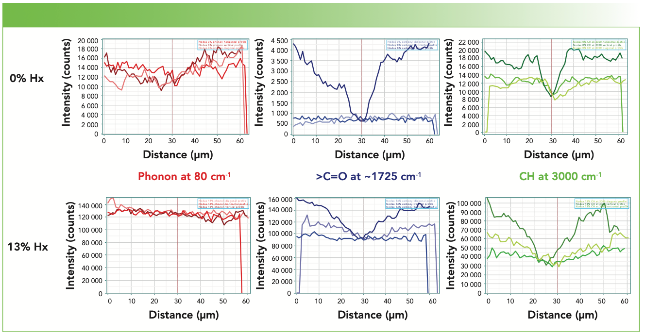
FIGURE 3: Spectra averaged over a thin slice of the spherulite maps for high crystallinity (0%) and low crystallinity (13%) Nodax. The spectra were scaled to the maximum intensity at approximately 2930 cm-1.
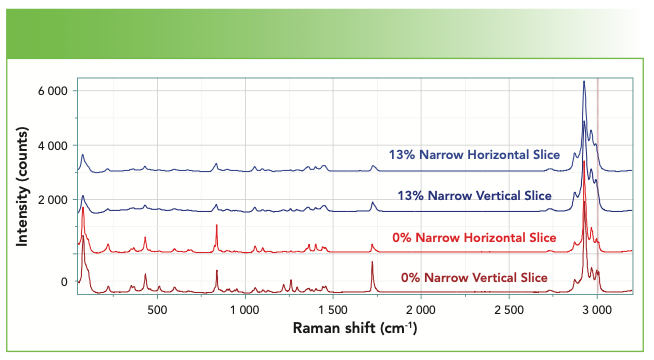
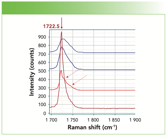
Figure 3 shows the spectra (from top to bottom) of narrow horizontal and vertical slices of the low crystallinity samples, and then horizontal and vertical slices of the high crystallinity samples. To validate the conjecture in the previous paragraph, I show the carbonyl region expanded in Figure 4. Note that the spectra of the low crystallinity material at the top shows only approximately a 30% increase in intensity in the vertical slice, but in the spectra of the high crystallinity material at the bottom, there is a very large increase in the crystalline component for the vertical profile. In addition, I have placed arrows to indicate some subtle features buried in the broad amorphous bands.
FIGURE 4: The carbonyl region from the previous slide is expanded to indicate better the differences in behavior.
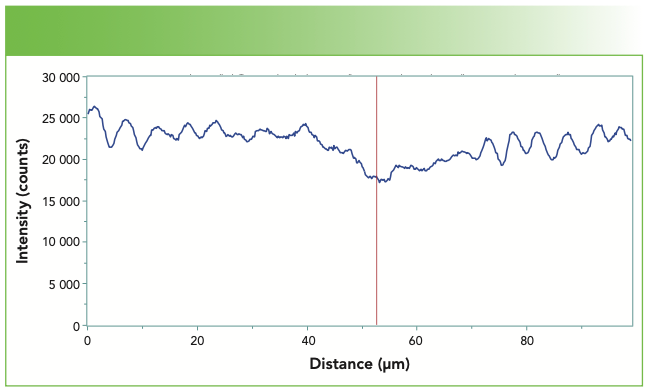
I am currently recording line profiles with a higher groove density grating and more closely spaced data points. Under these conditions, we are clearly able to correlate the bands in the micrograph to differences in the Raman spectra. Figure 6 shows one such scan, revealing the variation in the intensity of the carbonyl band in a horizontal cut through the center of a spherulite of the pure PHB sample. What is curious is that the 0% sample does not show the banding in the micrograph recorded between crossed polars. By systematically measuring radial slices of spherulites, we should be able to understand what the role of polymer orientation has to do with the bands seen in the micrographs.
FIGURE 5: Expansion of the spectra of Figure 3 in the CH region. In particular the relative intensities of the anomalously high CH bands (2997 and 3009 cm-1) can be compared.
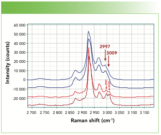
FIGURE 6: Intensity of the carbonyl band as a function of distance along a horizontal cut through a spherulite center in a pure PHB sample (0% Hx).
Summary
This project started because I wanted to compare spectra of different PHBHx samples with different concentrations of Hx. Because I need to eliminate polarization and orientation effects on relative intensities to compare samples, it occurred to me that mapping a spherulite and calculating the average spectrum would eliminate these effects. This idea stunningly opened up new possibilities. Not only can I see intensity ripples as a function of radial distance, but by comparing spectra between horizontal and vertical cuts I can start analyzing the polarization and orientation behavior. Clearly, there is a lot of information coming from these measurements that we will be able to exploit in a more complete analysis of the spectra of this material.
References
(1) Liu, C.; Noda, I.; Martin, D. C.; Chase, D. B.; Ni, C.; Rabolt, J. F. Growth of Anisotropic Single Crystals of a Random Copolymer, Poly[(R)-3-hydroxybutyrate-co-(R)-3-hydroxyhexanoate] Driven by Cooperative –CH···O H-Bonding. Polymer 2018, 154, 111–118. DOI: 10.1016/j.polymer.2018.08.046.
(2) Sato, H.; Dybal, J.; Murakami, R.; Noda, I.; Ozaki, Y. Infrared and Raman Spectroscopy and Quantum Chemistry Calcula- tion Studies of C–H···O Hydrogen Bondings and Thermal Behavior of Biodegradable Polyhydroxyalkanoate. J. Mol. Struct. 2005, 744–747, 35-46, DOI: 10.1016/j.molstruc.2004.10.069
(3) Discussion with Isao Noda
(4) Adar, F.; Street, R.; Noda, I. Isothermal Crystallization of Polyhydroxyalkanoate (PHA) Utilizing Raman Spectroscopy to Follow Chain Packing As Well As Molecular Motion, Spectrochim. Acta, Part A, Accepted September, 2022.
Fran Adar is the Principal Raman Applications Scientist for Horiba Scientific in Edison, New Jersey. Direct correspondence to: SpectroscopyEdit@mmhgroup.com. ●


Chinese Researchers Develop Dual-Channel Probe for Biothiol Detection
April 28th 2025Researchers at Qiqihar Medical University have developed a dual-channel fluorescent probe, PYL-NBD, that enables highly sensitive, rapid, and selective detection of biothiols in food, pharmaceuticals, and living organisms.
New Study Reveals Insights into Phenol’s Behavior in Ice
April 16th 2025A new study published in Spectrochimica Acta Part A by Dominik Heger and colleagues at Masaryk University reveals that phenol's photophysical properties change significantly when frozen, potentially enabling its breakdown by sunlight in icy environments.
Advanced Raman Spectroscopy Method Boosts Precision in Drug Component Detection
April 7th 2025Researchers in China have developed a rapid, non-destructive Raman spectroscopy method that accurately detects active components in complex drug formulations by combining advanced algorithms to eliminate noise and fluorescence interference.