Tuning the Photoluminescence of Graphene Oxide
Special Issues
The emission wavelength of graphene oxide is tuned through photothermal reduction and the spectral shift monitored using photoluminescence spectroscopy to achieve the desired emission.
The emission wavelength of graphene oxide is tuned through photothermal reduction and the spectral shift monitored using photoluminescence spectroscopy to achieve the desired emission.
Graphene has received tremendous attention due to its remarkable electrical, thermal, and mechanical properties. However, the valence and conduction bands of graphene touch, and pure graphene, therefore, has zero bandgap, and does not exhibit photoluminescence (PL). When oxidized to form graphene oxide (GO), the π electron network of graphene is disrupted, which induces a bandgap, and the material becomes photoluminescent. This opens the door to optoelectronic applications, and is investigated using PL spectroscopy in this application note.
Experimental
An aqueous dispersion of GO (PlasmaChem GmbH) was diluted to 0.02 mg cm-3 and the PL measured using the FLS1000 Photoluminescence Spectrometer equipped with double excitation and emission monochromators and PMT-900 and PMT 1700 photomultiplier tube detectors.
Results
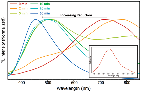
The high scatter and low quantum yield of GO makes it a challenging sample for most PL spectrometers to measure because the monochromators are unable to completely remove the scattered excitation light. The FLS1000 can be equipped with double monochromators, which increases the stray light rejection of the monochromator from 1:1×10⁵ to 1:1×10¹⁰ and ensures the spectra of even the most weakly emitting and highly scattering samples can be measured accurately. The PL spectrum of GO was measured using double monochromators and extends from 350 nm to 1250 nm with a peak at 710 nm (Figure 1 inset).

Figure 1: Change in the PL spectra of GO with increasing photothermal reduction. λex = 300 nm, Δλex = 5 nm, Δλem = 5 nm. Inset: complete spectrum.
To tune the emission wavelength of GO, it was photothermally reduced by exposure to a Xenon lamp and the PL spectra measured after different exposure times (Figure 1). As the exposure time is increased, the PL blue shifts with a maximum at 450 nm after 60 min. The blue shift arises from the relative intensity of the 450-nm peak, increasing at the expense of the 710-nm, which reveals that the peaks correspond to two chemically distinct species, with the 710-nm being converted to the 450-nm species during reduction.
Conclusion
The PL of GO was investigated using the FLS1000 Photoluminescence Spectrometer. Pristine GO was found to exhibit broad PL with a maximum at 710 nm. By photothermally reducing the GO and monitoring the change in emission with the FLS1000, the PL could be tuned from 710 nm to 450 nm.
References
(1) A. Sehrawat, P. Sehrawat, S. S. Islam, P. Mishra, and S. Ahmad, Sci. Rep.8, 3537 (2018).
(2) C. Chien, S. Li, W. Lai, Y. Yeh, H. Chen, I. Chen, L. Chen, K. Chen, T. Nemoto, S. Isoda, M. Chen, T. Fujita, G. Eda, H. Yamaguchi, M. Chhowalla, and C. Chen, Angew. Chem. Int. Ed. 51, 6662 (2012).

Edinburgh Instruments
2 Bain Square, Kirkton Campus
Livingston, EH54 7DQ, United Kingdom
tel. +44 (0) 1506 425 300
Website: www.edinst.com

New Study Reveals Insights into Phenol’s Behavior in Ice
April 16th 2025A new study published in Spectrochimica Acta Part A by Dominik Heger and colleagues at Masaryk University reveals that phenol's photophysical properties change significantly when frozen, potentially enabling its breakdown by sunlight in icy environments.
Advanced Raman Spectroscopy Method Boosts Precision in Drug Component Detection
April 7th 2025Researchers in China have developed a rapid, non-destructive Raman spectroscopy method that accurately detects active components in complex drug formulations by combining advanced algorithms to eliminate noise and fluorescence interference.