The 2019 Emerging Leader in Molecular Spectroscopy Award
Spectroscopy
Ishan Barman, this year’s Emerging Leader in Molecular Spectroscopy award recipient, leads a research team combining spectroscopy, imaging, and chemometrics to seek greater understanding of the pathological changes of human cells and tissues.
This year's molecular spectroscopy award recipient, Ishan Barman, is an energetic and accomplished young researcher in biomedical Raman spectroscopy. With a combination of spectroscopy, imaging, and chemometrics, his work unites structural and molecular information to provide greater detail to our understanding of pathological changes in cells and tissues.
Ishan Barman is an associate professor at Johns Hopkins University in Baltimore, Maryland. He is the 2019 winner of the Emerging Leader in Molecular Spectroscopy Award, which is presented by Spectroscopy magazine. This annual award recognizes the achievements and aspirations of a talented young molecular spectroscopist who has made strides early in his or her career toward the advancement of molecular spectroscopy techniques or applications. The winner must be within 10 years of receiving his or her highest academic degree in the year the award is presented. The award will be presented to Barman at the SciX Conference, which will be held October 13–18, 2019, in Palm Springs, California, where he will give a plenary lecture and be honored in an award symposium.
Barman received his PhD in 2011 from the Massachusetts Institute of Technology, in Cambridge, Massachusetts. Following post-doctoral research at the Laser Biomedical Research Center (LBRC) at MIT from 2011 to 2013, he then became an assistant professor at Johns Hopkins University, in mechanical engineering with a joint appointment in the Department of Oncology in 2014. In 2018, he added another joint appointment, in the Department of Radiology and Radiological Science. He has since been promoted to associate professor.
Barman has published more than 75 peer-reviewed papers, holds 9 patents and patent applications, and has presented more than 150 papers and talks at scientific conferences. His work has been extensively featured in leading scientific media (Technology Review, Physics Today, Physics World, C&E News) and in popular media outlets (Wall Street Journal, CNN Newsroom with Ali Velshi). He has received several awards and scholarships, including the National Institute of Health (NIH) Director’s New Innovator Award; the Outstanding Young Engineer award from the Maryland Academy of Sciences; an Emerging Investigator award from the Royal Society of Chemistry; the Dr. Horace Furumoto Innovations Young Investigator Award from the American Society for Laser Medicine and Surgery; and the Tomas A. Hirschfeld Award from the Federation of Analytical Chemistry and Spectroscopy Societies (FACSS). In total, he has received more than 30 grants, fellowships, and awards. With an impressive h-index of 29, Barman’s research encompasses medical and pharmaceutical applications by exploring new interdisciplinary research questions at the interface of biomedicine and engineering.
When he was a student at MIT, Barman’s efforts focused on addressing the many challenges that impede optical spectroscopy-based noninvasive blood glucose detection. As a faculty member at Johns Hopkins, his research program has expanded to include the development of spectroscopic imaging for tissue analysis, novel surface-enhanced Raman scattering (SERS) probes for liquid biopsy and high-precision in vivo cancer imaging, and new single-cell analysis platforms with combined capture, manipulation and sensing capabilities. In recent years, his group has published peer-reviewed papers in Cancer Research, Nature Materials, Nano Letters, Angewandte Chemie Intl. Ed., Chemical Science, Small, Nanoscale, ACS Sensors, ACS Photonics, Analytical Chemistry, and Accounts of Chemical Research.
Professor Rohit Bhargava, the director of the Cancer Center at Illinois and the Founder endowed professor in engineering at the University of Illinois at Urbana-Champaign, has known Barman for more than a decade, since Barman was a graduate student at MIT. “Ishan is simply brilliant,” said Bhargava. “He has such a great understanding of spectroscopy and science, and is always so wonderful to talk to and discuss new ideas.”
Manoharan Ramasamy, the Global Lead for the Process Analytics Center of Excellence for Small Molecules at Merck, has also been impressed by Barman’s work. “In a very short span of a few years and at this very young age, Ishan has significantly advanced the field of science in many areas,” he said. “His work has been published in highly cited, major refereed journals, and he has been awarded with many patents and recognized with a number of awards.” Ramasamy also described Barman’s developments as “groundbreaking.”
Even with a crowded research schedule, Barman continues volunteer work at a prolific pace. He is a volunteer in scientific societies and groups, a conference session organizer (FACSS/SciX), and an active technical reviewer for more than 50 scholarly journals.
Designing Optical Pathophysiologic Sensing Tools
Barman’s work in Raman spectroscopy focuses on translating new photonic tools from conceptualization through each aspect of design and fabrication, typically for in vivo measurements. Once measurements are obtained, he uses machine learning to determine differential molecular markers that enable objective disease detection and outcome prediction. Yukihiro Ozaki, professor emeritus at Kwansei Gakuin University in Japan, commented that in the rapidly expanding field of molecular spectroscopy, Barman stands out in terms of ingenuity and scholarly depth. “I am deeply impressed by Dr. Barman’s creativity, his broad skill set, and his willingness to explore new interdisciplinary research questions at the boundaries of biomedicine and engineering,” Ozaki wrote in a letter supporting Barman’s nomination for the award. “His work is innovative and inventive, and has transcended traditional disciplinary boundaries.”
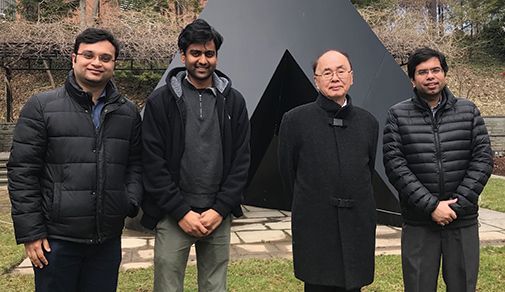
Figure 1: At Johns Hopkins University. Left to right: Soumik Siddhanta, a post-doctoral researcher; Santosh Paidi, a doctoral student; Professor Yukihiro Ozaki, and Barman (photo courtesy of Y. Ozaki).
As an MIT doctoral student, Barman was guided by the late Professor Michael Feld and by Dr. Ramachandra R. Dasari, the Associate Director of the Laser Biomedical Research Center (LBRC) and the George R. Harrison Spectroscopy Laboratory at MIT. There, he developed techniques for sensitivity and specificity improvements in clinical Raman measurements by designing a turbidity correction framework to mitigate sampling differences between patients. He also developed a spectroscopic concentration correction model to compensate for the difference in concentration of metabolites between blood and interstitial fluid compartments. Noninvasive glucose concentration measurements are plagued by the physiological time lag between interstitial fluid (ISF) glucose and blood glucose. This lag is problematic for spectroscopic techniques, which measure the glucose in the ISF, not the blood. Because blood glucose is used for calibrating spectroscopic instruments and is the important glucose level for clinical decision-making, there are major problems associated with the lag. To resolve this problem, Barman and colleagues developed a dynamic concentration correction (DCC) scheme; using the mass transfer of glucose between ISF and blood, they were able to ensure greater consistency between blood measurements and spectral measurements (1,2). This correction allows one to compute the expected ISF glucose level from the blood glucose levels, to provide more accurate spectroscopic calibration.
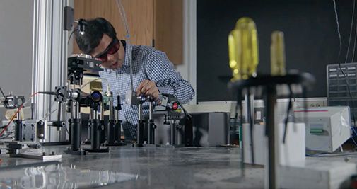
Figure 2: Barman making optical bench adjustments for Raman spectroscopic measurements (photo courtesy of I. Barman).
Barman also developed innovative data processing improvements for Raman, such as support vector machines (SVM), to account for nonlinear effects in the spectra–concentration relationship. Dasari was very impressed with the work Barman did with him at MIT, and the work he has done since. “His research output has been staggering with numerous quality publications,” Dasari said. “His work is highly valued in the field and is well cited.”
Decoding the Molecular Pathology of Cancers
Barman has maintained a principal focus on Raman spectroscopy-based tissue analysis that will permit disease detection prior to morphologic manifestation, focus target searches in pharmacological research, and enable patient stratifications for more effective therapy. His recent work has led to increased understanding of cancer etiology relating to metastasis. His work to integrate spectroscopic measurement with biopsy needles provides a new tool with the potential for real time cancer detection (3). This optical sensor is designed to be used to simultaneously identify microcalcification status and diagnose any underlying breast lesions in real time.
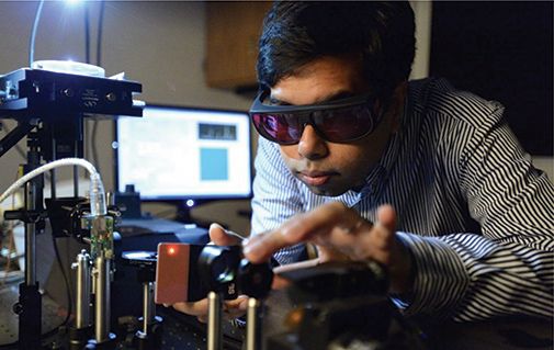
Figure 3: Barman homing-in on cancer cell detection using his custom Raman microscope (photo courtesy of I. Barman).
Barman has also designed and produced customized SERS probes exhibiting near single-molecule sensitivity for use in liquid biopsy platforms for diagnosis as well as for therapy response monitoring. His laboratory is further engineering targeted nanoprobes for high-precision in vivo imaging of cancers to provide real-time assessment of localized tumor burden and micrometastatic satellite lesions. In what has been referred to as a milestone paper written in 2017, Barman and colleagues introduced Raman spectroscopy and chemometric techniques for detection of changes in organs of future metastasis that are actively modified by the primary tumor before metastatic spread has even occurred. The study reported the use of these techniques in identifying premetastatic molecular adaptations in lungs-prior to observation of corresponding morphological changes-in response to primary breast tumors in mouse models (4). This approach shows the potential for revealing new drugable targets to block metastasis, and may be used to guide treatment regimens that achieve maximum cancer cell kill and arrest premetastatic adaptations.
In related work that year, label-free Raman spectroscopy was used to identify spectral bands for defining organ-specific cancer cell metastases. Decision algorithms applied to the Raman spectra were able to discriminate the unique biological cell types despite their isogenic profile. The findings of this work provided strong evidence that metastatic spread generates tissue-specific adaptations at the molecular level within cancer cells (5).
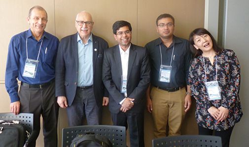
Figure 4: Left to Right: Igor Lednev of the University at Albany SUNY, Valery Tuchin of Saratov State University, in the Russian Federation, Ishan Barman, Hemanth Noothalapati, of Shimane University in Japan., and Ms. Miyuki Yamamoto. Photo taken at the Japan-Taiwan Medical Spectroscopy International Symposium and 14th Annual Meeting of the Japan Association of Medical Spectroscopy, on Awaji Island, Japan, December 4–7, 2016. The photo was taken following a traditional Japanese tea ceremony conducted by Ms. Yamamoto (photo courtesy of I. Lednev).
“His contributions to biomedical optics and spectroscopy have been fundamental and substantial, and he has led the technological development and clinical translation of several noninvasive or minimally invasive optical tools,” commented Dasari of MIT, who added that Barman exhibits a rare combination of creativity, diligence, and maturity. “These characteristics place Barman as a top individual in his peer group in biophotonics and molecular spectroscopy,” he said.
Raman Spectroscopic Determination of Long-Term Glycemic Markers for Diabetes Monitoring
Another area of focus for Barman has been diabetes research. In 2012, Barman was able to demonstrate glycated albumin detection and quantification using Raman spectroscopy without the addition of reagents (6). Glycated albumin is an important marker for monitoring the short-term glycemic history of diabetics.
Barman and coworkers also demonstrated the potential of Raman spectroscopy for quantitative detection of hemoglobin A1c (HbA1c), without using any external molecular probes or dyes, a test that reveals the average level of blood sugar in a patient over the preceding few months (7). The technique harnessed for this test is called drop coating deposition Raman(DCDR) spectroscopy. The causality behind making this measurement is a series of subtle but highly reproducible changes in the Raman blood spectra related to nonenzymatic glycosylation (glycation) of the hemoglobin molecule. These measurable spectral changes provide accurate determination of glycated and nonglycated hemoglobin when Raman spectra are analyzed using chemometric methods.
Fusion of Raman Spectroscopy and Optical Microscopy: A Multimodality Paradigm for Cell and Tissue Analysis
Chemical imaging of cancer cells is another challenge that Barman has taken on. For this work, Barman and coworkers have developed a multimodal microscopy system (MMS), incorporating confocal Raman, confocal reflectance, and quantitative phase microscopy (QPM) into a single imaging instrument (8). Confocal reflectance and quantitative phase microscopy show detailed spatial morphology as images, and confocal Raman microscopy provides the molecular details. By combining these different microscopic imaging modes, it is possible to acquire detailed morphological and chemical information without using external stains or molecular probes.
This work was extended in the development of a portable, optical fiber probe–based spectroscopic tissue scanner for quantitative diagnostic imaging of tissue during surgical procedures (9). The scanner prototype was tested in human specimen for intraoperative breast cancer margin assessment. Spectroscopic techniques used for the tissue scanner included both diffuse reflectance spectroscopy (DRS) and intrinsic fluorescence spectroscopy (IFS). The instrument was designed for hyperspectral imaging capabilities such that the DRS and IFS spectra are collected for each scanned image pixel.
Other cancer detection research of Barman’s group has involved the development of a plasmon-enhanced Raman spectroscopic assay featuring nanostructured biomolecular probes and spectroscopic imaging (10). This system is tailored for multiplexed detection of breast cancer biomarkers, such as cancer antigen (CA) 15-3, CA 27-29, and cancer embryonic antigen (CEA). The tags used for the SERS measurements are functionalized with monoclonal antibodies to enable detection of CA15-3, CA27-29, and CEA. These biomarkers are detected using antibody–antigen interactions, resulting in a functional sandwich spectro-immunoassay device.
Michael Walsh, an assistant professor at the University of Illinois at Chicago, is among those impressed by Barman’s work in cancer diagnostics. “The paper that Ishan published this year in Cancer Research (11) is a landmark paper that brings a significant amount of attention to the spectroscopy field from the wider cancer community,” he said. “This paper demonstrates the important role that spectroscopy will have in the future of precision medicine.”
Translational Endeavors and Broader Impact
Barman has also worked to develop simple and cost-effective substrates for SERS detection that are synthesized from silver ink on paper (12). Paper-fluidics assays have emerged as promising biodetection platforms owing to their salient advantages in terms of ease of manufacture, storage, transport and biodegradability. The reported ink-based coating technique ensures uniform absorption for strong and reproducible plasmonic enhancement for SERS measurements. The simple fabrication process combined with the control over analyte flow patterns offers significant boost for development of paper-based SERS analytical devices, especially for use in resource-limited settings.
Success in SERS applications depends on the specific nanomaterial substrates (or tags) used to induce the enhancement effect. Barman and his team leveraged silver-core, gold-shell nanoparticles to differentiate between closely related human and murine antibody drugs in solution with very high specificity and accuracy (13). Notably, the biologic sample set included antibodies belonging to the same isotypes that have the same amino acid sequence in their constant region, which constitutes the major part of the structure.
Quantitative determination of aggregation and particle formation for antibody–drug conjugate (ADC) therapeutics with label-free Raman spectroscopy also has been recently addressed by Barman (14). Characterization of protein aggregation is of particular significance, as aggregates may lose the intrinsic pharmaceutical properties as well as engage with the immune system instigating undesirable downstream immunogenicity. In this work, label-free Raman spectroscopy is used in combination with a support vector machine (SVM)-based regression model to provide fast and accurate analysis (via chemometric prediction) for a wide range of protein aggregates. The proposed Raman spectroscopy-based method shows strong potential for stability testing and lot release as part of a quality control and manufacturing strategy of these ADCs to ensure safe and efficacious therapeutic material for human patients. It is essential that the chemical subtleties be known so that the end products are precisely consistent, both chemically and physically; this level of quality control enables production of biopharmaceutical products exhibiting consistent clinical and toxicological activity.
Specialized Data Analysis and Chemometrics
An underlying enabling development in Barman’s work has been the application of a variety of chemometric techniques; these techniques have allowed Barman to advance the field of spectroscopic measurements. His group has also used chemometric methods to analyze laser-induced breakdown spectroscopy (LIBS) data for material identification including development of a genetic algorithm-derived classifier that demonstrates statistically significant improvement in classification accuracy over more traditional approaches with an order of magnitude fewer discrimination feature (factor) requirements (15).
The Characteristics and Fruit of Enthusiasm
Taking on the challenges of biomedical applications of Raman spectroscopy and achieving such valuable advances doesn’t just require intellect; other personal qualities play an essential role. One such quality is enthusiasm. “In my numerous interactions with [Barman], what stands out is his clarity of thought and energy in taking on the challenging problems,” said Ozaki. “He brings a special level of enthusiasm to everything he does.” Ozaki recalled that when he and Barman first met, Barman had just read one of Ozaki’s articles on identifying primary algebraic structures that describe the chemical reaction system in terms of spectroscopic observables. “Within ten minutes, he came up with so many new ideas, and he was peppering me with many questions,” said Ozaki. “It was amazing and exciting. We wrote an article a couple of years later that was based on this conversation. I always feel energized after speaking with him!”
Another valuable quality for such work is having a mindset for interdisciplinary study. “What I have come to appreciate most is Ishan’s ability to seamlessly work across multiple disciplines and make fundamental contributions at each step,” said Igor K. Lednev, a professor at the University at Albany, SUNY. As examples, Lednev described other less noted, yet extremely important, contributions that Barman and his research group have made, such as the development of real-time chemical imaging methods for the diagnosis of middle ear pathology, and the development by Barman and collaborators of an inexpensive multiwavelength fluorescence imaging otoscope using readily available components. “His exceptional productivity, coupled with the quality of his work in next generation diagnostic oncology, has been widely recognized,” Lednev concluded.
Another valuable quality is the ability to work with others-both colleagues and students. “Ishan has excellent interpersonal skills,” noted Dasari. “Ishan’s infectious personality and people skills bring together groups of any size and help them work effectively together. He also displays great patience and understanding in mentoring junior researchers.”
Future Prospects
Given his success to date, Barman’s prospects for the future are especially bright. Barman focuses his work at the interface of medicine and vibrational spectroscopy, and he is ready to make substantial contributions that directly address patient quality of care in cancer and other diseases. “I am confident Barman will provide some truly practical and robust solutions that are actually helpful to cancer patients as well as making important advancements in the field of spectroscopy,” said Bhargava.
Ozaki agreed. “Given his record of diverse and impactful contributions, he is well poised to be a leader in biomedical optics and spectroscopy for years to come,” he said, “I also expect that Barman will be instrumental in nurturing the next generation of biomedical engineers and scholars.”
Dasari echoed those comments. “I simply do not know any limits to his ability to enter a field, master its frontier, and roll those frontiers back to open new applied domains,” he said.
References
(1) I. Barman, C.R. Kong, G.P. Singh, R.R. Dasari, and M.S. Feld, Anal. Chem. 82(14) 6104–6114 (2010).
(2) N.C. Dingari, I. Barman, G.P. Singh, J.W. Kang, R.R. Dasari, and M.S. Feld, Anal. Bioanal. Chem. 400(9), 2871–2880 (2011).
(3) I. Barman, N.C. Dingari, A. Saha, S. McGee, L.H. Galindo, W. Liu, D. Plecha, N. Klein, R.R. Dasari, and M. Fitzmaurice, Cancer Res. 73(11), 3206–3215 (2013).
(4) S.K. Paidi, A. Rizwan, C. Zheng, M. Cheng, K. Glunde, and I. Barman, Cancer Res. 77(2), 247–256 (2017).
(5) P.T. Winnard Jr, C. Zhang, F. Vesuna, J.W. Kang, J. Garry, R.R. Dasari, I. Barman, and V. Raman, Oncotarget8(12), 20266 (2017).
(6) N.C. Dingari, G.L. Horowitz, J.W. Kang, R.R. Dasari, and I. Barman, PLoS One 7(2), e32406 (2012).
(7) I. Barman, N.C. Dingari, J.W. Kang, G.L. Horowitz, R.R. Dasari, and M.S. Feld, Anal. Chem. 84(5), 2474–2482 (2012).
(8) J.W. Kang, N. Lue, C.R. Kong, I. Barman, N.C. Dingari, S.J. Goldfless, J.C. Niles, R.R. Dasari, and M.S. Feld, Biomed. Opt. Express2(9), 2484–2492 (2011).
(9) N. Lue, J.W. Kang, C.C Yu, I. Barman, N.C. Dingari, M.S. Feld, R.R Dasari, and M. Fitzmaurice, PloS one, 7(1), e30887 (2012).
(10) M. Li, J.W. Kang, S. Sukumar, R.R. Dasari, and I. Barman, Chem. Sci. 6(7), 3906–3914 (2015).
(11) S.K. Paidi, P.M. Diaz, S. Dadgar, S.V. Jenkins, C.M. Quick, R.J. Griffin, R.P Dings, N. Rajaram, and I. Barman, Cancer Res.79(8), 2054–2064 (2019). doi: 10.1158/0008-5472.
(12) Z. Huang, S. Siddhanta, C. Zhang, T. Kickler, G. Zheng, and I. Barman, J. Raman Spectrosc. 48, 1365–1374 (2017).
(13) S.K. Paidi, S. Siddhanta, R. Strouse, J.B. McGivney, C. Larkin, and I. Barman, Anal. Chem. 88(8), 4361–4368 (2016).
(14) C. Zhang, J.S. Springall, X. Wang, and I. Barman, Anal. Chim. Acta,1081, 138–145 (2019).
(15) A.K. Myakalwar, N. Spegazzini, C. Zhang, S.K. Anubham, R.R. Dasari, I. Barman, and M.K. Gundawar, Sci. Rep. 5, 13169 (2015).
Jerome Workman, Jr. is the Senior Technical Editor for Spectroscopy and LCGC North America. Direct correspondence to jworkman@mmhgroup.com
New Study Reveals Insights into Phenol’s Behavior in Ice
April 16th 2025A new study published in Spectrochimica Acta Part A by Dominik Heger and colleagues at Masaryk University reveals that phenol's photophysical properties change significantly when frozen, potentially enabling its breakdown by sunlight in icy environments.
Advanced Raman Spectroscopy Method Boosts Precision in Drug Component Detection
April 7th 2025Researchers in China have developed a rapid, non-destructive Raman spectroscopy method that accurately detects active components in complex drug formulations by combining advanced algorithms to eliminate noise and fluorescence interference.