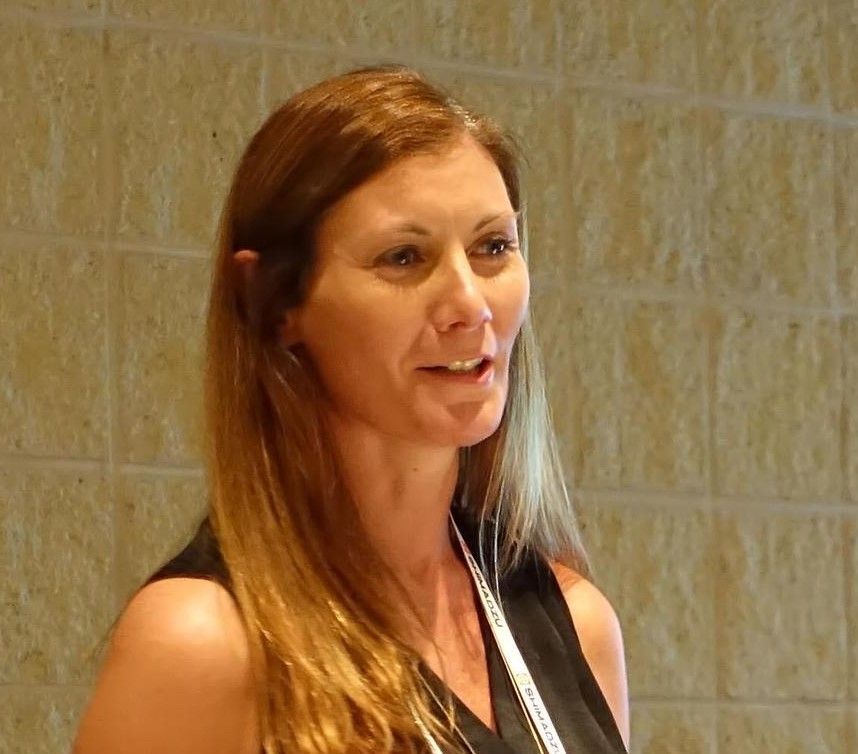
A Decade of Surface Enhanced Spatially Offset Raman Scattering (SESORS)
In 2011, Karen Faulds of the University of Strathclyde and her collaborators published a paper where surface enhanced spatially offset Raman spectroscopy (SESORS) imaging was first explored. Ten years later, Faulds co-authored a paper where SESORS signals could be detected from nanotags at depths down to 48 mm for the first time using a handheld spatially offset Raman (SORS) instrument. Faulds, who recently spoke to us about these papers and the advances in the science that made them possible, is the 2022 recipient of the RSC Analytical Division Mid-Career Award, presented annually for the most meritorious contributions to any area of analytical chemistry made by a mid-career scientist. This interview is part of an ongoing series of interviews with the winners of awards that are presented at the annual SciX conference, which will be held this year from October 2 through October 7, in Covington, Kentucky.
You first explored the concept of surface enhanced spatially offset Raman spectroscopy (SESORS) a little over a decade ago (1). What are some of the benefits of combining both surface enhanced Raman spectroscopy (SERS) and deep Raman techniques together?
We published the first papers on SESORS in 2010 and 2011 with Nick Stone and Pavel Matousek, where we demonstrated we could detect nanoparticles through up to 50 mm of tissue using a transmission-based approach. It is very challenging to use optical techniques to detect through tissue, due the absorbance and scattering of the tissue. Normal Raman will only penetrate to just below the subsurface, so it doesn’t give information about what is going on at depth. Matousek invented and developed the SORS technique in the early 2000’s where it became possible to detect Raman at the subsurface. However, the depth which signals can be detected down to was still limited by the fact that Raman is an intrinsically weak effect. Therefore, combining it with SERS, to give SESORS, allowed much deeper depths to be probed, since now a much larger Raman signal is being detected due to surface enhancement, enabling detection at much greater depths to become achievable.
Now, approximately ten years later, you published a paper outlining the depth prediction of nanotags in tissue using this process (2). How have the advances in the science between the two papers made it so that depth prediction became possible?
The original work we carried out (1) used a transmission approach which requires collection of the Raman scattering at the opposite side of the sample to the excitation source. We have been working to develop the SESORS approach using a SORS backscattering geometry where excitation and collection are spatially offset from each other (excitation and collection occurs on the same side of the sample), with a spatial offset. Since the first paper, we built our own simple, in-house SORS system (3) allowing us to control the optics and the spatial offset more easily, thereby giving us the ability to better understand and measure the scattering parameters. The recent work by Matthew Berry and Samantha McCabe (2) has allowed us to use a ratiometric approach, using SERS peak intensity ratioed with a tissue Raman band from the matrix, to allow us to develop a depth calibration model to predict the depth of nanoparticles in tissue. This work has also recently been applied to imaging and trying to understand the 3D location of nanoparticles buried at depth in tissue from the 2D image that is observed at the surface of the tissue when it is mapped (4). This results in linear offset-induced drag in the 2D surface image, so it does not accurately give the actual location of the nanoparticles due to the nature of the collection and the offset used. This experimental work maps theoretical work using Monte Carlo simulations carried out by Pavel’s group, and essentially it is getting to the point that the depth and location of nanoparticles at depth can be measured and predicted using SESORS. However, that said, it is very dependent on the tissue matrix, and if different tissue compositions, fat, tissue layers are present, this will affect the depth prediction model, so there is still some way to go in predicting depth and location in an in vivomodel!
Briefly discuss your overall discoveries concerning this measurement process and its implications. Which development was most critical for this to be developed—instrumental advancements or nanotag creation? Were you surprised by the results achieved?
Our focus has very much been on the chemistry aspects, and the development of different nanoparticles has allowed us to reach greater depths. There is now a portable SORS system on the market which we have also been using to make measurements. We have developed advances in the nanoparticles by introducing different Raman reporter molecules that are in resonance with the excitation wavelength to give surface enhanced resonance Raman scattering (SERRS) in combination with SORS to give SESORRS. We published the first paper on this in 2018 (5), which allowed us to detect nanoparticles at a depth of 25 mm through tissue, and in a tumor model through 15 mm of tissue, using the handheld SORS system. This was in collaboration with Mike Detty at the University at Buffalo, who very sadly recently passed away, who made wonderful red shifted dye molecules, which gave fantastic SERS responses at the NIR wavelengths required for use in tissue. Our recent work has used silica coated small aggregates of gold nanoparticles to give a large SERS response, while remaining small enough to still have potential use in vivo, that give us excellent SERS responses which have allowed detection up to 48 mm through tissue (2,4). Our chemistry expertise is definitely more focused on the nanoparticle design, and we leave the instrumental developments to the experts!
Are there any challenges or limitation involved with utilizing Raman measurements using nanotags? What options or alternative developments are available to overcome these challenges or to improve your approach?
The future potential utilization of SESORS for in vivo measurements is a challenge. The nanoparticles can be passivated and coated, which renders them non-toxic. However, a lot of work is focused on targeting nanoparticles to, for example, a tumor site, and this is potentially problematic, particularly if administered by systemic injection. The nanoparticles are known to accumulate in other parts of the body, such as the liver, spleen, and kidneys as well as at the tumor site, and this could lead to off target effects. Therefore, the use of localization of nanoparticles to allow deeper detection needs to be utilized in the right environment, with local injection being preferable to systemic, and thought is required where this approach would be most beneficial to a patient in terms of detection and treatment through photothermal heating or drug delivery.
What sort of response has your initial paper received from the research community back in 2011, and how does it compare with the response received for your recent paper?
The original work has obtained a lot of citations, and, since then, multiple researchers have started to work on SESORS approaches, and it is great to see some really nice work being published in the area, such as the continued lovely work by Stone and Matousek, as well as work by Bhavya Sharma on the application of SESORS in neurology, and by my former PhD student Fay Nicolson. I guess time will tell about the response to our new work, but it is certainly great that we can all get back out and about to conferences again to talk about our latest research and interact with the community again!
Are there any next steps planned regarding this research?
Oh yes! There are many things planned! But one of the immediate things we want to investigate is how different tissue types, with different fat, tissue, or blood perfusion, will affect these depth- location models, and whether it will ever be possible to apply them to in vivo[A1] models where the tissue architecture will vary between patients.
The RSC Analytical Division Mid-Career Award is presented annually for the most meritorious contributions to any area of analytical chemistry made by a mid-career scientist. Please comment on the meaning of being the 2022 recipient.
I am very honored and humbled that the research work carried out by my research group, past and present, is being recognized by this award. It is testament to all the hard work by the amazing scientists I have had the privilege to work with over the years. Although…mid-career? How did that happen? I still feel like I am just starting out!
References
(1) N. Stone, M. Kerssens, G. Rhys Lloyd, K. Faulds, D. Graham, and P. Matousek, Chem. Sci. 2, 776-780 (2011). DOI: 10.1039/c0sc00570c
(2) M.E. Berry, S.M. McCabe, N.C. Shand, D. Graham, and K. Faulds, Chem. Commun. 58, 1756 (2022). DOI: 10.1039/d1cc04455a
(3) S. Asiala, N.C. Shand, K. Faulds, and D. Graham, ACS Appl. Mater. Interfaces 9(30), 25488-25494 (2017). DOI: 10.1021/acsami.7b09197
(4) M.E Berry, S.M McCabe, S. Sloan-Dennison, S. Laing, N.C. Shand, D. Graham, and K. Faulds,ACS Appl. Mater. Interfaces 14(28), 31613–31624 (2022). DOI: 10.1021/acsami.2c05611
(5) F. Nicolson, L.E. Jamieson, S. Mabbott, K. Plakas, N.C. Shand, M.R. Detty, D. Graham, and K. Faulds, Chem. Sci. 9(15), 3788-3792 (2018). DOI: 10.1039/C8SC00994E
Karen Faulds

Karen Faulds is an expert in the development of surface enhanced Raman scattering (SERS) and Raman techniques for novel analytical detection strategies, in particular multiplexed bioanalytical applications. She has published over 160 publications and her Groups research has been recognized through multiple awards. She is a Fellow of the Royal Society of Chemistry, Society for Applied Spectroscopy and Royal Society of Edinburgh. She is Chair of the Infrared and Raman Discussion Group (IRDG), an elected member of the RSC Analytical Division Council and the Federation of Analytical Chemistry and Spectroscopy Societies (FACSS) Governing Board and a trustee of the Analytical Chemistry Trust Fund (ACTF). She is an Associate Editor for Analyst and co-Editor in Chief of RSC Advances and Analyst and advisory board for Chemical Society Reviews.
AI-Powered SERS Spectroscopy Breakthrough Boosts Safety of Medicinal Food Products
April 16th 2025A new deep learning-enhanced spectroscopic platform—SERSome—developed by researchers in China and Finland, identifies medicinal and edible homologs (MEHs) with 98% accuracy. This innovation could revolutionize safety and quality control in the growing MEH market.
New Raman Spectroscopy Method Enhances Real-Time Monitoring Across Fermentation Processes
April 15th 2025Researchers at Delft University of Technology have developed a novel method using single compound spectra to enhance the transferability and accuracy of Raman spectroscopy models for real-time fermentation monitoring.
Nanometer-Scale Studies Using Tip Enhanced Raman Spectroscopy
February 8th 2013Volker Deckert, the winner of the 2013 Charles Mann Award, is advancing the use of tip enhanced Raman spectroscopy (TERS) to push the lateral resolution of vibrational spectroscopy well below the Abbe limit, to achieve single-molecule sensitivity. Because the tip can be moved with sub-nanometer precision, structural information with unmatched spatial resolution can be achieved without the need of specific labels.