Ancient History Revealed Using Laser Light: Unraveling the Secrets of Roman Egyptian Blue
A study published in Scientific Reports has given intricate details into the production and composition of Roman Egyptian blue pigment. Using advanced Raman microspectroscopy, researchers explored pigment balls and murals from ancient Swiss cities, uncovering evidence of raw material provenance, crystal lattice disorder, and the formation of a copper-bearing green glass phase, revealing the sophisticated techniques employed by Roman artisans.
A recent study, using laser-based Raman spectroscopy, published in Scientific Reports by Petra Dariz and Thomas Schmid from the Federal Institute for Materials Research and Testing (BAM) in Berlin, Germany, delves into the complexities of Roman Egyptian blue pigment, revealing its production, composition, and historical significance.
Raman Spectroscopy Reveals Ancient History
Egyptian blue, a vivid blue pigment widely used during the Roman Empire, has captivated researchers for centuries due to its intricate manufacturing process and rich color. In their study, Dariz and Schmid conducted in-depth comparative analyses of Roman Imperial pigment balls and fragmentary murals from the ancient cities of Aventicum and Augusta Raurica, both ancient Roman cities located in modern-day Switzerland. These findings were built upon a prior investigation of trace compounds within Early Medieval Egyptian blue.
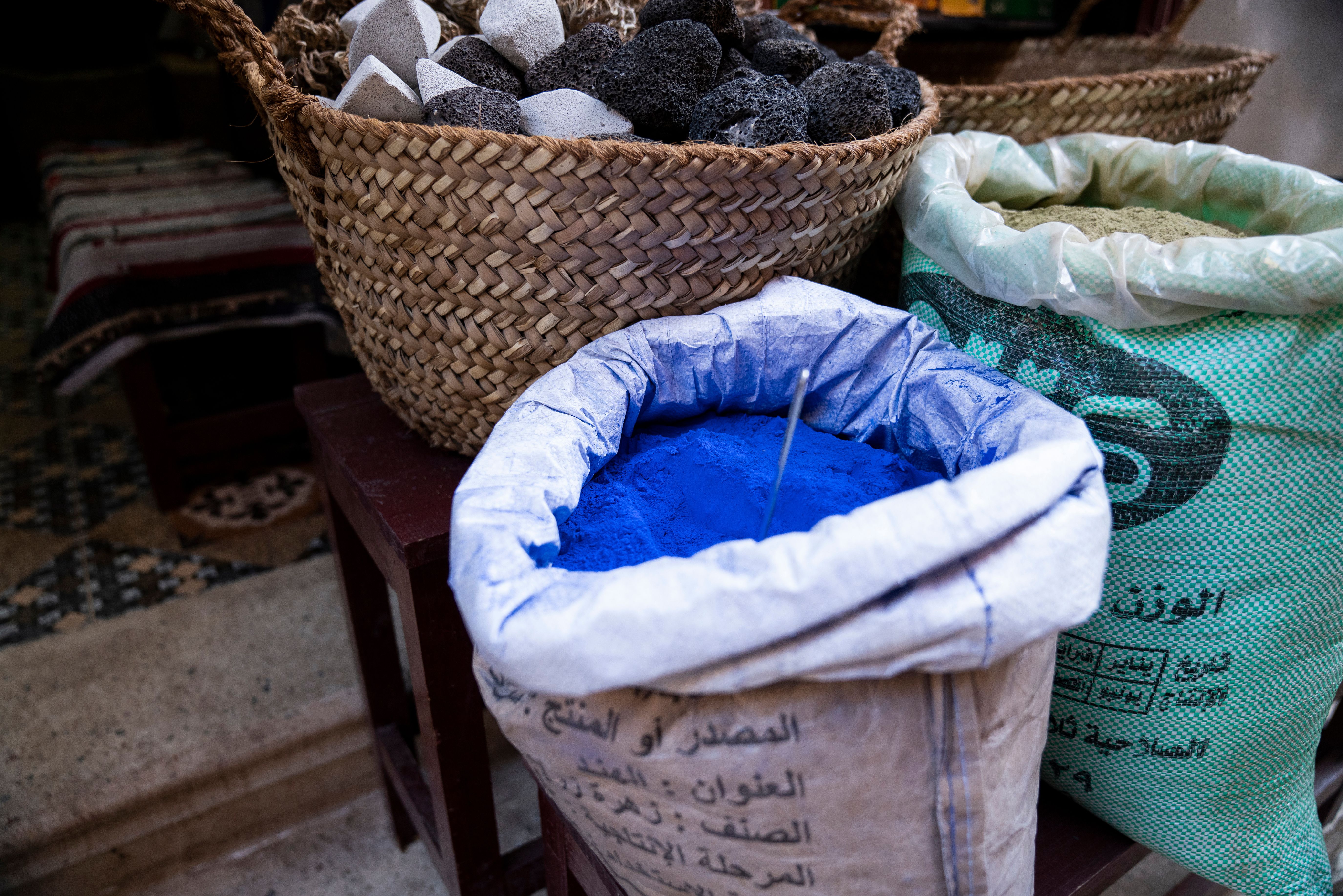
Blue Paint pigment at an Egyptian outdoor market | Image Credit: © Cavan - stock.adobe.com

The researchers employed Raman microspectroscopy to examine these artifacts, resulting in the identification of an array of associated minerals in the raw materials used for pigment synthesis. This led to confirmation of the use of quartz sand consistent with sediments from the Volturno River in Campania, Southern Italy, alongside roasted sulfidic copper ore and mixed-alkaline plant ash as a fluxing agent (1). This supports the existence of a centralized pigment production site in the northern Phlegrean Fields, aligning with historical accounts by Vitruvius (2) and Pliny the Elder (3), ancient Roman authors from the 1st century B.C. and 1st century A.D., respectively.
Volturno river waterfalls, scenic view in summer with green moss, Molise region, Italy | Image Credit: © cenz07 - stock.adobe.com

Raman microspectroscopic imaging was carried out utilizing a commercially available Raman microscope, employing a 532 nm continuous-wave (CW) laser for excitation at a maximum power of 40 mW on the sample surface, which was then moderated to 20 mW using a neutral density filter. This area-covering imaging approach led to the detection of 26 minerals, achieving sub-permille precision, in conjunction with the identification of the chromophoric compound cuprorivaite CuCaSi4O10 (1,4).
The study's significance extends beyond provenance. The Raman spectra uncovered insights into diverse process conditions and compositional irregularities, which affected the crystalline structure of cuprorivaite, the key chromophoric compound in the Egyptian blue pigment color. Additionally, the researchers identified the formation of a copper-bearing green glass phase, likely influenced by alkali flux concentrations. Despite solid-state reactions dominating the synthesis process, these findings indicate that localized melting contributed to glass phase creation (1).
During the Roman era, Egyptian blue was distributed in standardized balls of approximately 15 to 20 mm in diameter. The shade of blue was determined by the painter's choice of grain size. Vitruvius provided a glimpse into the manufacturing process, highlighting the use of sand, copper ore, and alkali flux (2). The study supports the monopoly of Egyptian blue production in the Gulf of Pozzuoli, providing a continuous trade route that persisted over centuries.
Rome, Italy: The Roman Forum. Old Town of the city | Image Credit: © krivinis - stock.adobe.com

The researchers also explored pigment balls and murals from Aventicum and Augusta Raurica, dating back to the first to third centuries A.D. By analyzing trace compounds, particularly those linked to quartz sand and the use of sulfidic copper ore, they reinforced the notion of a centralized pigment production site, further substantiating historical records.
These findings not only deepen our understanding of ancient pigment production and its provenance but also highlight the sophistication of Roman technological capabilities. The comprehensive insights gleaned from Raman microspectroscopy underscore the valuable role of modern scientific techniques in unraveling the mysteries of our artistic past.
Unveiling the Synthesis Process
The careful analysis conducted by Dariz and Schmid offers a notable glimpse into the synthesis process of Egyptian blue, an enigmatic pigment that adorned Roman artifacts. According to historical records, Egyptian blue was crafted in Alexandria and subsequently in Puteoli, with sand and copper ore forming the core ingredients. This research attests to this by identifying the presence of sulfidic copper ore, carefully roasted to yield copper oxide. This crucial step was essential for the pigment's characteristic blue hue (1).
View of Alexandria harbor, Egypt | Image Credit: © javarman - stock.adobe.com

Moreover, the inclusion of an alkaline flux, in the form of soda-rich or mixed-alkaline plant ash, was detected in the study. This finding aligns with the Raman spectra, revealing the presence of sulfate and phosphate salts of sodium, potassium, magnesium, and calcium. These components further illustrate the intricate chemistry and artistry involved in producing Egyptian blue.
Insights into Crystal Lattice Disorder
The research also explored the crystal lattice disorder within cuprorivaite, the central compound responsible for Egyptian blue's vibrant color. Raman spectra highlighted gradual peak shifts and changes in bandwidth, indicative of crystal lattice disorder arising from varying reaction conditions. This phenomenon, potentially due to insufficient reaction time, showcased the complex interplay of factors influencing the pigment's final properties.
Moreover, the study unveiled the formation of a copper-bearing green glass phase, a testament to the nuanced processes underlying Egyptian blue production. The emergence of this phase, influenced by alkali flux concentration, provided a unique perspective into the delicate balance required for achieving the desired color and texture of the pigment.
A Glimpse into History
The pigment balls and murals analyzed in this study, originating from Aventicum and Augusta Raurica, offer a rare glimpse into the Roman artistic legacy. Dated between the first and third centuries A.D., these artifacts illuminate the widespread use of Egyptian blue during the height of the Roman Empire. The thorough investigation of trace compounds in these artifacts corroborated historical accounts, pointing towards a monopoly of pigment production centered in the Gulf of Pozzuoli, as affirmed by ancient writers such as Vitruvius and Pliny the Elder.
As modern science continues to intertwine with archaeology and art history, the work of Dariz and Schmid exemplifies the impact of interdisciplinary research. Their study not only advances our understanding of ancient pigments and their production but also celebrates the intricate craftsmanship that has shaped our cultural heritage. Through their meticulous analysis, Dariz and Schmid have resurrected the vibrant hues that once adorned Roman artworks, offering a new lens through which to appreciate the artistic legacy of antiquity.
References
- Dariz, P.; Schmid, T. Raman Focal Point on Roman Egyptian Blue Elucidates Disordered Cuprorivaite, Green Glass Phase and Trace Compounds. Sci. Rep. 2022, 12, 15596. DOI: 10.1038/s41598-022-19923-w
- Vitruvius (Marcus Vitruvius Pollio). De architectura libri decem (Ten Books on Architecture); 1st century B.C., Book 7, Chapter 11. Available online at University of Chicago Library: http://penelope.uchicago.edu/Thayer/e/roman/texts/vitruvius/7*.html (accessed 2023-08-09).
- Pliny the Elder (Gaius Plinius Secundus). Naturalis Historia (Natural History); 1st century A.D., Translated by John Bostock and H.T. Riley. Available online at Perseus Digital Library: http://www.perseus.tufts.edu/hopper/text?doc=Perseus%3Atext%3A1999.02.0137 (accessed 2023-08-09).
- Jaksch, H.; Seipel, W.; Weiner, K. L. et al. Egyptian blue—Cuprorivaite a Window to Ancient Egyptian Technology. Naturwissenschaften 1983, 70, 525–535. DOI: 10.1007/BF00376668
AI-Powered SERS Spectroscopy Breakthrough Boosts Safety of Medicinal Food Products
April 16th 2025A new deep learning-enhanced spectroscopic platform—SERSome—developed by researchers in China and Finland, identifies medicinal and edible homologs (MEHs) with 98% accuracy. This innovation could revolutionize safety and quality control in the growing MEH market.
New Raman Spectroscopy Method Enhances Real-Time Monitoring Across Fermentation Processes
April 15th 2025Researchers at Delft University of Technology have developed a novel method using single compound spectra to enhance the transferability and accuracy of Raman spectroscopy models for real-time fermentation monitoring.
Nanometer-Scale Studies Using Tip Enhanced Raman Spectroscopy
February 8th 2013Volker Deckert, the winner of the 2013 Charles Mann Award, is advancing the use of tip enhanced Raman spectroscopy (TERS) to push the lateral resolution of vibrational spectroscopy well below the Abbe limit, to achieve single-molecule sensitivity. Because the tip can be moved with sub-nanometer precision, structural information with unmatched spatial resolution can be achieved without the need of specific labels.