Applications of Raman Spectroscopy in Breast Cancer Surgery: A SciX Interview with Ioan Notingher, Part 1
As a preview to the SciX 2024 conference, Spectroscopy sat down with Ioan Notingher to talk about his research.
The SciX 2024 Conference is set to take place from October 20 to 25th, 2024, in Raleigh, North Carolina. As a leadup to SciX, we sat down with Ioan Notingher of the University of Nottingham to talk about his research in optical microscopy and applying Raman spectroscopy to study cells and tissues (1).
Ioan Notingher is a physicist at the University of Nottingham's School of Physics and Astronomy, where he leads the Biophotonics Group. He graduated from Babes-Bolyai University in Romania and completed his PhD at London South Bank University, followed by postdoctoral research at Imperial College London and Edinburgh University (2). Appointed as a lecturer at Nottingham in 2006, his research focuses on optical microscopy and spectroscopy techniques for label-free molecular imaging of biomaterials, cells, and tissues (2).
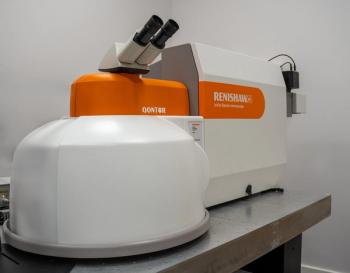
In a recent study, Notingher and his team explored the use of autofluorescence (AF) imaging combined with Raman spectroscopy for intra-operative assessment of sentinel lymph nodes (SLNs) in breast cancer surgery. SLN biopsy is a standard method for evaluating lymph node involvement in breast cancer, and positive results often require further surgery for axillary lymph node clearance (ALNC) (1). The researchers developed a non-destructive AF-Raman microscope to analyze SLN samples, using AF imaging to guide Raman spectroscopy measurements (1).
After optimizing the technique using data from 85 samples, they tested it on an independent set of 81 lymph nodes, comparing results with post-operative histology (1). The test set included 66 negative and 15 positive lymph nodes from 78 patients. The AF-Raman technique achieved an area under the receiver operating characteristic (ROC) curve of 0.93, with an overall detection accuracy of 94%, sensitivity of 80%, and specificity of 97% (1). One challenge was accurately identifying histiocyte-rich areas, which were underrepresented in the training data.
The study suggested that further refinement, including expanding the data set to incorporate more diverse samples, could improve the technique (1). If successful, AF-Raman could enable real-time detection of metastatic SLNs during surgery, potentially reducing the need for additional operations.
In part 1 of our conversation with Notingher, he answers the following question:
- On Tuesday October 22nd, you are set to give a talk titled, “Applications of Raman Microscopy in Breast Cancer Surgery: Towards Intra-Operative Assessment of Surgical Margins and Sentinel Lymph Nodes.” Can you offer our audience a preview of what they can expect to hear from your talk?
To view the rest of our coverage of the upcoming SciX 2024 Conference, click here:
References
- Barkur, S.; Boiter, R. A.; Mihai, R.; et al. Intraoperative Spectroscopic Evaluation of Sentinel Lymph Nodes in Breast Cancer Surgery. Breast Cancer Res. Treat. 2024, 207 (1), 223–232. DOI:
10.1007/s10549-024-07349-z - Chasse, J. Intraoperative Spectroscopic Evaluation of Sentinel Lymph Nodes in Breast Cancer Surgery: An Interview with William F. Meggers Award Winner Ioan Notingher. Spectroscopy. Available at:
https://www.spectroscopyonline.com/view/intraoperative-spectroscopic-evaluation-of-sentinel-lymph-nodes-in-breast-cancer-surgery-an-interview-with-william-f-meggers-award-winner-ioan-notingher (accessed 2024-10-03).
Newsletter
Get essential updates on the latest spectroscopy technologies, regulatory standards, and best practices—subscribe today to Spectroscopy.


