From the Bench to the Bedside: Infrared Spectroscopy and the Diagnosis and Treatment of Dry Eye and Cataracts
This article reviews the application of IR spectroscopy to study meibum and determine if any relationships exist between tear film stability and meibum lipid composition, environment, and conformation.
The focus of this review is the application of infrared (IR) spectroscopy to study human lens and tear lipids with minimal discussion of the role of lipids and the complex etiology of cataracts and dry eye. The power of IR spectroscopy to measure composition, conformation, structure, and environment of biomolecules was applied to diagnose cataracts and dry eye and follow the efficacy of treatment. Principal component analysis and other standard algorithms were used to analyze IR spectra.
A thin lipid layer on the surface of the tears is believed to contribute to tear stability (1,2). Most of the tear film lipid comes from the meibomian glands located in the eye lids (1–3). The major function of tears is to keep the underlying cornea clear and hydrated. Blinking spreads the tears across the surface of the cornea and the mechanical action of the blink expresses a lipid called meibum from the meibomian glands located in the eye lids on the lid margin (1,2). The meibum finds its way to the tear film surface where it forms a heterogeneous, duplex layer that is about 17 molecules thick (4). It is believed that the lipid layer decreases the surface tension of the tears, which allows them to spread, and lowers the evaporation rate of the underlying aqueous liquid. Shortly after blinking, if one does not blink again, the tear film "breaks up." In all cases of dry eye, the tear film is unstable and tear breakup times are shortened. Dry eye affects more than seven million people in the United States alone (5). The roles of lipids and the etiology of dry eye have been reviewed (1–3). This article reviews the application of IR spectroscopy to study meibum and determine if any relationships exist between tear film stability and meibum lipid composition, environment, and conformation (6–12).
Experimental
Meibum and tear lipids can be collected a number of ways, such as by using a spatula, microcapillary pipettes, lipid absorbent tape, or paper strips (3). For most of the studies discussed below, meibum was collected by squeezing the eyelid between two cotton swabs. About 0.5 mg of meibum is expressed on the eyelid surface and collected using a sterile spatula. The meibum is then transferred to a silver chloride window and transmission IR spectra are measured. In the studies reviewed, IR spectra were measured using a Fourier transform IR (FT-IR) spectrometer equipped with a mercury cadmium telluride (MCT) detector (Nicolet 5000 Magna Series, Thermo Fisher Scientific, Inc.). The meibum on the AgCl window was placed in a temperature-controlled IR cell. The cell was jacketed by an insulated water coil connected to a circulating water bath (model R-134A, Neslab Instruments). The sample temperature was measured and controlled by a thermistor touching the sample cell window. The water bath unit was programmed to measure the temperature at the thermistor and to adjust the bath temperature so that the sample temperature could be set to the desired value. The rate of heating or cooling (1 °C/15 min) at the sample was also adjusted by the water bath unit. Temperatures were maintained within ±0.01 °C. IR data analysis was performed using GRAMS/386 software (Galactic Industries).
Cataracts and Lens Lipids
The lens is located in the front of the eye behind the cornea and functions to focus light on the retina at the back of the eye. The lipids of the lens do not turn over, and most of the lipids made at birth are present at death (13). Light passing through a human lens traverses through thousands of cellular membranes that serve as impermeable barriers to cations as well as a matrix for proteins. The phospholipid composition of human lens membranes is unusual. The human lens contains dihydrosphingomyelin, found in significant quantities only in the lens. Lens membranes are some of the most saturated membranes in the human body, and contain extremely high levels of cholesterol, particularly near the center of the lens (10:1 mole:mole cholesterol to phospholipid). The composition of the human lens changes dramatically with age and cataract. The roles of lipids and the etiology of cataracts have been reviewed (13). IR spectroscopy was used to study lens membrane lipid conformation in relation to compositional differences between species, and the aging and cataractous human lens (13–34).
IR Spectroscopy and Lipid Conformation
Lipids can be fluid like olive oil, solid like butter, or somewhere in between. Fluid lipids are generally not completely liquid, but interact with one another and are said to be in a gel phase. Solid lipids are not completely solid and contain some mobility so they are said to be in a liquid crystalline phase. Lipids are generally composed of hydrocarbon chains containing methylene (-CH2-) moieties. Conformation is how molecules are arranged in space. The methylene moieties may arrange as gauche rotamers, prevalent in disordered lipids like olive oil, which is fluid at room temperature. The methylene moieties may also arrange as trans rotamers, more abundant in ordered lipids like butter, which is solid at room temperature (Figure 1). Gauche conformations may more properly be termed a synclinal alignment of groups attached to adjacent atoms (35) and trans rotamers may also be called antiperiplanar or anti.
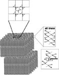
Figure 1: Multilamellar monolayers of ordered (solid or unmelted) wax molecules that are arranged in an orthorhombic crystal structure in which the hydrocarbon chain conformation is all trans. The hydrocarbon chains of fluid waxes contain gauche rotamers (top) orthorhombic or monoclinic packing. Adapted with permission from reference 11.
When lipids are in a "solid" gel phase, the hydrocarbons pack tightly together and van der Waals interactions between hydrocarbon chains are maximal. When lipids are in a fluid, liquid crystalline phase, gauche rotamers cause the hydrocarbon chains to bend (Figure 1). The surface area of fluid lipids is larger than that of ordered lipids and van der Waals interaction between chains is minimal. Thus, lipid hydrocarbon chain conformational order may be evaluated in terms of the amount of CH2 trans and gauche rotamers.
The phase of the lipid has a significant effect on the IR spectra, which can be used to quantify the conformational order of lipids (36). A change in phase from an ordered gel phase to a disordered liquid crystalline phase results in a decrease in the intensity of the IR CH2 stretching bands and both the symmetric and antisymmetric CH2 stretching band frequencies shift higher. The frequency of the CH2 symmetric stretch, ṽsym, is dependent on the amount of trans or gauche rotamers (36) and has been used to characterize lipid phase transitions to measure the trans rotamer content of lipid hydrocarbon chains with changes in temperature (36–41). The minimum and maximum ṽsym for completely ordered hydrocarbons (100% trans rotamers) and completely disordered (100% gauche rotamers) was calculated from the IR spectra of distearoylphosphatidylcholine at -50°C and IR spectra of a complete isomer mixture of hexane at 25 °C, respectively. Completely ordered lipids have a ṽsym of 2848 cm-1. A ṽsym of 2855.36 cm-1 was measured for completely disordered hydrocarbon chains (100% gauche rotamers) (7). Lipid–lipid interaction strength, the percentage of lipids undergoing a phase transition, how the melting of one lipid influences the melting of adjacent lipids (cooperativity), and the enthalpy and entropy of lipid phase transitions can be calculated by curve fitting lipid phase transitions measured using IR spectroscopy (7,8,11).
Although the focus of this review is the IR spectroscopy of lens and tear lipids, it should be noted that Raman spectroscopy has also been used to measure the hydrocarbon chain order of lens (25) and meibum (60) lipids. The 2886 cm-1 Raman band is a Fermi resonant band, sensitive to intrachain as well as interchain interactions (64). The intensity of this band decreases as intramolecular chain disorder increases (64). Also, as lateral packing interactions between chains are disrupted, the intensity of the 2886 cm-l band decreases. The peak height ratio I2886 cm-1/I2850 cm-1 may be used as an order parameter to determine the hydrocarbon chain fluidity of a membrane. Raman spectroscopy has been used to measure regional phospholipid, cholesterol, and protein composition and structure in the lens in situ (65).
Clinical Significance of Hydrocarbon Chain Conformation Measured with IR Spectroscopy
Cataracts and Lipid Conformation
Lipid conformational order at physiological temperatures measured using IR spectroscopy increased in the human lens with age and cataract formation (13). In other words, lens membrane lipids become stiffer with age and cataract formation. This could cause an increase in light scattering and perhaps influence the activity of proteins in the membrane. Plausible scenarios were suggested in which an increase in lens membrane stiffness with age could contribute to presbyopia or passive membrane permeability of cations and cataract. In studying lenses from a variety of species, it was found that the degree of lipid order increased linearly with sphingolipid content (Figure 2). Lens lipid saturation and order are related and it was proposed that the amount of ordered saturated sphingolipids and hence lipid order (13), could increase with age and cataract because of increased oxidation and subsequent degradation of glycerolipids by phospholipase A2. Humans may have adapted so that their lens membranes have a high sphingolipid content to confer resistance to oxidation, allowing these membranes to stay clear for a relatively longer time than is the case in many other species (13).
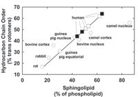
Figure 2: The relationship between lens sphingolipid content and hydrocarbon chain order. Hydrocarbon chain order reflects the structural stiffness of the hydrocarbon chain region of lipids in membranes. Clear human lens cortex and nucleus (closed square), cataractous human lenses (closed triangle). Adapted with permission from reference 13.
Dry Eye and Lipid Conformation Measured with IR Spectroscopy
With meibomian gland dysfunction (MGD), a major type of dry eye, hydrocarbon chains are more ordered (Figure 3b) (more trans rotamers), with stronger lipid–lipid interactions and a higher phase transition temperature (Figure 3a). It is interesting that the phase transition temperature of meibum from normal donors is close to the temperature of the surface of the eye. An increase in order with dry eye indicates there are compositional changes occurring. Lipid order may be important because it was observed that as the temperature of the eye lid was raised from 25 °C to 45 °C, lipid delivery to the margins increased (42) with hydrocarbon disorder (7) and meibum lipid hydrocarbon motion (6). Perhaps hydrocarbon chain order and motion could determine the delivery of meibum lipid from the meibomian glands to the lid margins and subsequently to the tear film. It is likely that meibum phase transitions are why warming the eye lid, one of the oldest treatments for dry eye is advantageous (43). It has been proposed that if a therapy could be developed that decreases the lipid phase transition temperature, the meibum would be more fluid and flow out of the meibomian gland.
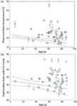
Figure 3: (a) The relationship between donor age and phase-transition temperature for meibum lipid from donors with meibomian gland dysfunction (MGD). (b) The relationship between donor age and hydrocarbon chain order at lid temperature, 33.4 °C, from meibum lipid from donors with MGD. Error bars are ± SE. Lines: linear fit and 95% confidence limits of data from meibum lipid donors without a history of dry eye symptoms. F = female; M = male; B = black; C = caucasian; H = hispanic. Adapted with permission from reference 11.
Like the lens membranes, discussed in the section above, lipid saturation and lipid order at physiological temperatures were linearly related to the lipid phase-transition temperature (Figure 4) of meibum. The phase-transition temperatures of the standard waxes were linearly related to the degree of saturation, and similar to the linear relationship between saturation and phase-transition temperature measured for human meibum, which is predominantly wax, and other natural membranes such as those of the lens. Meibum lipid order and phase-transition temperatures decrease with age (8), and this trend may be attributed to lipid saturation (44). The key to why babies have a more stable tear film than adults may be that their meibum is more ordered and saturated. Long-chain ordered saturated films are generally incompressible films and are better retardants of evaporation than unsaturated compressible films (45).
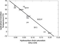
Figure 4: Relationship between phase-transition temperature and hydrocarbon chain saturation. Samples measured in this study: OO = oleyloleate, PO = palmityloleate, PP = palmitylpalmitate, SP = sterylpalmitate, PPPO = equimolar mixture of PP and PO, SPOO = equimolar mixture of SP and OO. Solid line: least-squares linear regression fit to all the data. HL = human lens lipid, ROS P = bovine rod outer segment plasma membrane, SR F = fast twitch rabbit muscle sarcoplasmic reticulum membrane, SR S = slow twitch rabbit muscle sarcoplasmic reticulum membrane. Adapted with permission from reference 11.
Meibum conformational order was used as a diagnostic marker for dry eye to follow the efficacy of treatment for dry eye (46). In a pilot clinical trial, topically applied azithromycin achieved clinical control or improvement in symptoms and signs of MGD (46):
"The correlation of improvement in phase transition temperature of the meibomian gland lipid with the determined percent trans rotamer composition of the lipid strongly suggests that the ordering of the lipid molecules is altered in the disease state (MGD) and that azithromycin can improve that abnormal condition toward normal in a manner that correlates with clinical response to therapy."
Lipid Packing Measured Using IR Spectroscopy
The splitting of CH2 bending and rocking bands in the IR spectra of many lipid types has been attributed to correlation field splitting and suggests that the hydrocarbon chains are arranged in an orthorombic or monoclinic geometry and orthorhombic–monoclinic packing geometry (47). The splitting of the CH2 bending band (Figure 5a) and rocking bands (Figure 5b) in spectra of meibum lipid at lower temperatures suggest that the hydrocarbon chains are arranged in an orthorombic or monoclinic geometry (Figure 1) (7).

Figure 5: IR spectra of meibum from a 53-year-old person: (a) CH2 bending band region, (b) CH2 rocking region. The band splitting is caused by packing changes. Adapted with permission from reference 7.
Carbonyl Environment
Almost all major lipid classes including wax and cholesteryl esters in tears have carbonyl moieties. Lipid IR carbonyl band frequency, 1750–1711 cm-1, is sensitive to its surrounding environment. When the carbonyl is in an environment with a stronger dielectric constant the carbonyl band frequency is lower, and the width of the band is greater (47). Based on the frequency and bandwidth of the IR carbonyl band of meibum, the carbonyl of meibum from children is in an environment with a stronger dielectric constant compared to that of the meibum from adolescents and adults (9).
Principal Component Analysis of IR Spectra
Principal component analysis (PCA) has been used to calculate all the possible variations in IR spectra (48). PCA is a type of chemometric study used to perform calculations on many types of measurements of chemical data, and over the past three decades there have been more than 19,000 health sciences publications involving PCA. As an example, PCA was recently used in forensics to predict the age of a person from the IR spectra of their fingerprint sebum (49). PCA can detect subtle changes in IR spectra that cannot be discerned easily, even by a trained spectroscopist, and these changes can be correlated to factors such as age and disease.
For PCA, a training set of IR spectra is needed in which the principal component is known. PCA produces spectra sometimes called eigenvectors that contain compositional and structural information about the changes that occur with the principal component (variable), MGD, or age in this case. If the PCA model is good, a reconstructed spectrum can be obtained by adding all the products of each score associated with an eigenvector times the eigenvector so that the reconstructed spectrum will be very similar to the original spectrum.
To choose the ideal number of eigenvectors, a prediction residual error of the sum of squares (PRESS) plot is used. The smaller the PRESS value the better the model is able to predict the concentrations of the calibrated constituents, age, and "meibomian gland dysfunction and normal scores." The chosen number of eigenvectors determined by the PRESS plots can be confirmed by plotting the eigenvector number versus the percent of total variance it accounts for.
To determine which eigenvectors are the most important to the modeling components, the scores for an eigenvector are plotted against the scores of another eigenvector. For instance, in plotting the scores of eigenvector 1 versus eigenvector 2, the three age groups are resolved (Figure 6d) and are classically separated by elliptical Mahalanobis boundaries.
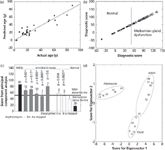
Figure 6: Cross validation was used for each spectrum in the training set of 20 spectra, removing a spectrum and corresponding age from the training set and using the remaining samples to perform the calibration calculation. 14 samples were not in the training set. (a) CH stretching and fingerprint regions were used to predict the age of the sample. Adapted with permission from reference 9. (b) A training set was used to discriminate between the spectra of meibum from normal donors (Mn) and the spectra of meibum from donors with meibomian gland dysfunction (Md). This shows that the infrared spectra must contain compositional and structural information about the changes that occur with meibomian gland dysfunction. A score above 50 (vertical line) is considered a sample with meibomian gland dysfunction. (
•) = Mn group, (Î) = Md group. Linear regression fit and (-). Adapted with permission from reference 10. (c) Data are from the FT-IR spectroscopy spectra of human meibum. PCA was used to analyze the CH and OH stretching region 3612â2490 cm-1 and the fingerprint region 1814â676 cm-1. Based on a training set of 77 IR spectra from donors with meibomian gland dysfunction (Md) and normal donors (Mn), scores were assigned to the spectra. A score greater than 59 indicates that the infrared spectra is similar to the spectra of Md. A score lesser than 59 indicates that the infrared spectra are similar to the spectra of Mn. Gray bars, azithromycin study; filled bars, data are from a larger. Adapted with permission from reference 50. *Statistically significant difference, P
New Study Reveals Insights into Phenol’s Behavior in Ice
April 16th 2025A new study published in Spectrochimica Acta Part A by Dominik Heger and colleagues at Masaryk University reveals that phenol's photophysical properties change significantly when frozen, potentially enabling its breakdown by sunlight in icy environments.