Combining SERS and Machine Learning to Advance Single-Cell Analysis
Researchers from Stanford University have combined surface-enhanced Raman spectroscopy (SERS) with machine learning (ML) to enable rapid, precise single-cell analysis, offering potentially transformative applications in diagnostics and personalized medicine.
Single-cell analysis is an important component in biological analysis. It studies cell-to-cell variation within a cell population in living organisms (1). When done correctly, single-cell analysis provides more information about each cell, which helps researchers learn more about its functions in the body. Single-cell analysis also helps researchers decipher gene expression profiles between individual cells and detect subpopulations of cells previously not uncovered (2).
A recent study explored this topic in more detail. Researchers from Stanford University examined a new approach to single-cell analysis by integrating surface-enhanced Raman spectroscopy (SERS) with machine learning (ML) (3). Their study, which was published in the journal ACS Nano, demonstrates how SERS and ML can advance clinical diagnostics.By playing to the strengths of what SERS and ML has to offer, this method can provide precise, interpretable insights into the transcriptome, proteome, and metabolome (3).
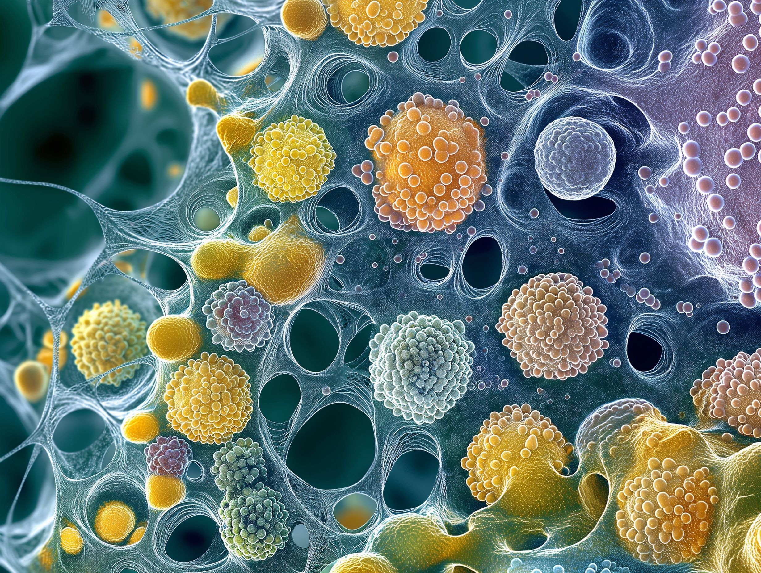
Human skin cells under a microscope. | Image Credit: © mirifadapt - stock.adobe.com

SERS is a sensitive technique that amplifies Raman scattering signals using nanostructured surfaces (3,4), Because of its high sensitivity and selectivity, SERS has long been recognized for its potential applications in high-sensitivity molecular analysis, surface and interface chemistry, and nanotechnology (3,4). However, its broader adoption has been hindered by challenges in spectral interpretation and reproducibility. This research leverages cutting-edge nanophotonics—specifically plasmonics, metamaterials, and metasurfaces—to enhance Raman scattering (3). These advancements not only increase the strength of the Raman signals, but they also enable rapid, label-free spectroscopic analysis (3).
An unique element to this study is integrating ML into their method to assist with spectral data interpretation. ML algorithms, including neural networks, gradient-based methods, and transfer learning, have exhibited the potential to extract biologically relevant information from complex Raman spectra (3).
The research team showed what their approach could do through single-cell Raman phenotyping. These included three main things: bacterial antibiotic susceptibility; stem cell analysis; and cancer diagnostics. For bacterial antibiotic susceptibility, researchers showed that SERS-ML can accurately predict the effectiveness of antibiotics on bacterial cells, a critical advance in combating antibiotic resistance (3). For stem cell analysis, the team showed that their method could profile expression markers, allowing them to gain more insights into stem cell differentiation (3). And finally, the researchers demonstrated how SERS-ML can enable the detection of cancerous cells at the molecular level, offering potential for early diagnosis and personalized treatment plans (3).
Despite the potential of the technique, some challenges remain. Instrumentation costs, the need for standardized protocols, and the risk of algorithmic biases in ML models must be addressed to ensure reproducibility and equitable applications (3). Moreover, interdisciplinary collaboration between spectroscopists, data scientists, and biologists will be essential to maximize the impact of SERS-ML technology (3).
By merging nanophotonics and machine learning, the Stanford University researchers have laid the groundwork for a new era of single-cell analysis that is both rapid and highly interpretable (3). This approach could transform fields ranging from drug development to personalized medicine, emphasizing the growing synergy between spectroscopy and computational technologies (3).
Combining SERS and ML can help provide insights into the molecular level of cells. As these technologies continue to advance, their impact on science and medicine is likely to be transformative, offering a deeper understanding of biological complexity and a pathway to innovative solutions in healthcare (3).
References
- Hodzic, E. Single-cell Analysis: Advances and Future Perspectives. Bosn. J. Basic Med. Sci. 2016, 16 (4), 313–314. DOI: 10.17305/bjbms.2016.1371
- Thermo Fisher Scientific, Single Cell Analysis. Thermo Fisher. Available at: https://www.thermofisher.com/us/en/home/life-science/cell-analysis/single-cell-analysis.html (accessed 2024-12-12).
- Zhang, Y.; Chang, K.; Ogunlade, B.; et al. From Genotype to Phenotype: Raman Spectroscopy and Machine Learning for Label-Free Single-Cell Analysis. ACS Nano 2024, 18 (28), 18101–18117. DOI: 10.1021/acsnano.4c04282
- Han, X. X.; Rodriguez, R. S.; Haynes, C. L.; et al. Surface-enhanced Raman Spectroscopy. Nat. Rev. Methods Prim. 2021, 1, 87. DOI: 10.1038/s43586-021-00083-6
AI-Powered SERS Spectroscopy Breakthrough Boosts Safety of Medicinal Food Products
April 16th 2025A new deep learning-enhanced spectroscopic platform—SERSome—developed by researchers in China and Finland, identifies medicinal and edible homologs (MEHs) with 98% accuracy. This innovation could revolutionize safety and quality control in the growing MEH market.
AI-Driven Raman Spectroscopy Paves the Way for Precision Cancer Immunotherapy
April 15th 2025Researchers are using AI-enabled Raman spectroscopy to enhance the development, administration, and response prediction of cancer immunotherapies. This innovative, label-free method provides detailed insights into tumor-immune microenvironments, aiming to optimize personalized immunotherapy and other treatment strategies and improve patient outcomes.