Early Signs of Brain Injury Now Identifiable Using Raman-Based Detection System
In a new article published in Science Advances, researchers from the University of Birmingham and the University of Cambridge developed a Raman spectroscopy-based system for detecting early signs of traumatic brain injury (TBI) in patients (1).
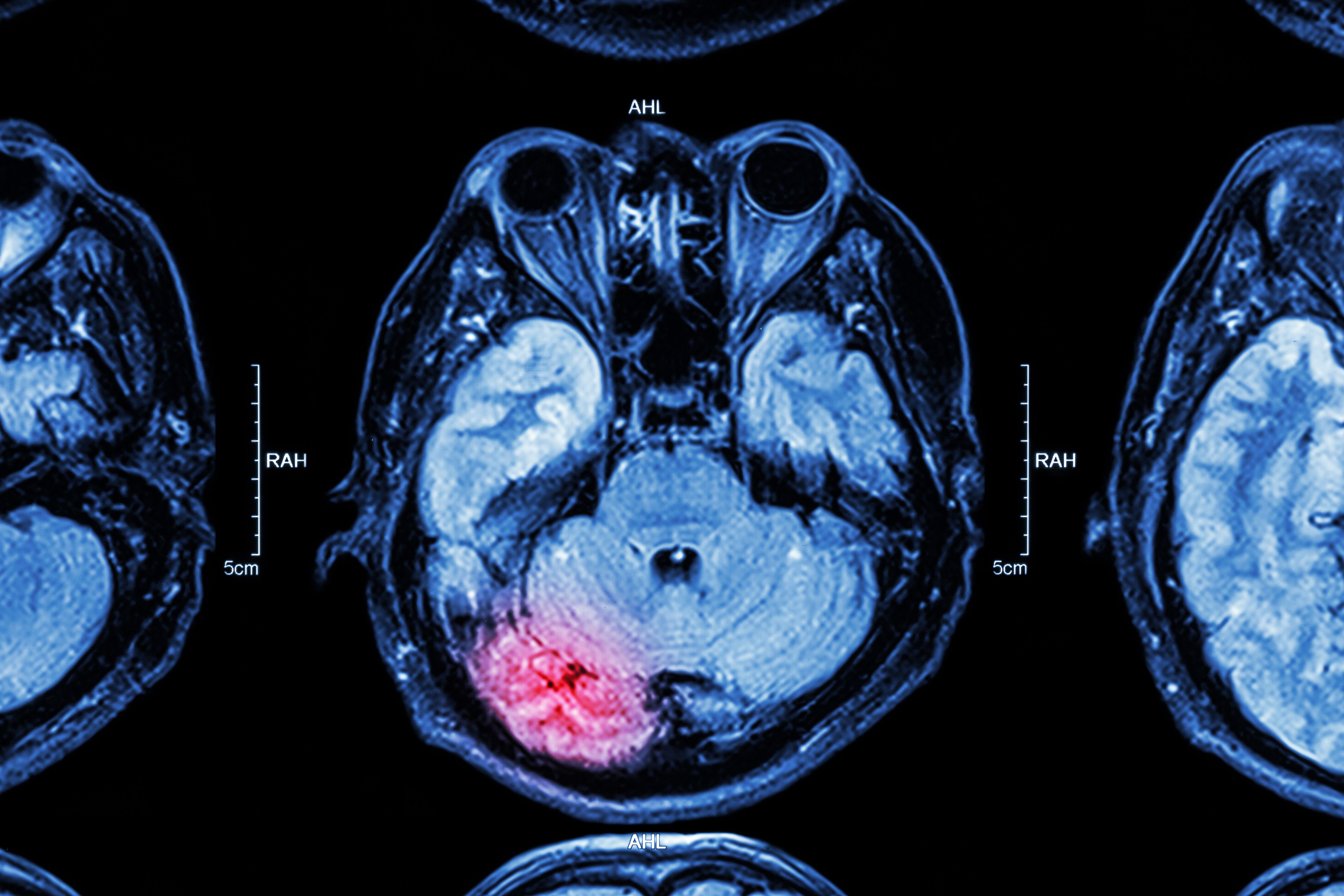
MRI of brain : brain injury | Image Credit: © stockdevil - stock.adobe.com

Traumatic brain injury (TBI) affects 135 million people globally, yet very few people show clinical systems in early stages of TBI. Brain tissue lacks regenerative capacity, and TBI can lead to long-term and persistent deficits. Critical decisions for TBI patients must be made within the first hour after trauma, and with the dangers of misdiagnosis or treatment delay, TBI can be difficult to treat. As such, early diagnosis is crucial for improving patient outcomes.
There is currently no point-of-care technology for the quantitative assessment of TBI, at least with enough sensitivity or timeliness to properly aid early diagnosis efforts. To rectify this, the scientists developed laser-based spectroscopic technology meant to analyze the neuroretina and optic nerve of the eye as a representative of brain tissue. This is meant to provide a window into the brain’s biochemistry, specifically keeping the central nervous system in continuity. “By targeting CNS biochemical changes, we reduce the need to filter out confounding (non-CNS) compounds, measuring the brain side of the blood-brain barrier,” the scientists wrote in the study (1).
At the back of the eye is a small part of the brain that is barely covered, used to carry visual information from the retina to the brain. This is the area that was analyzed using multiplex resonance Raman spectroscopy, which was targeted at different TBI biomarkers, or molecular fingerprints, as disease and injury proxies. Markers for brain health include structural changes in brain-specific lipids and biochemicals, which can stem from local tissue damage or the presence of certain neuromarkers in the CNS. These neuromarkers have demonstrated strong correlation with injury severity, specifically with acute TBI.
The system, called EyeD, functions with Raman spectroscopy and fundus imaging of the optic nerve, which isolates Raman and white light paths. A detector collects Raman signals, and data is classified using artificial neural network algorithms that provide dimensionality reduction, feature extraction, and multiclass detection. This allows for the underlying chemical differences between classes to be identified, providing accurate classification for simultaneously rich-information and high-classification specificity. This system rapidly distinguishes TBI from control groups of tissue samples, enabling important steps for applying this technology in real-world environments.
Using their findings, the scientists designed a portable device for eye-safe data acquisition. This handheld technology can greatly improve the speed and cost of TBI diagnosis. Furthermore, it can be integrated with technology like smartphone cameras and AI diagnostic support. The EyeD technology has potential to offer critical clinical insight in different medical scenarios. There is still work to be done, but this system could revolutionize how TBI is diagnosed and treated, thus improving health care savings and saving lives in the process.
Reference
(1) Banbury, C.; Harris, G.; Clancy, M., et al. Window into the mind: Advanced handheld spectroscopic eye-safe technology for point-of-care neurodiagnostic. Sci. Adv. 2023, 9 (46). DOI: https://doi.org/10.1126/sciadv.adg5431
Nanometer-Scale Studies Using Tip Enhanced Raman Spectroscopy
February 8th 2013Volker Deckert, the winner of the 2013 Charles Mann Award, is advancing the use of tip enhanced Raman spectroscopy (TERS) to push the lateral resolution of vibrational spectroscopy well below the Abbe limit, to achieve single-molecule sensitivity. Because the tip can be moved with sub-nanometer precision, structural information with unmatched spatial resolution can be achieved without the need of specific labels.
New Multi-Spectroscopic System Enhances Cultural Heritage Analysis
April 2nd 2025A new study published in Talanta introduces SYSPECTRAL, a portable multi-spectroscopic system that can conduct non-invasive, in situ chemical analysis of cultural heritage materials by integrating LIBS, LIF, Raman, and reflectance spectroscopy into a single compact device.