Emerging Raman Techniques for Rapid Noninvasive Characterization of Pharmaceutical Samples and Containers
Spatially offset and transmission Raman spectroscopy enable chemical characterization of diffusely scattering samples at depths not accessible by conventional Raman methods.
Raman spectroscopy has recently seen major advances in the area of deep noninvasive characterization of diffusely scattering samples stemming from the emergence of spatially offset Raman spectroscopy and the associated renaissance of transmission Raman spectroscopy. These techniques permit detailed chemical characterization of diffusely scattering samples at depths not accessible by conventional Raman methods. This article reviews this newly emerging field focusing on recent developments with relevance to pharmaceutical analysis, including volumetric quantification of pharmaceutical formulations and noninvasive detection of counterfeit drugs in plastic containers.
A wide range of analytical applications in the pharmaceutical arena require rapid and noninvasive chemical characterization of intact formulations. Examples include the quantification of pharmaceutical capsules and tablets (both bare and coated), monitoring the progress of reactions through reactor windows, the identification of incoming materials through unopened packaging, and the detection of counterfeit medicine through unopened glass and opaque plastic bottles.
Raman spectroscopy holds a great potential in this area, largely because of its high chemical specificity (surpassing that of near-infrared [NIR] absorption spectroscopy and being comparable to that of mid-infrared [MIR]) (1) and ability to probe samples in the presence of water (unlike for terahertz and MIR absorption spectroscopies). A drawback of the Raman technique is that it is restricted to samples that do not exhibit strong fluorescence emission within the Raman detection range. However, in many situations, this issue can be overcome by using NIR laser excitation and avoiding the population of emissive electronic states (2).
Raman spectroscopy is based on the inelastic scattering of photons on a quantum system yielding typically red-shifted photons that result from the exchange of energy with the sample components. An example of this process would be exchanging a quantum of energy with a molecular or crystal lattice and activating its vibrational motions. Red-shifted photons typically are detected using a spectrograph and a charge-coupled device (CCD) detector. The Raman spectral profile (band positions, relative intensities, and bandwidths) can be used as a fingerprint of sample chemical constituency or crystalline structure (for example, in detecting the presence of polymorphs). Being a linear technique, a mixture of sample subcomponents is represented through a linear combination of Raman spectra of the individual mixture components, and consequently, relative sample concentrations can be derived readily from the Raman spectra assuming there is no chemical interaction between the sample components.
To date, Raman spectroscopy has been used widely in analyzing samples through transparent containers such as glass bottles. However, with some containers, such as green glass bottles, the technique is hampered by intense fluorescence emission from the container, which reduces the sensitivity of the technique. When probing opaque packaging and products (that is, through diffusely scattering materials such as white or colored plastic bottles, powders, or tablets), conventional Raman spectroscopy generally has been confined to near-surface analysis. For example, with pharmaceutical powders and tablets, its penetration depth has been restricted to 1–2 mm (3). This limitation is associated with the prevalent optical measurement geometry, so-called backscattering collection geometry, used in a vast majority of commercial Raman instruments (2). This arrangement typically results in the subsurface layer signal being overwhelmed by much stronger Raman and fluorescence signals originating from the surface layers of the sample, thus hampering accurate subsurface quantitative analysis.
Two principal variants of Raman spectroscopy have emerged in recent years as promising practical tools for deep noninvasive probing of opaque samples such as powders and opaque bottles (4,5): spatially offset Raman spectroscopy (SORS) (6), in which the Raman spectra of individual sublayers within a multilayered system can be isolated allowing, for example, the content of opaque plastic bottles to be interrogated, and transmission Raman spectroscopy, which provides volumetric (bulk averaged) information on potentially inhomogeneous sample compositions such as pharmaceutical tablets and capsules (3).
Spatially Offset Raman Spectroscopy
In SORS, Raman spectra are collected from regions that are spatially offset (by a distance Δs) on the sample surface from the laser incident zone (see Figure 1). Such Raman spectra contain differing relative contributions from layers at different depths within the sample (for example, the container wall and its powder content). This is a direct consequence of a lateral spread of photons on the sample surface originating from deeper areas of the sample, which is brought about by photon diffusion. Fluorescence originating from surface layers of the sample, such as a container wall, tablet coating or capsule shell material, also can be suppressed effectively in the SORS technique.

Figure 1: A schematic illustration of conventional backscattering Raman, SORS, and transmission Raman spectroscopy.
Raw SORS spectra typically can contain residual surface-layer Raman components, and these can be removed using additional data manipulation. For a two-layer system, this can be achieved using a simple scaled subtraction of two SORS spectra acquired at different spatial offsets (for example, at the zero and nonzero spatial offsets [6]), canceling the residual contribution from the surface layer. This approach can be automated.
An alternative optical arrangement is a variant of SORS called inverse SORS (7,8). In a reversal of the traditional SORS geometry the laser light is brought onto the sample surface in a ring shape and Raman light is collected through a group of optical fibers confined within the center of the illumination ring zone. The radius of the ring-shaped beam approximately equates to the spatial offset, and this can be varied to optimize the probing conditions to a particular sample layer. Inverse SORS is beneficial when probing sensitive samples that require analysts to adhere to illumination intensity limits, such as with photosensitive samples and biological materials or when probing in explosive environments. The much lower intensities are achieved by spreading the laser beam over a wider illumination area compared with traditional SORS.
In pharmaceutical settings, SORS is beneficial in interrogating the contents of opaque plastic containers, colored glass bottles, and fouled reactor windows. A special collection and illumination configuration geometry can be arranged that permits the probing of both transparent and diffusely scattering containers. In this configuration, a tilted laser beam is used and passed through the container or vessel wall spatially offset from the collection zone at an angle to intersect the Raman collection zone located below the wall within the probed medium (9). When such a setup is presented with a diffusely scattering sample (for example, an opaque plastic bottle), the system acts as a conventional SORS setup, as the laser photons quickly lose memory of their direction of travel in the diffusely scattering medium (for example, a plastic container wall). This configuration has an extra advantage in that it also suppresses the fluorescence and Raman signals that originate from the container even when it is transparent (for example, as with green glass), as the spatially offset laser beam can be set to intersect the container wall out of sight of the Raman collection system.
As with conventional Raman spectroscopy (or UV, visible, NIR, MIR, and terahertz absorption spectroscopy), the SORS technique is limited to nonmetallic containers. In addition, the SORS methods are restricted to probing samples that do not excessively fluoresce or materials that do not highly absorb laser or Raman photons (for example, black materials). However, fluorescence originating from the packaging or other surface layers can be effectively reduced with SORS.
Another benefit is that the SORS technique does not require a priori knowledge of the composition of any layer in the sample. The layers themselves do not need to be parallel or of uniform thicknesses; a change in the relative intensities of signals from individual layers with spatial offsets is sufficient to produce SORS spectra that can be used to decompose Raman spectra into pure layer components.
Transmission Raman Spectroscopy
In transmission Raman spectroscopy, the laser beam is incident on the sample at one side and the Raman signal collected from the opposite side (see Figure 1). This configuration can be considered a special case of SORS, as the illumination and collection points are displaced to the extreme (3). Although the transmission Raman technique was demonstrated in the early days of Raman spectroscopy (10), its benefits to probing pharmaceutical samples were not recognized and exploited. These include explicit volumetric sample information that effectively reduces subsampling errors (11) accompanied by the suppression of fluorescence emanating from surface layers (for example, a coating or capsule shell [3,12]). The use of this method in practical pharmaceutical applications is also facilitated by recent advances in the performance of Raman instruments.
The methods discussed above for deep Raman spectroscopy have been demonstrated in a wide range of disciplines including biomedical (4,5,8,13), pharmaceutical (14), and security screening (9). Several application examples are given below to illustrate the versatility of the techniques and the way they can be deployed in pharmaceutical settings.
Quality Control in Pharmaceutical Manufacture
Volumetric Quantification of Pharmaceutical Products in Quality Control: In a number of analytical applications in pharmaceutical environments there is a need to quantify rapidly the bulk content of intact pharmaceutical products such as tablets and capsules. The need for such applications in process monitoring and control is emphasized by the FDA's Process Analytical Technology (PAT) initiative. A traditional method of choice in quality control is high performance liquid chromatography (HPLC), which is slow, costly, and involves sample destruction through the inherent dissolution step. Therefore, it also suffers from an inability to characterize solid state properties such as polymorphic forms contained within the sample. Although NIR absorption spectroscopy can provide a rapid nondestructive alternative to HPLC, it can only be used in certain situations because of its limited chemical specificity (15,16). In the past, the NIR method has been shown to be able to reach accuracy of quantification on par with, and in some cases even higher than, conventional Raman spectroscopy. A major drawback of NIR, however, stems from its restricted chemical specificity and strong dependence on a sample's physical characteristics, such as particle size. This often leads to poor robustness and a need for frequent, costly, and time-consuming recalibration. Conventional Raman cannot fulfill the key requirements primarily because of the aforementioned subsampling, which precludes the characterization of deep areas of a sample and is of particular importance to capsules and coated tablets. These problems are largely resolved with transmission Raman spectroscopy (3).
The gross volumetric probing capability of transmission Raman spectroscopy is illustrated in Figure 2, where a standard paracetamol tablet of 3.9 mm thickness with a simulated impurity layer of a 2 mm thickness of trans-stilbene powder was probed in both the conventional and transmission Raman spectroscopy configurations. The impurity layer was located either at the front or the back of the tablet. In this experiment, conventional Raman spectroscopy suffered from severe subsampling and could only yield the Raman signature of the surface layer in either sample orientation. In contrast, the transmission geometry provided a Raman spectrum comprising a mixture of the tablet and the "impurity" signals.
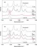
Figure 2: The Raman spectra obtained from a two-layer sample (a paracetamol tablet and a 2-mm-thick trans-stilbene "impurity" layer) using (a) conventional backscattering geometry and (b) transmission geometry. The measurements are performed at two sample orientations, with paracetamol (P) at the top and bottom of the trans-stilbene (T) cell as indicated.
In recent research, Johansson and colleagues (17) established the ability of the transmission Raman technique to provide quantitative information about binary formulations within tablets and capsules. The study achieved a relative root mean square error of prediction for active pharmaceutical ingredient (API) concentration of ±2.2 % and ±3.6%, respectively, with a 5-s acquisition time. The measurements also demonstrated the robustness of the calibration model, which provided a satisfactory prediction accuracy for a relatively low number of calibration points (3).
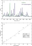
Figure 3: Example of the quantification of a binary mixture of powders in glass vials consisting of two components, acetaminophen and caffeine, using PLS. (a) Transmission Raman spectra of the two pure components: The spectra were baseline corrected and normalized to the most intense bands. (b) Prediction of the concentration of formulations from transmission Raman spectra through glass vials using PLS.
A similar study was also performed by Eliasson and colleagues (18) using transmission Raman spectroscopy on white capsules containing production-relevant formulations prepared in a laboratory. The capsules generated interference that hampered the use of other PAT-compatible optical spectroscopic methods such as NIR absorption and conventional Raman spectroscopy. These signals were effectively suppressed by a factor of ~30 by the transmission Raman approach. The study was performed on 150 capsules and the measured relative root mean square error of API prediction was ±1.2 % with a 5-s acquisition. Figure 3 illustrates conceptually the performance of transmission Raman spectroscopy on a binary mixture (acetaminophen and caffeine) measured through a clear vial. In a similar manner, it is also possible to characterize the polymorphic content through the low-wavenumber region of the Raman spectrum typically dominated by crystal lattice phonon mode vibrations. This is exemplified on two pharmaceutical samples, paracetamol and aspirin (see Figure 4).

Figure 4: Comparison of transmission Raman spectra of (a) aspirin and (b) paracetamol tablets with their typically intense low wavenumber polymorph regions.
Probing of Opaque Plastic and Colored Bottles
Noninvasive Detection of Counterfeit Drugs (SORS): Another important analytical application is the noninvasive detection of counterfeit drugs through plastic bottles and blister packs. This is a major issue representing a significant health threat to society (19). Recently, Eliasson and Matousek (14) demonstrated that SORS can provide an effective method for interrogating chemical signature of the content of unopened opaque plastic containers yielding considerably higher sensitivity than that available with conventional Raman spectroscopy. The same technique also can be used to inspect transparent colored containers. As such, the SORS method is well suited for screening for the presence of counterfeit drugs or for confirming the identity of a chemical held inside a container without opening it. Similarly, the technique permits the probing of the inner content of the reactor through fouled windows while reducing the effect of the fouling on the acquired Raman signal, which typically would yield skewed quantification results.
The conceptual demonstration of these applications is given in Figure 5 where aspirin tablets are held inside a white plastic pharmaceutical bottle and monitored noninvasively. An attempt at obtaining spectra using conventional Raman spectroscopy resulted in spectra dominated by Raman signals originating from the container wall that overwhelmed the Raman signal of the content (in this specific case aspirin). In contrast, SORS, following a scaled subtraction of two SORS spectra obtained at different spatial offsets, provided a clean Raman spectrum of the tablets held inside the bottle and permitted their chemical constituency to be characterized accurately. The experiments were performed using a continuous diode laser (830 nm, 250 mW) with an acquisition time of 1 s.
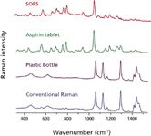
Figure 5: Noninvasive Raman spectra of aspirin tablets measured through a white, diffusely scattering plastic container. Conventional Raman and SORS raw data are shown offset together with the reference Raman spectrum of aspirin. The processed SORS spectrum matches well that of paracetamol, and the conventional Raman spectrum is dominated by Raman signals originating from the bottle wall.
Conclusions
The advent of SORS and the renaissance of transmission Raman spectroscopy have stimulated the development of new analytical capabilities suitable for pharmaceutical environments. These include the rapid quantification of intact pharmaceutical tablets and capsules in quality control and the authentication of pharmaceutical products and counterfeit medicine.
In general, these methods offer great benefits in pharmaceutical production applications. Because of their throughput advantages and the nondestructive nature of their deployment, they also offer prospects for controlling and adjusting pharmaceutical manufacturing processes at key points to optimize the process for higher accuracy and reduced wastage. The techniques are also beneficial in continuous manufacture. The techniques have the potential to displace NIR absorption spectroscopy and HPLC in a number of analytical applications in pharmaceutical settings in the coming years.
Pavel Matousek is the Chief Scientific Officer with Cobalt Light Systems, Start Electron, Harwell Science & Innovation Campus, Oxfordshire, OX11 0QR, United Kingdom, and an STFC Fellow with the Central Laser Facility, Science and Technology Facilities Council, Rutherford Appleton Laboratory, Oxfordshire, OX11 0QX, United Kingdom. Dr. Matousek can be contacted via e-mail at pavel.matousek@stfc.ac.uk
Fiona Thorley is a Research Investigator with SSCI, A division of Aptuit, 111 Milton Park, Abingdon, Oxfordshire, OX14 4RZ, United Kingdom.
Ping Chen is a Research Investigator with SSCI, A division of Aptuit, 3065 Kent Avenue, West Lafayette, IN 47906. Michael Hargreaves is a Senior Scientist, Craig Tombling is the Chief Operating Officer, Paul Loeffen is the Chief Executive Officer, Matthew Bloomfield is an Applications Manager, and Darren Andrews is the Business Development Director with Cobalt Light Systems, Start Electron, Harwell Science & Innovation Campus.
References
(1) A. Heinz, C.J. Strachan, K.C. Gordon, and T. Rades, J. Pharm. Pharmacol. 61, 971 (2009).
(2) M.J. Pelletier, Analytical Applications of Raman Spectroscopy (Blackwell Science, Oxford, United Kingdom, 1999).
(3) P. Matousek and A.W. Parker, Appl. Spectrosc. 60, 1353 (2006).
(4) N.A. Macleod and P. Matousek, Appl. Spectrosc. 62, 291A (2008).
(5) Emerging Raman Applications and Techniques in Biomedical and Pharmaceutical Fields, P. Matousek and M.D. Morris, Eds. (Springer, Heidelberg, 2010).
(6) P. Matousek, I.P. Clark, E.R.C. Draper, M.D. Morris, A.E. Goodship, N. Everall, M. Towrie, W.F. Finney, and A.W. Parker, Appl. Spectrosc. 59, 393 (2005).
(7) P. Matousek, Appl. Spectrosc. 60, 1341 (2006).
(8) M.V. Schulmerich, K.A. Dooley, M.D. Morris, T.M. Vanasse, and S.A. Goldstein, J. Biomed. Opt. 11, 060502 (2006).
(9) C. Eliasson, N.A. Macleod, and P. Matousek, Anal. Chem. 79, 8185 (2007).
(10) B. Schrader and G. Bergmann, Zeitschrift fur Analytische Chemie Fresenius 225, 230 (1967).
(11) J. Johansson, S. Pettersson, and S. Folestad, J. Pharmaceut. Biomed. 39, 510 (2005).
(12) P. Matousek and A.W. Parker, J. Raman Spectrosc. 38, 563 (2007).
(13) P. Matousek and N. Stone, Analyst 134, 1058 (2009).
(14) C. Eliasson and P. Matousek, Anal. Chem. 79, 1696 (2007).
(15) D.E. Pivonka, J.M. Chalmers, and P.R. Griffiths, Applications of Vibrational Spectroscopy in Pharmaceutical Research and Development (John Wiley & Sons, Chichester, 2007).
(16) R. Salzer and H.W.Siesler, Infrared and Raman Spectroscopic Imaging (Wiley-VCH, Weinheim, 2009).
(17) J. Johansson, A. Sparen, O. Svensson, S. Folestad, and M. Claybourn, Appl. Spectrosc. 61, 1211 (2007).
(18) C. Eliasson, N.A. Macleod, L. Jayes, F.C. Clarke, S. Hammond, M.R. Smith, and P. Matousek, J. Pharm. Biomed. Anal. 47, 221 (2008).
(19) R. Mukhopadhyay, Anal. Chem. 79, 2622 (2007).

Nanometer-Scale Studies Using Tip Enhanced Raman Spectroscopy
February 8th 2013Volker Deckert, the winner of the 2013 Charles Mann Award, is advancing the use of tip enhanced Raman spectroscopy (TERS) to push the lateral resolution of vibrational spectroscopy well below the Abbe limit, to achieve single-molecule sensitivity. Because the tip can be moved with sub-nanometer precision, structural information with unmatched spatial resolution can be achieved without the need of specific labels.
Tomas Hirschfeld: Prolific Research Chemist, Mentor, Inventor, and Futurist
March 19th 2025In this "Icons of Spectroscopy" column, executive editor Jerome Workman Jr. details how Tomas B. Hirschfeld has made many significant contributions to vibrational spectroscopy and has inspired and mentored many leading scientists of the past several decades.


