Equipment Roundup: Refeyn Ltd.’s MassFluidix HC Upgrade Enhances High-Concentration Analysis
In this Equipment Roundup, the editors of Spectroscopy feature Refeyn Ltd.’s upgraded MassFluidix HC system, optimizing efficiency and quality of mass photometry experiments.
In this Equipment Roundup, the editors of Spectroscopy highlight the newest products from instrument manufacturers around the world. Do you have a new product that you’d like featured in a future installment of Spectroscopy’s Equipment Roundup? Send a summary and photo of the product to John Chasse, Managing Editor of Spectroscopy, at jchasse@mjhlifesciences.com
Refeyn Ltd., an Oxford, UK corporation specializing in the development, production, and distribution of mass photometry technologies for industry and academia, recently announced the release of a powerful expansion of its consumables portfolio (1,2).
The company states that this introduction of next-generation microfluidic chips and improvements to Refeyn’s microfluidic system, as well as a new protein calibration standard, will save users time, while ensuring reproducibility across experiments and laboratories, and supporting a broader range of applications (1).
According to the Refeyn website (2), mass photometry is a revolutionary new way to analyze molecules that enables the accurate mass measurement of single molecules in solution, in their native state, and without the need for labels. This approach presents new possibilities for bioanalytics and research into the functional aspects of biomolecules, including molecular mass and binding phenomena.
The technique builds on the principles of interference reflection microscopy (3) and interferometric scattering microscopy (4). It was developed by the research group of Professor Philipp Kukura of the Physical and Theoretical Chemistry Laboratory at Oxford University. They used carefully controlled illumination, a novel spatial-filtering strategy in the detection beam path (5) and careful image analysis to demonstrate that the minute amount of light scattered by single molecules can be reliably detected and, more importantly, correlates directly with molecular mass (6).
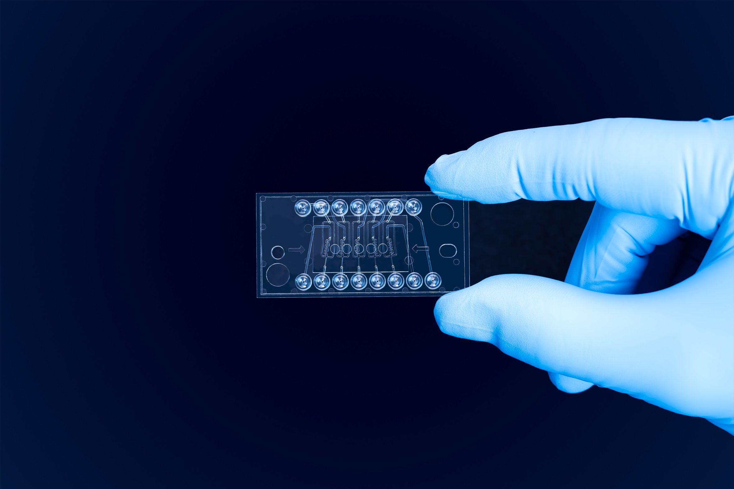
To enhance the rapid dilution of highly concentrated samples and improve insight into low-affinity and transient biomolecular interactions, Refeyn reports that several updates have been made to their popular MassFluidix HC microfluidics system. A key addition to this system is a second-generation microfluidic chip that now enables mass photometry measurements in five channels and features an even faster sample dilution of under 37 milliseconds. Usage is simplified with a channel redesign that removes the need for a bubble trap and introduces Luer connectors which enable easy connection of tubing to the chip. Furthermore, these ready-to-use chips are also now individually packaged, allowing customers to open just one at a time as needed (1).
Further improving ease-of-use of the MassFluidix HC, software integration means that the rapid dilution system can now be readily operated directly via Refeyn’s AcquireMP mass photometer control software. A new automatic microfluidic chip channel detection feature within this software also simplifies workflows by ensuring correct positioning of the chip for each measurement (1).
The upgraded system’s rapid dilution allows the characterization of samples at high concentration. This is particularly useful as a quality check for techniques that require higher concentrations, such as cryoEM and crystallography, as well as for detecting and determining stoichiometry for weak affinity protein-protein interactions. This has implications for traditionally difficult-to-characterize sample types, such as PROTACs and multi-component complexes; in addition to more common applications, such as antibody-receptor binding, and as an orthogonal technique to other binding characterization techniques (1).
Refeyn’s new MassFluidix second generation microfluidic chip enables rapid sample dilution and mass photometry measurement in each of its five channels. Photo courtesy of Refeyn Ltd.

References
1. Refeyn MassFluidix HC Home Page. https://www.refeyn.com/post/massfluidix-hc-upgrade-enhances-high-concentration-analysis(accessed 2024-10-16).
2. Refeyn Home Page. https://www.refeyn.com (accessed 2024-10-16).
3. Verschueren, H. Interference Reflection Microscopy in Cell Biology: Methodology and Applications. J. Cell Science 1985, 75 (1), 279–301. DOI: 10.1242/jcs.75.1.279
4. Ortega-Arroyo, J.; Kukura, P. Interferometric Scattering Microscopy (iSCAT): New Frontiers in Ultrafast and Ultrasensitive Optical Microscopy. Phys. Chem. Chem. Phys. 2012, 14, 15625–15636. DOI: 10.1039/C2CP41013C
5. Cole, D.; Young, G.; Weigel, A.; Sebesta, A.; Kukura, P. Label-Free Single-Molecule Imaging with Numerical-Aperture-Shaped Interferometric Scattering Microscopy. ACS Photonics 2017, 4, 211–216. DOI: 10.1021/acsphotonics.6b00912
6. Young, G.; Hundt, N.; Cole, D.; Fineberg, A.; Andrecka, J.; Tyler, A.; Olerinyova, A.; Ansari, A.; Marklund, E. G.; Collier, M. P.; Chandler, S. A.; Tkachenko, O.; Allen, J.; Crispin, M.; Billington, N.; Takagi, Y.; Sellers, J. R.; Eichmann, C.; Selenko, P.; Frey, L.; Riek, R.; Galpin,M. R.; Struwe, W. B.; Benesch, J. L. P.; Kukura, P. Quantitative Mass Imaging of Single Biological Macromolecules. Science 2018, 360, (6387), 423–427. DOI: 10.1126/science.aar5839
Best of the Week: AI and IoT for Pollution Monitoring, High Speed Laser MS
April 25th 2025Top articles published this week include a preview of our upcoming content series for National Space Day, a news story about air quality monitoring, and an announcement from Metrohm about their new Midwest office.
LIBS Illuminates the Hidden Health Risks of Indoor Welding and Soldering
April 23rd 2025A new dual-spectroscopy approach reveals real-time pollution threats in indoor workspaces. Chinese researchers have pioneered the use of laser-induced breakdown spectroscopy (LIBS) and aerosol mass spectrometry to uncover and monitor harmful heavy metal and dust emissions from soldering and welding in real-time. These complementary tools offer a fast, accurate means to evaluate air quality threats in industrial and indoor environments—where people spend most of their time.