Fluorescence Microscopy: A Conversation with Joseph Black Award Winner Mathew Horrocks
“At our core, we are passionate about unraveling the mysteries of biology using advanced imaging techniques. We specialize in single-molecule and super-resolution imaging, employing these powerful tools to delve into the intricate complexities of biology, with a focus on neuroscience and neurodegenerative disorders.“
– The Edinburgh Single-Molecule Biophysics Group website
University of Edinburgh School of Chemistry, Edinburgh, UK
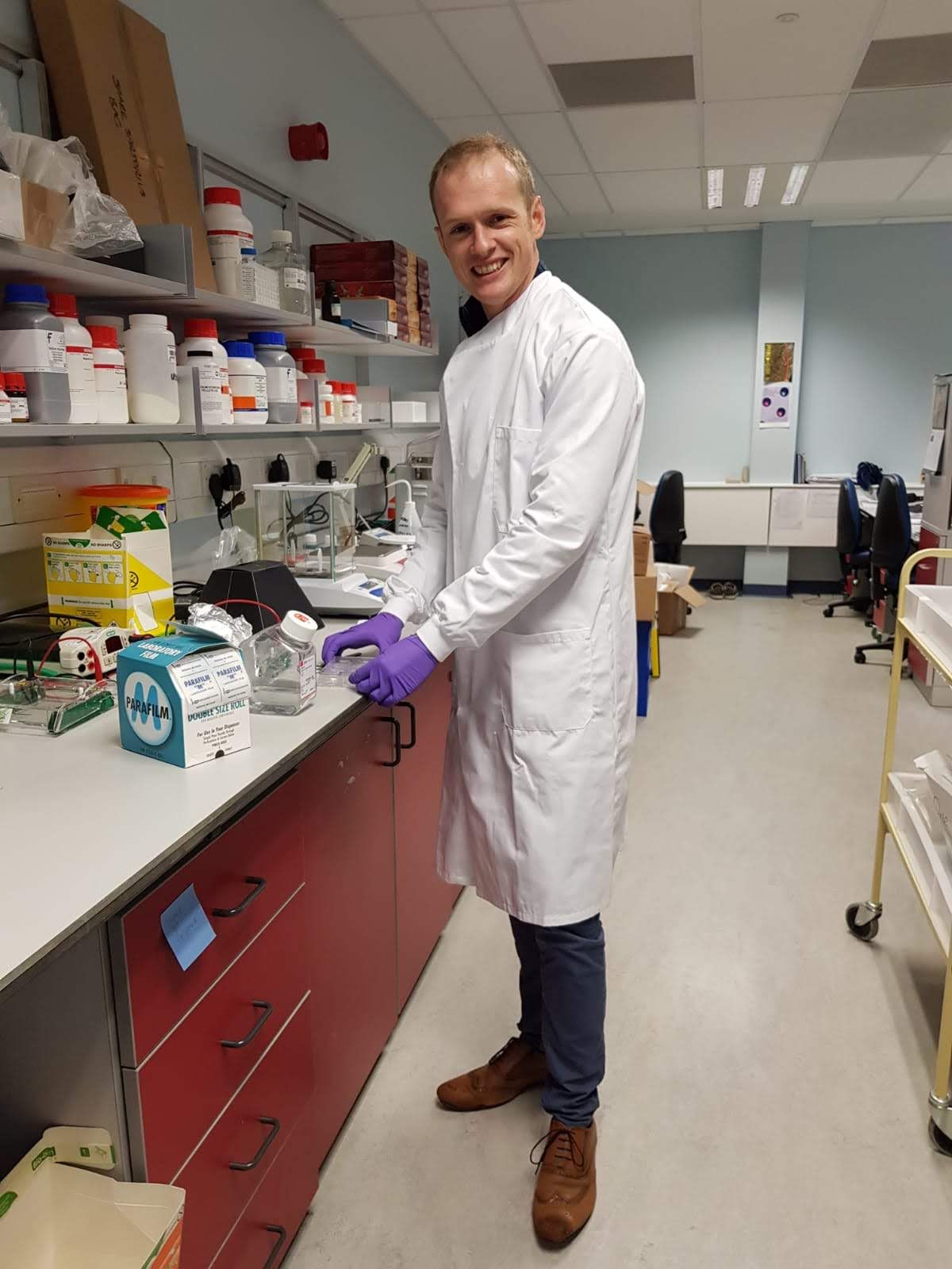
Mathew Horrocks | Image Credit: © Mathew Horrocks

Mathew Horrocks, team leader of the group, is the 2023 recipient of The Joseph Black Prize, awarded by the Royal Society of Chemistry for the most meritorious contributions to any area of analytical chemistry made by an early career scientist. Horrocks shared his thoughts about his current work developing and using single-molecule and super-resolution microscopy techniques to study amyloid oligomers and their commonality regarding a variety of neurodegenerative disorders with Spectroscopy.
Your research focuses on the development and use of single-molecule and super-resolution microscopy techniques to study amyloid oligomers and their commonality regarding a variety of neurodegenerative disorders. Why do you think that there is this commonality?
Interestingly, any protein can form amyloid structures. Indeed, this is the most thermodynamically stable form of a protein. Fortunately, our cells have developed mechanisms to prevent this from happening. As cells age, however, these mechanisms grow weaker, and the cell is less able to prevent amyloid formation. As neurons have a long lifetime, they are particularly susceptible to this process, and so it’s not surprising that protein aggregation is common to many neurodegenerative diseases. There are also common features shared between the proteins that have a high propensity to aggregate.
Could you elaborate on the microfluidic platform you developed for studying protein aggregates in Parkinson's disease?
Although our microscopy approaches allow individual molecules to be visualized, this can sometimes lead to data acquisition rates being low. We cannot increase the concentration of the analyte, as this would swamp the microscope, preventing single-molecule detection, and so instead, we needed to reduce the time required to detect each molecule. We achieved this using a simple one-channel microfluidic device. Rather than waiting for molecules to diffuse through our probe volume, we used the device to rapidly flow the molecules through it, therefore increasing the detection rate by an order of magnitude.
What were the key findings of your research on visualizing oligomers in human biofluids? How does this contribute to our understanding of Parkinson's disease?
Currently, there are no biochemical diagnostics for neurodegenerative disorders, and instead they are clinically diagnosed, with confirmation made post-Mortem. We therefore wanted to see if it was possible to visualize the protein oligomers within biofluids, as this would give us a window into what is happening in the brain. We were able to adapt our single-molecule approaches to achieve this, and surprisingly, we found that protein aggregates are present in samples from both healthy controls and Parkinson’s patients, however, the levels were higher in the latter group. This suggests that protein aggregation is likely a normal phenomenon in all people, but that it is above a tolerated threshold in patients living with the disorder. We’ve now looked in samples from patients who are part of drug trials, and interestingly, we see that the levels of oligomers decrease. This is an exciting finding, as it supports the hypothesis that the oligomers are important disease-causing agents.
You have assembled a network of interdisciplinary collaborators to assist in your efforts? Can you briefly discuss the strengths and experience that this team brings to the table?
Yes, I am extremely lucky to work with a highly interdisciplinary group of researchers and collaborators. As for the collaborators, we gain clinical insights into the disorders from consultant neurologists and biomedical researchers, we get novel probes from chemists, and get the latest microscopy techniques from physicists. I’ve found that working with a multidisciplinary team has led to the group being strong team players- each member has their own strengths, and they work together to pool their expertise. For example, we have some people who are great at coding and love analyzing data, and they tend to share their algorithms with others in the group. We also have others who are great at working with cells, and they often share these with other team members.
Were there any limitations or challenges you encountered in your work?
Yes, we are often limited by the reagents available to study these species. The microscopy approaches we have developed give us the ability to look at individual molecules at the nanometer length scale, however, to do this, they need to be tagged. The oligomers are often at very low (picomolar) concentrations, and so the tags need to be highly specific and bind with high affinity. There has been a lack of such tags in the field. We have been extremely lucky to have forged a collaboration with UCB Biopharma, who have provided us with a multitude of high affinity antibodies, revolutionizing our research abilities. I first conceptualized our most recent paper on detecting oligomers with confocal microscopy in 2012 but had to wait 10 years for the antibodies enabling this to be available!
Can you please summarize the feedback that you have received regarding this work?
People are usually very excited by our techniques. Unlike other detection approaches, such as ELISA, which only generate a number, we can directly see the species that cause the damage in these disorders, and furthermore we can see their different structures at the nanometer length scale.
Do you believe that this method can easily translate to the detection of other diseases, and, if so, which diseases do you believe would result in the most beneficial translations?
Yes, while we have focused on neurodegenerative diseases, there are many other disorders that are caused by protein misfolding and aggregation, for example Type II diabetes, and some forms of cancer. As most of the other disorders can be diagnosed more easily, our approaches are still most beneficial to neurodegenerative diseases, which are still difficult to diagnose.
What are the next steps in this research?
Our immediate plans are to use the approaches to look for oligomers in more easily accessible biofluids. While we have shown that it’s possible to specifically detect aggregates in CSF, this biofluid is collected via a lumbar puncture, which is not a pleasant procedure. What we really want to be able to do is detect them in blood, saliva, stool, or urine. This would pave the way for a widespread screening program.
What does your being named the recipient of the Black Prize mean to you professionally? Personally?
It is a huge honor to be the recipient of the prize. Professionally, it raises awareness of the work my team do, and has already led to us being invited to several conferences. On a personal level, it’s great to have our efforts in this area recognized, and it’s really been a team effort over the last decade or so.
About the Interviewee
Mathew Horrocks was born and brought up in Halifax, West Yorkshire, before studying Chemistry at Oriel College, University of Oxford. He did his master’s project with Professor Mark Wallace, where he was first introduced to single-molecule techniques. Following this, he moved to the University of Cambridge to work with Professor Sir David Klenerman, developing microscopy techniques to study the protein aggregates formed in neurodegenerative disorders, such as Parkinson’s and Alzheimer’s disease. Following a brief stint researching in New South Wales, Australia, Mathew returned to Cambridge in 2016 to take up a Junior Research Fellowship at Christ’s College, and a Herchel Smith Fellowship at the University of Cambridge. He moved to the University of Edinburgh in 2018 to take up a post as Lecturer (Assistant Professor) in the School of Chemistry, where he also established the ESMB Group. He was promoted to Senior Lecturer (Associate Professor) in 2022, and currently leads a team of 13 researchers dedicated to the advancement and application of single-molecule and super-resolution techniques. Their collective efforts are focused on unraveling various biological questions, encompassing subjects such as the molecular underpinnings of memory storage, how proteins misfold, and the mechanisms behind mitochondrial dysfunction in cardiovascular diseases.
New Study Reveals Insights into Phenol’s Behavior in Ice
April 16th 2025A new study published in Spectrochimica Acta Part A by Dominik Heger and colleagues at Masaryk University reveals that phenol's photophysical properties change significantly when frozen, potentially enabling its breakdown by sunlight in icy environments.
Tracking Molecular Transport in Chromatographic Particles with Single-Molecule Fluorescence Imaging
May 18th 2012An interview with Justin Cooper, winner of a 2011 FACSS Innovation Award. Part of a new podcast series presented in collaboration with the Federation of Analytical Chemistry and Spectroscopy Societies (FACSS), in connection with SciX 2012 ? the Great Scientific Exchange, the North American conference (39th Annual) of FACSS.
New Fluorescence Model Enhances Aflatoxin Detection in Vegetable Oils
March 12th 2025A research team from Nanjing University of Finance and Economics has developed a new analytical model using fluorescence spectroscopy and neural networks to improve the detection of aflatoxin B1 (AFB1) in vegetable oils. The model effectively restores AFB1’s intrinsic fluorescence by accounting for absorption and scattering interferences from oil matrices, enhancing the accuracy and efficiency for food safety testing.