Fluorescent Probe Created to Better Analyze Chemicals Behind Depression
In a recent study out of Fujian Normal University and the Second Affiliated Hospital of Fujian University of Traditional Chinese Medicine in Fuzhou, China, scientists created a dual-response fluorescent probe to analyze the vesicular exocytosis of noradrenaline (NE) in a cell culture environment. With this fluorescent probe system, they hoped to gain insight into the pathological mechanisms of depression disorder and ease the diagnosing and tracking of the disease. Their findings were published in Spectrochimica Acta Part A: Molecular and Biomolecular Spectroscopy (1).
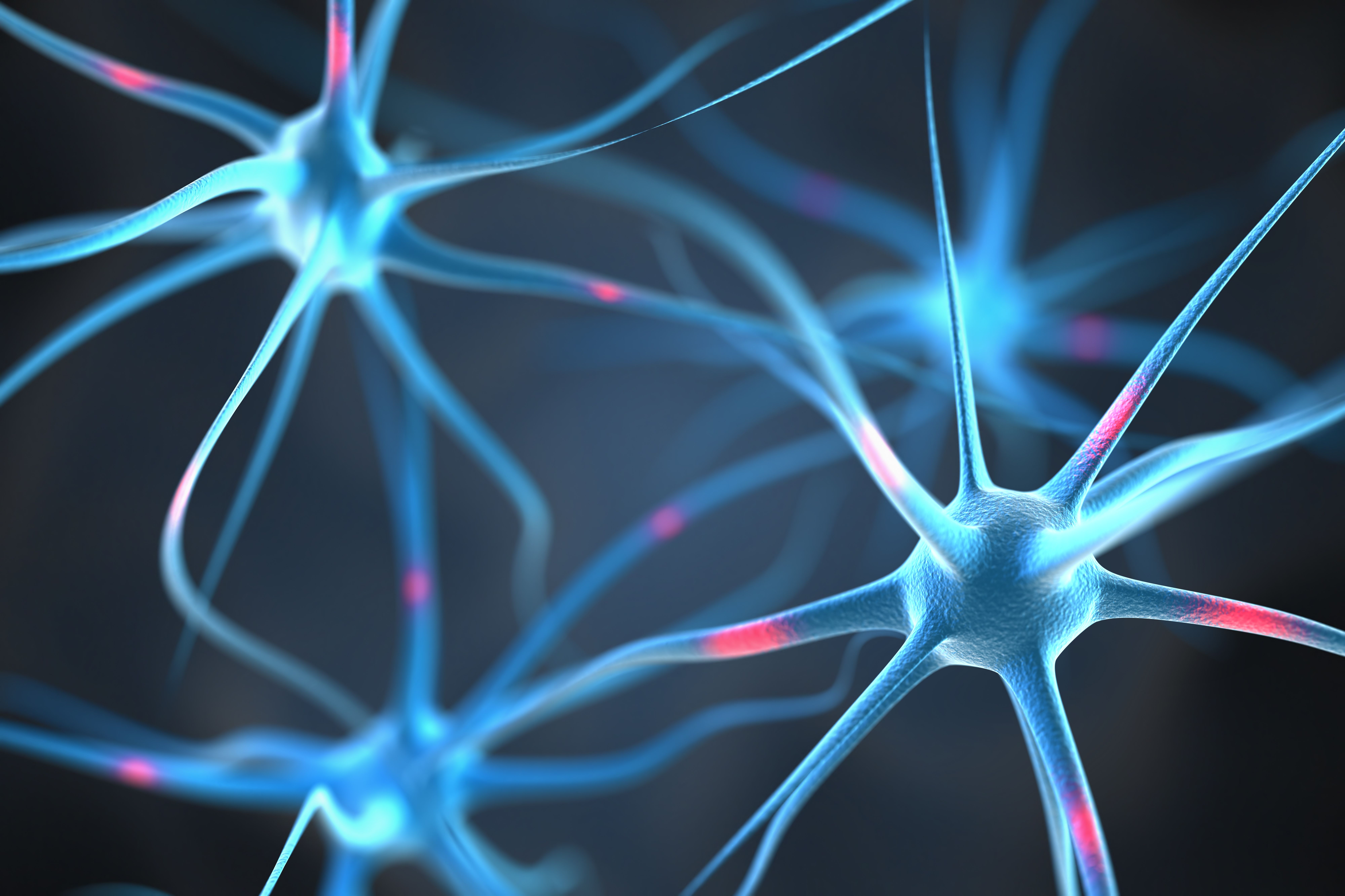
Neurons in the brain | Image Credit: © Leigh Prather - stock.adobe.com

According to the World Health Organization, approximately 3.8% of the world’s population, or approximately 280 million people, suffer from depressive disorder (2). This disorder can affect people from all walks of life, with more than 700,000 people dying from suicide every year; in fact, suicide is said to be the fourth leading cause of death in 15–29-year-olds. Symptoms of the condition can include sadness, loss of pleasure, sleep disorders, and inappetence. However, the disorder’s complicated pathogeny has led to a rudimentary knowledge of the condition. To reliably measure this condition, tools would need to dynamically observe the course of depression in real-time; this would allow for the disease to be properly diagnosed and monitored, while also allowing for the development of treatment strategies.
Noradrenaline (NE), being one of the key catecholamine neurotransmitters in the central and sympathetic nervous system, has critical influence on how and when depression occurs. Typically, depression is related to the vesicular exocytosis of NE rather than the intracellular concentration of NE. The former process typically relies on calcium ions, which are also used to reduce viscosity and enable smooth vesicle movements. This led to the inference that cell viscosity influences NE exocytosis, meaning that developing a method for simultaneously detecting NE and viscosity would be highly beneficial for depression research.
Read More: Early Signs of Brain Injury Now Identifiable Using Raman-Based Detection System
Following this inference, the scientists designed and synthesized a dual-response fluorescent probe made of a rhodol-isophorone and a thioester group (RHO-DCO-NE). This probe, according to the scientists, can simultaneously detect NE concentration and viscosity levels, all with negligible crosstalk between the two channels. The rhodol-isophorone derivative serves as a fluorescent core and works as a viscosity-sensitive unit, where a rotatable unsaturated double bond remains. As for the thioester group, it is used as an NE-specific responsive group. Using this probe, the scientists studied the effects of viscosity changes on the NE release of PC12 and corticosterone-induced PC12 cells. The experiment’s data showed that by reducing viscosity levels, the release of NE of depression cell models can be accelerated. These insights provide new revelations into how the pathological mechanisms of depression function, which can potentially help in properly diagnosing and treating the disorder.
References
(1) Xiong, X.; Qiu, J.; Fu, S.; Gu, B.; Zhong, C.; Zhao, L.; Gao, Y. A Dual-Response Fluorescent Probe for Norepinephrine and Viscosity and Its Application in Depression Research. Spectrochim. Acta Part A: Mol. Biomol. Spectrosc. 2024, 315, 124270. DOI: 10.1016/j.saa.2024.124270
(2) Depressive Disorder (Depression). World Health Organization 2023. https://www.who.int/news-room/fact-sheets/detail/depression (accessed 2024-4-18)
New Study Reveals Insights into Phenol’s Behavior in Ice
April 16th 2025A new study published in Spectrochimica Acta Part A by Dominik Heger and colleagues at Masaryk University reveals that phenol's photophysical properties change significantly when frozen, potentially enabling its breakdown by sunlight in icy environments.
Tracking Molecular Transport in Chromatographic Particles with Single-Molecule Fluorescence Imaging
May 18th 2012An interview with Justin Cooper, winner of a 2011 FACSS Innovation Award. Part of a new podcast series presented in collaboration with the Federation of Analytical Chemistry and Spectroscopy Societies (FACSS), in connection with SciX 2012 ? the Great Scientific Exchange, the North American conference (39th Annual) of FACSS.
New Fluorescence Model Enhances Aflatoxin Detection in Vegetable Oils
March 12th 2025A research team from Nanjing University of Finance and Economics has developed a new analytical model using fluorescence spectroscopy and neural networks to improve the detection of aflatoxin B1 (AFB1) in vegetable oils. The model effectively restores AFB1’s intrinsic fluorescence by accounting for absorption and scattering interferences from oil matrices, enhancing the accuracy and efficiency for food safety testing.