Key LC–MS Techniques Benefit Life Sciences Researchers
Special Issues
State-of-the-art mass spectrometry (MS) techniques of growing importance to life sciences research now include not just liquid chromatography (LC)–MSn (n = 2–11), but also LC–matrix-assisted laser desorption ionization-time-of-flight (MALDI-TOF), LC-MALDI-TOF-TOF, electrospray ionization (ESI)-TOF, and LC-Fourier transform (FT) MS.
Scientists often select the newest generation of innovative matrix-assisted laser desorption ionization-time-of-flight (MALDI-TOF) and MALDI-TOF-TOF mass spectrometers to take advantage of their extraordinary research capabilities. For example, an important MALDI-TOF and MALDI-TOF-TOF application is high-success protein identification from 2D gels. Here, MALDI-TOF-TOF is the foremost component of new proteomics analysis systems for peptide mass fingerprinting, peptide fragment analysis, and posttranslational modifications (PTM) analysis. The newest generation of ultrahigh-performance TOF-TOF mass spectrometers enables high-throughput protein identification by MALDI-TOF peptide mass fingerprinting, immediately followed by more detailed, high-caliber protein characterization using MALDI-TOF-TOF tandem mass spectrometry (MS-MS) on the same prepared sample. Comprehensive MS-MS information is delivered to TOF-TOF users from rather minute sample amounts within just a few seconds. In addition, new T3-sequencing capabilities allow top-down sequence analysis on even many intact proteins (1).

Figure 1
The newest generation of top TOF-TOF instrumentation provides scientists with two MS-MS methods for de novo sequencing. For highest specificity, laser-induced decomposition with post-fragmentation acceleration TOF-TOF MS is the method of choice, providing clear spectra with readily available amino acid and sequence-specific ions such as i-, a-, b- and y-ions. For in-depth analysis of selected peptides, high-energy collision-induced dissociation (CID) is very valuable as well.
Regarding protein identification and quantitation by LC-MALDI, new advanced approaches provide unique, intelligent LC workflow for LC–MALDI experiments. With new isotope coded protein label (ICPL, Toplab GmbH, Martinsried, Germany) triplex technology now available, three independent entire proteomes can be quantitated even simultaneously. Low-abundance proteins are identified and can be characterized in detail using MALDI-TOF-TOF, in which identifying and characterizing low-abundance proteins are considered the "holy grail" of proteomics.
Sophisticated workflows further enhance results through a synergistic data-dependent combination with LC–electrospray ionization (ESI)-MS-MS data. LC–ESI-MALDI-MS-MS wizard-driven workflows utilize complementary information from the two ionization techniques in a single eclectic experiment, providing much more useful in-depth knowledge and a new level of insight. Quantitative expression proteomics experiments with various isotope-labeling techniques including ICPL, isotope coded affinity tag (ICAT, University of Washington, Seattle, Washington), and isotope tagging for relative and absolute protein quantitation (iTRAQ, Applera Corp., Norwalk, Connecticut) are now being realized as well. Flexible quantitation software is becoming future proofed, allowing scientists to incorporate new or unique isotope labels.

Figure 2
Meanwhile, many major developments have been taking place concerning advanced biomarker panel discovery, validation, and identification. Here again, MALDI-TOF-TOF plays a commanding role, providing seamless integration into clinical proteomics solutions for high-resolution peptide and protein putative biomarker panel discovery, identification, and validation. Generally, such solutions are approved for research only, and are not yet in the clinic — the next big step.
Another rapidly developing MS method is advanced MALDI molecular imaging. A recent innovation in quantitative proteomics in this field is 2D peptide imaging of cells or tissue extracts as an application of MALDI molecular imaging. Sliced tissue sections can now be scanned with a very small laser focus and analyzed rapidly at 200 Hz, and with very high levels of sensitivity. This approach is fully supported by new imaging software, avant-garde algorithms, and workflows. A simple click on any spot on a color map of cancerous tissue, for example, provides the research scientist with all the detailed MS data supporting that colored spot, whether they are protein, peptide, or drug metabolite data. Visualization of the spatial distribution of proteins, drug candidate compounds, and biomarkers is a compelling tool in the exciting fields of putative biomarker panel evaluation and drug development. Coupled with wizard-driven workflow, MALDI-TOF MS provides a fast, meritable screening tool for direct analysis from tissue.
There is also breaking scientific news in electrospray-based MS. Research scientists now select ESI-quadrupole (Q) TOF mass spectrometers for accurate mass or exact mass measurements in straightforward, unambiguous formula determination of small molecules; metabolic studies; analysis of complex mixtures; in-depth investigation of proteins, including quality control of intact proteins; de novo sequencing, and the study of noncovalent complexes.
Advanced micro TOF quadruple-based ESI mass spectrometers now uniquely offer three dimensions of identification information simultaneously on all results: precise mass, MS-MS, and isotopic analysis for unprecedented certainty in discovery science. With quite compact footprints, the new micro ESI TOF benchtop instruments regularly outperform the earlier floor-standing ESI-Qq-TOF systems. Advanced electrospray orthogonal TOF mass spectrometer designs are particularly well equipped for the requirements of metabolomics, proteomics, and drug development. Easy formula determination of small molecules, metabolic studies, analysis of complex mixtures, digests, and in-depth evaluation of intact proteins are key applications. Next-generation ion funnel ESI sources now provide scientists with impressive improvements in sensitivity. Such electrospray ion sources, with their advanced optics, dramatically improve ion transmission. Research scientists worldwide are taking advantage of new high-performance Q-q-front ends that feature quadrupole mass filters and a quadrupole collision cell for accumulation of parent and fragment ions before mass analysis. The entire range of fragment ions is available at increased sensitivity for the TOF mass analysis.
Unequaled mass accuracy has become available over a wide dynamic range. For example, ESI micro TOF-quadrupole spectrometers can reach a mass accuracy of better than 3 ppm, using innovative detection technologies. Such instruments can take advantage of the stability of micro TOF spectrometers over an exceptionally wide dynamic range, without tedious recalibration routines. Mass position is kept stable for hours during temperature changes and over a wide dynamic range with significant variations in sample concentration, a critical consideration for research scientists. As continues to be the case with LC–nuclear magnetic resonance (NMR)–MS(n) , such ESI TOF-quadrupole mass spectrometers are being closely coupled to top-of-the-line NMR spectrometers (now reaching up to 950 Mhz) to provide more in-depth useful knowledge.
Using the most advanced algorithms, scientists now compare true isotopic patterns against their measured spectra. Sophisticated software determines the correlation between measured and theoretical isotopic patterns, leading to results with significantly increased confidence. This combination drastically reduces the number of possible formula candidates and leads to unequivocal determination of elemental compositions of compounds.
Bringing together the features of micro TOF spectrometers (the excellent mass accuracy on fragments and superb focus resolving power exceeding 15,000 FWHM — with flexible data-dependent MS-MS strategies), the new ESI micro TOF quadrupole-type mass spectrometers allow de novo sequencing of peptides even for PTMs. Such solutions produce confident results in cutting-edge proteomics research, closely complementing the accurate mass and structural analysis of small molecules that other MS instrumentation enables.
ESI-ion trap MSn provides breakthrough performance for proteomics, drug discovery, LC–MS, and MSn research. LC–MSn systems bring MS-MS specificity to routine work and provide high-performance MSn for a broad range of applications. Radical new high-capacity ion-trap technology takes ion traps to a whole new level. High-capacity traps are now fully integrated into proteomics solutions suites, combining automated sample preparation, nanoflow separation systems, advanced MS, and bioinformatics. Importantly, high-capacity ion traps now available feature the very latest implementations of electron transfer dissociation (ETD), a fine fragmentation technique that preserves PTMs.
Modern functional proteomics demands a step beyond just protein identification. It is becoming more and more essential to unravel the many modifications proteins undergo, triggering their biological activity. Unique capabilities for PTM discovery are now available. With advanced ETD, scientists are equipped for specific information-rich PTM analysis using the new generation of highest capacity ion traps. ETD techniques allow peptide and protein fragmentation while preserving modifications such as phosphorylation or glycosylation. This result gives researchers easy access to protein sequencing and simultaneous identification of type and location of various PTMs.
ETD MS-MS spectra of peptides can now be collected on-the-fly during LC–MS-MS runs using the new higher capacity ion traps with PTM discovery capabilities. Because of its nonergodic nature, ETD typically creates very clean MS-MS spectra with intact PTMs. The new advanced ETD implementations provide complete amino acid series, without the low-mass cut-off traditionally encountered in ion-trap MS-MS. The combination with the superior mass accuracy of the highest capacity ion traps enables powerful de novo sequencing capabilities. Taking advantage of the special modes of the high-capacity ion traps, advanced ETD offers a unique and powerful system for single run auto-LC–MS-MS-MS ETD experiments specifically for phosphorylation analysis. The combination of high-capacity ion trap acquisition speed and sensitivity with new information-rich ETD MS-MS capability redefines ion trap standards for PTM analysis and de novo sequencing.
In addition to all this good news for the life sciences, hybrid quadrupole-hexapole (Q-h) FT superconducting magnet mass spectrometers have now become available commercially up through 15-T magnetic fields for on-the-fly ultrahigh-resolution LC–MS–FT-MS analysis. The FT-MS architecture provides flexibility and performance for the most challenging problems and is seen as synonymous with superior performance. Q-h FT-MS combines the power of FT-MS with the latest quadrupole instrumentation (complete with linear ion-trapping modes) and multipole collision cell for a quantum leap in analytical capability. The unique geometry provides facile and efficient precursor mass selection with collision-induced dissociation in the Q-h "front end" to the FT-MS system for on-the-fly ultrahigh-resolution LC–MS–FT-MS analysis. Moreover, the Q-h "front end" to the FT-MS architecture provides added functionality for the enrichment of low-abundance species via mass selective accumulation. Futuristic features include LC–MS-MS with data-dependent acquisition, using unified software solutions. Data can now be acquired faster than one spectrum per second, with resolving power of greater than 100,000, ideal for LC–FT-MS and LC–MS–FT-MS research.
Scientists can perform comparative analysis of complex proteomes for biomarker discovery by employing differential isotopically labeled acetylation (data to be published) followed by LC–LC–FT-MS on a state-of-the-art 12-T FT-MS. The analysis could be performed even more precisely on an ultrahigh-resolution 15-T FT-MS system.
A fundamental challenge in proteomics has been to discover disease biomarker panels by comparing complex proteomes. Gel-based approaches are quite useful but can be limited by sample complexity or by the nature of proteomes compared (for example, highly hydrophobic membrane-associated proteins). Multidimensional LC can overcome some of these problems but can be limited by mass tagging strategies using reagents, which can be expensive or hard to synthesize, or which can target reactive side chains not present in all peptides. Scientists have developed an exceptional technique for comprehensive binary proteome comparison that employs isotopically labeled acetic anhydride to label all peptides in a sample, coupled to multidimensional LC separation with the ultrahigh mass accuracy and resolving power of the 12-T FT-MS mass spectrometer for sample analysis.
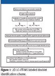
Protein samples are digested with trypsin and the peptides are mass tagged by labeling with either acetic anhydride or hexadeuteroacetic anhydride. The peptides from each sample now differ in mass by increments of 3.019 Da. Equivalent amounts of sample peptides are mixed and separated by high performance liquid chromatography (HPLC), either one-dimensional (1-D) reversed-phase LC, or 2-D LC employing a strong cation exchange (SCX) column and a C18 column. Online MS spectra typically are acquired with 12-T FT-MS mass spectrometers, after which the spectra are analyzed to identify the monoisotopic peaks. A list is further filtered to locate labeled and unlabeled doublets spaced by the proper mass difference. The intensity ratios of the peptides are then used to quantify the relative abundance of the proteins in the original samples.
With scientists' goal of comprehensive proteomic comparison, their first need is to compare the numbers of peptide pairs detected in traditional 1-D reversed-phase LC–FT-MS with the number of peptide pairs detected in 2-D LC–FT-MS. To accomplish this, sophisticated software has been written to detect and quantify these peptide doublets in a completely automated manner.
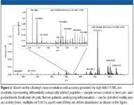
With a 1-D LC–FT-MS system to compare different plasma proteomic samples in cystic fibrosis research at the University of North Carolina, Chapel Hill, a spectrum analysis algorithm successfully identified 3770 peptide doublets. Of these doublets, 2370 (63%) had intensity ratios of ≤ twofold, 1726 (46%) had intensity ratios of ≥ threefold, 1182 (31%) had ratios > fivefold, and 657 (17%) had intensity differences > 10-fold. In contrast, 2-D LC–FT-MS analysis revealed nearly three times as many (9274) peptide doublets. Interestingly, the relative proportions of peptide doublets falling into each category of fold-difference were similar, but not identical: ≤ twofold, 5352, (57%); ≥ threefold, 3964 (43%); > fivefold, 2762 (30%); >10-fold, 1753 (19%).
A monoisotopic peak-picking algorithm is capable of analyzing overlapping isotope patterns that are often present because the label adds only three mass units to the analyte. Furthermore, the scientists' clever algorithm takes into consideration multiple labeling, multiple charging (changing the observed mass spacing), and the mass error of the instrument during the doublet identification. Clearly, coupling the high resolution and mass accuracy of FT-MS with a mass-tagging approach that potentially labels all peptides in a sample will allow scientists to interrogate complex protein samples more thoroughly for subtle, yet biologically relevant differences in protein expression. Furthermore, this approach should allow scientists to compare relative fold differences of multiple peptides from a single protein, thereby increasing the accuracy of the observed ratios. This approach has been applied successfully to the discovery of protein biomarkers in plasma for early detection of lung infection in cystic fibrosis patients.
With all these exceptional enabling life sciences tools now in their toolbags, research scientists worldwide are diving much deeper into the proteomes.
Acknowledgments
The above cystic fibrosis study was funded by a grant from the Cystic Fibrosis Foundation (CFFTIBORCHE05U0), by a grant from the North Carolina Biotechnology Center (NCBC 2005-IDG-1015), by a grant from the National Institutes of Health (1-S10-RR019889-01), and by a gift from an anonymous donor to support research in proteomics at UNC.
Michael Easterling, J. Paul Speir, and Michael Willett are with Bruker Daltonics, Billerica, Massachusetts. Cameron O. Scarlett is with University of North Carolina, Chapel Hill, North Carolina, and University of Wisconsin, Madison, WIsconsin. Severine A. Ouvry-Patat is with University of North Carolina, Chapel Hill, North Carolina. Ryan M. Danell is with Danell Consulting, Greenville, North Carolina. Christoph H. Borchers is with University of North Carolina, Chapel Hill, North Carolina, and University of Victoria — Genome British Columbia Proteomics Centre.
References
(1) D. Suckau, A. Resemann, M. Schuerenberg, P. Hufnagel, J. Franzen, and A. Holle, Analytical and Bioanalytical Chemistry, Springer Berlin, 376(7), 952–965 (2003).
(2) C.O. Scarlett, R.M. Danell, M. Easterling, J.P. Speir, and C.H. Borchers, paper presented at the 54th ASMS Conference on Mass Spectrometry and Allied Topics, Seattle, Washington May 28–June 2, 2006.

New Study Reveals Insights into Phenol’s Behavior in Ice
April 16th 2025A new study published in Spectrochimica Acta Part A by Dominik Heger and colleagues at Masaryk University reveals that phenol's photophysical properties change significantly when frozen, potentially enabling its breakdown by sunlight in icy environments.