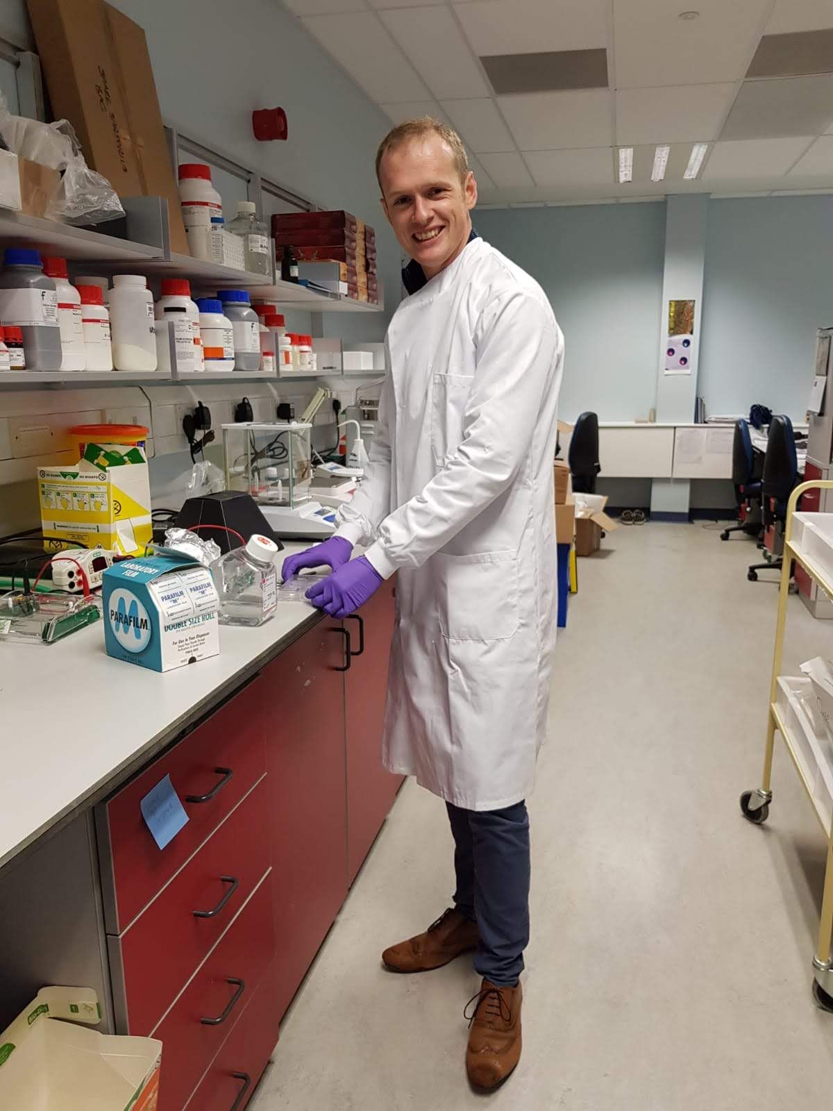
Mathew Horrocks Wins Joseph Black Award at SciX Conference
Horrocks and his research group focus on single-molecule and super-resolution microscopy.
Mathew Horrocks is the winner of the 2023 Joseph Black Award

Mathew Horrocks, senior lecturer at the University of Edinburgh School of Chemistry, is the winner of the 2023 Joseph Black Award. Horrocks was presented with the award, which honors contributions to any area of analytical chemistry made by an early career scientist, at the SciX conference in Sparks Nevada on Tuesday.
During an award symposium, Horrocks discussed his research on single-molecule and super-resolution microscopy approaches to help visualize individual protein aggregates in complex biological samples (1). This method is particularly helpful for neurodegenerative disorders, such as Parkinson’s and Alzheimer’s disease, which are characterized by the misfolding and aggregation of soluble monomeric protein into insoluble amyloid fibrils.
Super-resolution microscopy refers to a group of analysis techniques that surpass the traditional diffraction limit of light microscopy, allowing researchers to observe cellular structures and molecules at nanometer-scale resolutions.
“Currently, there are no biochemical diagnostics for neurodegenerative disorders, and instead they are clinically diagnosed, with confirmation made post-Mortem,” Horrocks told Spectroscopy in a previous interview(2). “We therefore wanted to see if it was possible to visualize the protein oligomers within biofluids, as this would give us a window into what is happening in the brain.”
Horrocks studied chemistry at Oriel College at the University of Oxford, where he was first introduced to single-molecule techniques. In 2016, he took a junior research fellowship at Christ’s College and a Herchel Smith fellowship at the University of Cambridge. Then in 2018 he moved to the University of Edinburgh to head the Edinburg Single-Molecule Group, where his team focuses on developing single-use molecule and super-resolution approaches to study biological systems, with a particular focus on neuroscience and neurodegeneration.
Horrocks and his research group have also applied spectroscopic approaches to understanding how the healthy brain functions. They invited a new super-resolution microscopy technique that works in live cells. In the future, the group plans on use their approaches to look for oligomers in more easily accessible biofluids, Horrocks previously told Spectroscopy.
“While we have shown that it’s possible to specifically detect aggregates in cerebrospinal fluid (CSF), this biofluid is collected via a lumbar puncture, which is not a pleasant procedure,” he said. “What we really want to be able to do is detect them in blood, saliva, stool, or urine. This would pave the way for a widespread screening program.”
References
- Horrocks, M. Visualizing Neuroscience at the single-molecule level. In SciX Book of Abstracts, Sparks, Nevada, October 10, 2023; https://scixconference.org/scix2023program
- Chasse, J. Fluorescence Microscopy: A Conversation with Joseph Black Award Winner Mathew Horrocks. https://www.spectroscopyonline.com/view/fluorescence-microscopy-a-conversation-with-joseph-black-award-winner-mathew-horrocks (accessed 2023-09-29).
Best of the Week: AI and IoT for Pollution Monitoring, High Speed Laser MS
April 25th 2025Top articles published this week include a preview of our upcoming content series for National Space Day, a news story about air quality monitoring, and an announcement from Metrohm about their new Midwest office.
LIBS Illuminates the Hidden Health Risks of Indoor Welding and Soldering
April 23rd 2025A new dual-spectroscopy approach reveals real-time pollution threats in indoor workspaces. Chinese researchers have pioneered the use of laser-induced breakdown spectroscopy (LIBS) and aerosol mass spectrometry to uncover and monitor harmful heavy metal and dust emissions from soldering and welding in real-time. These complementary tools offer a fast, accurate means to evaluate air quality threats in industrial and indoor environments—where people spend most of their time.