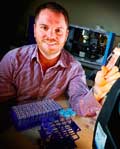
Matthew Baker, Winner of Spectroscopy’s Inaugural Emerging Leader in Molecular Spectroscopy Award, Focuses on Clinical IR and Raman Applications
Spectroscopy is proud to have created a new award, the Emerging Leader in Molecular Spectroscopy Award. As its name implies, the award recognizes a young scientist, and it is designed to encourage the next generation of molecular spectroscopists. Matthew Baker, the winner of the inaugural Emerging Leader in Molecular Spectroscopy Award, is a senior lecturer in chemistry at the University of Strathclyde, in Glasgow, Scotland. At Strathclyde, Baker leads research to advance the application of analytical chemistry to real-world problems in a variety of areas, including the biomedical, clinical, defense, and security fields. His main focus is the development of spectroscopic and spectrometric molecular pathology, disease diagnosis, and the detection of pathogenic bacteria and toxic chemicals. In particular, Baker has pioneered the use of vibrational spectroscopy for clinical diagnostics.

Spectroscopy is proud to have created a new award, the Emerging Leader in Molecular Spectroscopy Award. As its name implies, the award recognizes a young scientist, and it is designed to encourage the next generation of molecular spectroscopists. Matthew Baker, the winner of the inaugural Emerging Leader in Molecular Spectroscopy Award, is a senior lecturer in chemistry at the University of Strathclyde, in Glasgow, Scotland. At Strathclyde, Baker leads research to advance the application of analytical chemistry to real-world problems in a variety of areas, including the biomedical, clinical, defense, and security fields. His main focus is the development of spectroscopic and spectrometric molecular pathology, disease diagnosis, and the detection of pathogenic bacteria and toxic chemicals. In particular, Baker has pioneered the use of vibrational spectroscopy for clinical diagnostics.
Baker will be presented with the award at a plenary lecture during the SciX conference in Minneapolis, Minnesota. The session will take place at 8:00 am, on Monday, September 19. In an award symposium later that day, he will also give a presentation titled “Serum Spectroscopic Diagnostics: The Future for Clinical Diagnostics?”
Baker recently spoke to Spectroscopy about his scientific background, interests, and recent work.
Where or how did your interest in analytical chemistry and molecular spectroscopy begin?
It really started when I was looking for a placement for work experience when I was 15 or 16. I had always known I had wanted to work in a lab and in some form of chemistry but all my school offered, when I said I wanted to be a chemist, was a position in Boots (the UK version of Walgreens). Luckily, my dad was a mechanical & electrical engineer for a local lab that was being built for a contract manufacturer, Universal Products Ltd., that made everything from cosmetics to pharmaceuticals, and I got a work experience position there. After my work experience, I wrote to the managing director and got a part time job that turned into sponsorship through university. This job involved 18 k sponsorship over 4 years, a job every summer and I also did my yearlong industrial placement there. Also, the managing director sat down with me and provided mentorship and guidance, particularly on accounts and expenses for university. I worked there as my placement year in industry for my master’s degree in chemistry while at the University of Manchester Institute of Science and Technology (UMIST). My placement advisor was Dr. Peter Gardner (now Professor). We got chatting about his final-year project on the use of infrared (IR) microspectroscopy for the analysis of prostate cancer tissue, and it kind of all carried on from there.
What would you consider to be the greatest advances in clinical IR and Raman spectroscopy over the past 10 years?
The methodological and technological development over the past 10 years for IR and Raman has been superb. I am very interested in seeing how developments in high-resolution spectroscopic imaging such as tip-enhanced Raman spectroscopy (TERS) and nanoIR develop as these could be excellent tools across the biosciences. However, the major advance I feel has been the furthering of the combination of data analysis regimes in line with the ability to collect relatively large volumes of data. Through this advancement we will be able to develop impactful and useful tools across many industries and domains.
You have done some significant work pioneering the use of vibrational spectroscopy for clinical diagnostics. How did you get started on creating a vibrational spectroscopy method for brain tumor diagnosis that is suitable for clinical applications? What were the biggest challenges? What benefits does it bring to the field?
I started working on serum spectroscopy in Manchester to look at prostate cancer analysis, but we couldn’t crack it. Following that effort, I spent some time in Europe and the United States on a fellowship based on tissue and cellular analysis and then moved to work for the Defence Science and Technology Laboratory, which is part of the UK Ministry of Defense. I wanted to move back to academia and found an academic position and essentially I got the chance to think about the use of spectroscopy for clinical diagnostics and then decide where to start.
I thought the best chance for spectroscopy in the clinic would be in either intra-operative analysis or biofluid analysis, so when I returned to academia I offered to give talks to the local clinical research consortia, focusing on hard-to-detect chronic (for example, brain cancer) and infectious diseases (such as sepsis), and from there the research kicked off.
The biggest challenge really was getting the methodology proven for effective and repeatable analysis; now the applications are expanding. We have recently proven the ability to quantify small molecules in complex matrices such as serum.
Can you describe how are you expanding your work with vibrational spectroscopy using novel light sources to enable rapid, high-throughput imaging?
The advent of the use of quantum cascade lasers (QCLs) as IR sources has opened up the ability to perform high-throughput analysis and imaging of samples using discrete frequencies and with high source brilliance. Professor Rohit Bhargava has pioneered this area for biomedical spectroscopy, but essentially the complete IR spectrum is not required to accurately describe the sample under analysis and using the complete spectrum actually undermines the sensitivity and specificity of detection when performing disease-based studies (1). Using QCLs we have shown that we can perform a rapid image collection of pertinent frequencies that accurately describes our serum disease set based on cancer type. Using the imaging modality opens up the ability to collect a large number of spectra rapidly, perform quality control using the image-based approach, and potentially diagnose 16 patients within 2 min (2).
Your work focuses on the application of analytical chemistry to real-world problems in the biomedical, clinical, defense, and security domains, with a focus on pathology, disease diagnosis, and the detection of pathogenic bacteria and toxic chemicals. What specific developments are you hoping to achieve with spectroscopy in these areas?
The research in all these areas is based on the aim to understand the composition and behavior of molecules within complex matrices linked to real-world detection challenges. Essentially, I want to develop spectroscopic tools that can be used in the field or in the clinic where they are needed, where there are pressures such as dirty or harsh environments, or where the sample that you want to use is complex. For instance, with the serum work we are trying to develop a spectroscopic approach for the rapid analysis and detection of disease that uses very simple sample preparation to develop a spectroscopic tool that can be easily translated into current clinical practice without adding to the workload of already overburdened healthcare providers.
What research are you most proud of thus far?
Apart from the biofluid research, which is of course ongoing, a project of which I am proud, in terms of the solution provided, is my work on the detection of chemical warfare agents during my research at the Defence Science and Technology Laboratory. The main method for environmental detection of chemical weapons was via the extraction of the chemical from the soil; however, based upon the molecular makeup of the chemical, the extraction isn’t very efficient. During this research we contaminated a mustard seed with a chemical warfare agent, grew the seed, and then simply harvested the plant and ground it with ethanol. We then analyzed the alcohol extract via liquid chromatography–mass spectrometry (LC–MS) and gas chromatography–mass spectrometry (GC–MS) (with derivatization) and proved we could detect the intact chemical up to 45 days following contamination, providing a time capsule to detect use of chemical warfare agents through environmental analysis. This work was published (3–8).
You cofounded the UK Clinical Infrared and Raman Spectroscopy (CLIRSPEC) Network and from that, the International Society for Clinical Spectroscopy emerged. You also developed the CLIRSPEC summer school, and are involved in Raman4Clinics. Why did you think these organizations were needed? What have they been able to accomplish so far, and what do you envision for their future?
These organizations have been set up with the main aim of championing the translation of promising spectroscopic technology into the clinic. The CLIRSPEC network brings together leading international academics, industrialists, and clinicians to achieve a critical mass across the disciplines to help achieve this. It is now an international professional society with a range of members whose main vehicle is the successful SPEC conference series. The next conference will be held in Glasgow in 2018. So far we have been able to act as a focal point for the community, disseminating research and working on suggested protocols for reporting results. Importantly, we have also been reaching out to other domains to learn about the challenges associated with the translation to the clinic.
The CLIRSPEC summer school is an important date in the CLIRSPEC calendar where postgraduate students across the disciplines associated with clinical spectroscopic research, such as medicine, biology, computation, physics, and chemistry, come together on the shores of Lake Windermere in the UK for a mixture of lectures and student-led complex problem solving in clinical spectroscopy. The next one will be held in Windermere, July 4–7, 2017.
In the future, we aim to become more international and support the translation of spectroscopy to the clinic across the globe.
In 2015, you organized the world’s first Twitter poster contest. How did that project get started? What kind of turnout and engagement did you get from the participants?
I was thinking one day about how to use Twitter as more of a scientific tool, and the idea of a Twitter-based poster conference arose. Following that I emailed the Royal Society of Chemistry (RSC) to see if they wanted to organize it with me, because the impact of the poster conference via a professional body would be far greater than if I were organizing it alone. The RSC was interested and the event was organized with several analytical journals (Analyst, Analytical Methods, and JAAS). What was really important in the success of the first conference was the involvement of world-leading scientists on the scientific committee. The committee was hugely supportive and their publicizing and interaction on the day of was what truly made the conference a success. During the first conference we had 1734 tweets and 378 contributors with more than 1.5 million impressions in total (see http://f1000research.com/articles/4-798/v1). This year we held the second conference, organized by myself, Professor Craig Banks, Dr. Edward Randviir, and Dr. Sam Illingworth, again supported by an excellent scientific committee. We had more interaction than last year (including Tweets from five different continents) and hope to expand this conference for next year and in the future.
The participant engagement in both events (in 2015 and 2016) was superb with many posters being presented as well as many presenters engaging with the posters all over twitter and using the hashtag #RSCAnalyticalPoster.
What kind of research is your group currently involved in?
The group is set up basically as an experiment, with different projects focusing on developing each part. There are projects working on sample preparation via microfluidics, and analysis-be it spectroscopy or spectrometry-followed by data analysis. We are working on developing methodologies for the detection and identification of diseases.
Your work has been published a lot (about 53 articles and 18 classified reports) and you have given 54 presentations at national and international conferences. How do you balance working on new or cutting-edge research and the demands of teaching, supervising PhD students, as well as giving lectures and writing papers to share with your peers?
It is very much a team effort. I work with great collaborators across the world, inspiring students, and excellent postdocs. Overall, I describe academia as a lifestyle as opposed to a job, and it is one I truly enjoy. It isn’t hard to keep a lot of enthusiasm and ambition when you are working with enthusiastic and engaged undergraduate and postgraduate students.
What do you plan to focus on next? Is there one big problem in molecular spectroscopy that you really want to tackle?
My major focus is the translation of spectroscopy to the clinic and as such I want to move forward with appropriate clinical trials to prove the spectroscopic technology and get it to patients to be used in every day clinical practice and actually make an impact in the real world.
References
- K. Yek, S. Kenkel, J.N. Liu, and R. Bhargava, Anal. Chem.87, 485–493 (2014).
- C. Hughes, G. Clemens, B. Bird, T. Dawson, K.M. Ashton, M.D. Jenkinson, A. Brodbelt, M. Weida, E. Fotheringham, M. Barre, J. Rowlette, and M.J. Baker, Scientific Reports6, Article Number 20173 (2016).
- M.J. Baker, M.R. Gravett, F.B. Hopkins, D.G. Cerys Rees, J.R. Riches, A.J. Self, A.J. Webb, and C.M. Timperley, OPCW Today 3(1), 27–36 (2014).
- M.R. Gravett, F.B. Hopkins, A.J. Self, A. Webb, C.M. Timperley, and M.J. Baker, Proceedings of the Royal Society A 470 (20140076).
- M.R. Gravett, F.B. Hopkins, M.J. Main, A.J. Self, C.M. Timperley, A.J. Webb, and M.J.Baker, Analytical Methods 5, 50–53 (2013).
Nanometer-Scale Studies Using Tip Enhanced Raman Spectroscopy
February 8th 2013Volker Deckert, the winner of the 2013 Charles Mann Award, is advancing the use of tip enhanced Raman spectroscopy (TERS) to push the lateral resolution of vibrational spectroscopy well below the Abbe limit, to achieve single-molecule sensitivity. Because the tip can be moved with sub-nanometer precision, structural information with unmatched spatial resolution can be achieved without the need of specific labels.
Tomas Hirschfeld: Prolific Research Chemist, Mentor, Inventor, and Futurist
March 19th 2025In this "Icons of Spectroscopy" column, executive editor Jerome Workman Jr. details how Tomas B. Hirschfeld has made many significant contributions to vibrational spectroscopy and has inspired and mentored many leading scientists of the past several decades.