Molecular Characterization of Gadolinium-Doped Zinc Telluride Films by X-ray Photoelectron Spectroscopy
Spectroscopy
Zinc telluride films doped with gadolinium (ZnTe:Gd)-made by laser ablation and deposition-have been characterized by X-ray photoelectron spectroscopy (XPS) to determine the molecular species of the elements in the material and their presence as intentionally formed contaminants.
Zinc telluride films doped with gadolinium (ZnTe:Gd)-made by laser ablation and deposition-have been characterized by X-ray photoelectron spectroscopy (XPS) to determine the molecular species of the elements in the material and their presence as intentionally formed contaminants. The chemical shifts and the shapes of the zinc, gadolinium, and tellurium photoelectron lines have been studied and compared to those of appropriate model compounds that represent potential contaminants occluded in the final product that forms on the subsequent deposition of the laser-ablated plume–derived material.
Zinc telluride is a semiconductor with well-characterized properties (1–3) and uses such as optoelectronic devices (4), thin film solar materials (5), and other applications. In conjunction with cadmium to form cadmium zinc telluride (CZT), it is valuable for X-ray and gamma–ray detections materials (6) and electronic films and junctions (7). The use of semiconductors in different types of nuclear radiation detection instrumentation necessitates that some area of its surface must be used in conjunction with electrical contacts. Buildup of oxides and oxy-telluro-species on the surfaces of tellurides, for example, can cause insulating effects, modifying the material's performance as a semiconductor for the intended applications, while the bulk materials themselves may possess very small contaminant molecular inclusions that are not intentionally present. These inclusions are introduced into the material matrix during the high-temperature growth of growing bulk crystals.
The current study is a characterization of one material in this class using X-ray photoelectron spectroscopy (XPS) to profile possible extraneously formed gadolinium, zinc, and tellurium molecules that can be observed both as possible dopants in the material by formation at high temperatures and the formation of surface species because of atmospheric oxidation resulting from handling in the open air. The zinc telluride doped with gadolinium (ZnTe:Gd) studied here was designed and fabricated as a material with proven molecular semiconductor properties that are suitable for model semiconductor radiation detection materials. The samples here additionally incorporate gadolinium, which possesses a large neutron capture cross-section that is useful for neutron-related detection technology.
XPS (8,9) is ideal for such studies since, being surface-sensitive and relatively nondestructive, the technique can yield information about both the parent materials themselves in a highly pure, bulk state and any products that form on the surface. The photoemission of electrons, using soft X-rays (typically, Al Kα [1486.6 eV] or Mg Kα [1253.6 eV] for most commercial instruments), is conducted under ultrahigh vacuum (UHV) conditions. Figure 1 shows a typical survey XPS spectrum (0–1400 eV) for elemental copper that reflects the impingement of Al Kα (1486.6 eV) X-rays on the surface of the sample, followed by the emission of near-surface electrons of different kinetic energy from the electronic levels. The accompanying photoelectron lines, converted to binding energies (essentially, ionization energies) shown on the x-axis, correspond to the different levels of copper. By studying the various lines at different binding energies, the spectroscopist can assign them to different elements contained in a sample, along with-under optimum conditions-oxidation states, magnetic states, and electronic spin (diamagnetic versus paramagnetic) states. By using very good standards, one can extract semiquantitative data for elements in a sample in addition to the qualitative existence of each element.
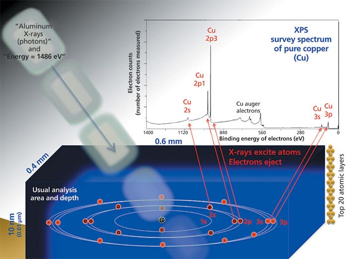
Figure 1: Basic conditions and processes for experimental X-ray photoelectron spectroscopy. (Adapted from original drawing by B. Vincent Crist.)
Experimental
Synthesis of Materials
ZnTe:Gd materials (Gd = 10%) were made by pulsed laser deposition (PLD) as previously described (10,11) using appropriate targets and depositing the desired material on a quartz substrate.
Instrumentation
Spectra were obtained using a Physical Electronics Model 5400 photoelectron spectrometer (equipped with an Al Kα [1486.6 eV] anode) with the samples undergoing no pretreatment after fabrication and then being mounted in a holder. All samples were handled openly in air during all experimental operations, with the intentional purpose of introducing atmospheric related surface species. Surface charge compensation and spectral calibration were achieved by assigning a binding energy of 284.8 eV to the adventitious carbon 1s photoelectron line.
Results and Discussion
Zinc telluride and telluride compounds have been extensively characterized by XPS to determine the chemical states and concomitant shifts of the elements present in the materials. Additionally, chemical shifts of gadolinium (12–14), zinc (15–17), and tellurium (18–20) lines in various compounds have previously been reported in the literature. However, a characterization of ZnTe:Gd, fabricated in the manner here-using pulsed laser ablation and deposition (PLD)-by XPS has not been previously reported. The ZnTe:Gd material can be seen as an experimental model to analyze for possible contaminant compounds in radiation detection semiconductors that have been discussed previously (21,22).
The PLD synthetic approach has several advantages for making molecular defect model materials for semiconductors and related materials useful in nuclear radiation detection materials. It is an energetic technique that vaporizes initial reactant materials to temperatures of several thousand degrees, followed by cooling and condensation of the vapor species, making it a method by which all possible chemical reactions that can occur in a vapor phase will do so. When the process is conducted in air such as here, atmospheric reactants such as CO2 and H2O may enter the mechanism of formation of the initial products observed. Secondary reactions such as chemisorption of CO2 and H2O with the cooled products may form simple adsorbates or actual molecular surface products that model the handling of the materials in air. Surface products such as these may have effects related to the radiation detection material-electrical contact interface.
X-Ray Pholoelectron Lines of Carbon and Oxygen
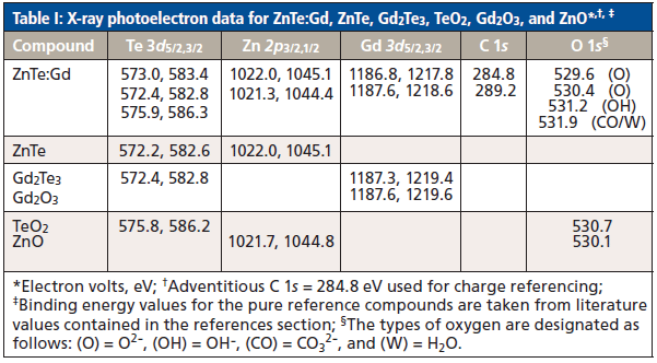
The carbon 1s region contains the adventitious carbon line at 284.8 eV and a line at 289.2 eV (Table I). The 289.2 line is in good agreement with that of CO2 from the atmosphere, either as an adsorbate or as a carbonate (23,24). There is no evidence of any appreciable carbide species, whose carbon 1s line is considerably lower than the 284.8 eV adventitious carbon line used as the surface charge calibrant. Of course, other products not identified or discussed here might exist at such low levels, they are essentially contained in the baseline. Zinc and gadolinium carbonates are distinct, molecular species that can combine with other atmospheric molecules to form new phases. Gadolinium carbonate hydrates (25) and hydroxy zinc carbonate (mineralogically, hydrozincite) (26) are excellent examples of secondary reaction products. Thus, the carbonate, hydroxy, and water attributed oxygen lines in Table I may also contain slight contributions from other formed carbonate–water–hydroxy complexes that cannot be completely detected with, for example, the carbonate functionality being generically assigned because of photoelectron line characteristics such as the carbon 1s line discussed above. Multiple hydroxide species also are difficult to differentiate, since even simple, molecular hydroxides fall in a very small range of binding energies (27).
The oxygen 1s spectrum consists of a quartet of peaks (Table I). The presence of multiple oxygen species is not surprising in light of both the fabrication technique of the samples and their casual handling in ambient, moist air. The lines at 529.6 and 530.4 eV are representative of ZnO and TeO2, while the peaks at 531.2 and 531.9 are indicative of a water-produced dissociated OH- species and H2O molecules, respectively.
X-Ray Photoelectron Lines of Gadolinium
The two sets of Gd 3d5/2,3/2 core-levels in ZnTe:Gd occur at 1187.6 and 1218.6 eV and 1186.8 and 1217.8 eV, with a corresponding spin-orbit splitting of 31.0 eV (Figure 2). Data from the zinc and tellurium lines strongly suggest that gadolinium is only present in the forms of Gd2Te3 and Gd2O3. Additionally, the 3d peaks are very broad, with a full width at half maximum (FWHM) of 4.8 eV when fit to two chemical states. While the broadness of the lines may indicate an insulating, charging surface, it is in agreement with previous studies that have observed FWHMs of pure gadolinium compounds of 4.4 and 5.3 eV (28,29). Other formed species such as those discussed above are not conducting and also might contribute to an insulation-based broadness, including multiple, simple adsorbed CO2 and H2O to form weak complexes. The presence of a satellite structure near 1196 eV is also observed here and has been previously recorded and explained in detail (29).
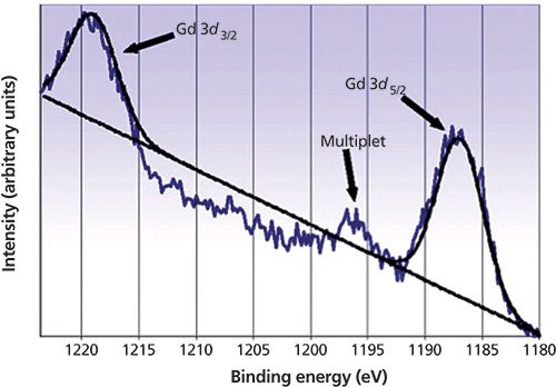
Figure 2: X-ray photoemission lines of gadolinium 3d3/2,5/2 for ZnTe:Gd films.
X-Ray Photoelectron Lines of Zinc
Figure 3 shows the Zn 2p emission lines with a splitting of 23.1 eV as observed in previous studies (15–17). Curve fitting indicates that zinc is present in only two chemical states-ZnTe and ZnO, with two lines with values of 1022.0 and 1021.3 eV, respectively, for the 2p3/2 energy level representing them. Both ZnTe and ZnO are semiconductors (30) rather than insulators and should not appreciably contribute to the observed line broadening of the spectral peaks studied here. They are very stable binary Group 16 compounds of zinc(II), the only stable oxidation state of zinc.
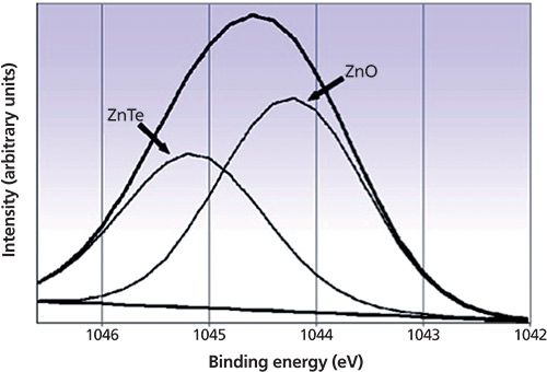
Figure 3: X-ray photoemission lines for the zinc 2p1/2 level and their assignments for molecular components in ZnTe:Gd films.
X-Ray Photoelectron of Tellurium
The 3d photoemission lines of Te are illustrated in Figure 4. The 3d3/2 and 3d5/2 peaks are separated by 10.4 eV. This spin-orbit splitting is in agreement with previously reported numbers for GdTe and other tellurium compounds (18–20). Curve fitting to both of the 3d level peaks shows that tellurium is present in three chemical states-ZnTe, Gd2Te3, and TeO2, with 3d5/2 energy levels of 573.0, 572.4, and 575.9 eV, respectively. Again, these energy level values correspond to previous studies on tellurium compounds (18–20). The presence of TeO2, the most stable oxide of tellurium, on the surface might be important for the indicated insulating effects previously discussed in this paper, along with the possibility of oxyanion species such as tellurites and tellurates. The binding energies for these latter two species, however, occur at higher values (20) than any observed in the present studies and would have to exist in minute amounts in the present materials.
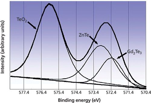
Figure 4: X-ray photoemission lines for the tellurium 3d5/2 level and their assignments for molecular components in ZnTe:Gd films.
Conclusions
X-ray photoelectron spectroscopy is an effective method for detecting different chemical states and molecular components of solid samples such as films. By use of an analysis of the spectra of the various elements in the sample, one can detect different functional species and groups identified with that element. The films that have condensed from very high temperature vapors exhibit discrete, molecular contaminant species. Further, the technique indicates that the films studied here contain species that are consistent with those observed in much larger bulk samples made by other synthetic routes driven by high temperature effects. The existence of hydroxide and carbonate functional groups, along with water, on the surface are most likely because of the samples interacting with the open atmosphere after initial formation, since all are unstable in the extreme temperatures produced in the synthetic laser plume.
Acknowledgments
This work was supported by the U. S. Department of Energy under Contract No. DE-AC0205CH11231 and the Center for Science and Engineering Education (CSEE) at Lawrence Berkeley National Laboratory. The authors wish to thank Beverly Wilgus for her efforts with the artwork related to Figure 1.
References
(1) M. Cornet, J. Degert, E. Abraham, and E. Freysz, Optics Lett. 39(20), 5921–5924 (2014).
(2) M. Bazarganipour and M. Salavati-Niasari, Micro & Nano Lett. 7(5), 388–391 (2012).
(3) M.A.M. Seyam, J. Alloys Compounds 541, 448–453 (2012).
(4) R.E. Nahory and H.Y. Fan, Phys. Rev. 156(3), 825–833 (1967).
(5) N.A. Shah and W. Mohmood, Thin Solid Films 544, 307–312 (2013).
(6) T.E. Schlesinger, J.E. Toney, H. Yoon, E.Y. Lee, B.A. Brunett, L. Franks, and R.B. James, Mater. Sci. Engineer. Reports 32(4–5), 103–189 (2011).
(7) T.L. Chu, S.S. Chu, C. Ferekides, and J. Britt, J. Appl. Phys. 71(11), 5635–5640 (1992).
(8) P. van der Heid, X-ray Photoelectron Spectroscopy: An Introduction to Principles and Practices (Wiley, 2011).
(9) D.L. Perry in Applications of Analytical Techniques to the Characterization of Materials , D.L. Perry, Ed. (Plenum Press, 1991), pp. 1–23.
(10) D.L. Perry, A.C. Thompson, R.E. Russo, X.-L. Mao, and K. Chapman, Appl. Spectrosc. 51(12), 1781–1783 (1997).
(11) D.L. Perry, X. Mao, and R.E. Russo, J. Mater. Res. 8(9), 2400–2403 (1993).
(12) K. Wandelt and C.R. Brundle, Surf. Sci. 157, 162–182 (1985).
(13) Y.-li Li, N.-fu Chen, Jian-ping Zhou, S.-lin Song, L.-feng Liu, Z.-gang, Yin, and C.-lin Cai, J. Cryst. Growth 265, 548–552 (2004).
(14) B.A. Orlowski, S. Mickievicius, V. Osinniy, A.J. Nadolny, B. Taliashvili, P. Driazwa, T. Story, R. Medicherla, and W. Drube, Nucl. Instr. Methods Phys. Res. B 238, 346–352 (2005).
(15) J.-C. Dupin, D. Gonbeau, P. Vinatier, and A. Levasseur, Phys. Chem. Chem. Phys. 2, 1319–1324 (2000).
(16) C.J. Vesely and D.W. Langer, Phys. Rev. B 4(2), 451–461 (1971).
(17) G.D. Khattak and M.A Salim, J. Electron Spectrosc. Relat. Phenom. 123, 47–55 (2002).
(18) R.G. Musket, Surf. Sci. 74, 423–436 (1978).
(19) W.E. Swartz, Jr., K.J. Wynne, and D.M. Hercules, Analyt. Chem. 43, 1884–1887 (1971).
(20) M.K. Bahl, R.L. Watson, and K.J. Irgolic, J. Chem. Phys. 66, 5526–5535 (1977).
(21) D.L. Perry and L.A. Franks, Proceed. of SPIE , 7079, 70791G (2008).
(22) K.G. Anand, Internat. J. Engineer. Manage. Sci. 4(2), 113–120 (2013).
(23) S.L. Wu, Z.D. Cui, F. He, Z.Q. Bai, S.L. Zhu, and X J. Yang, Mater. Lett. 58, 1076–1081 (2004).
(24) J.K. Heuer and J.F. Stubbins, Corrosion Sci. 41, 1231–1243 (1999).
(25) A. Sungur and M. Kizilyalli, J. Less-Common Metals 93, 419–423 (1983).
(26) S. Ghose, Acta Cryst. 17, 1051–1057 (1964).
(27) N.S. McIntyre and M.G. Cook, Analyt. Chem. 47(13) 2208–2213 (1975).
(28) D. Raiser and J.P. Deville, J. Electron Spectrosc. Relat. Phenom . 57, 91–5 (1991).
(29) J. Chung, J. Korean Phys. Rev. 38, 744–748 (2001).
(30) M. Afzaal, M.A. Malik, and P. O'Brien, New J. Chem. 31, 2029–2040 (2007).
Dale L. Perry is a senior scientist in chemistry with the Lawrence Berkeley National Laboratory, University of California, in Berkeley, California. Zhixun Ma is with PPG Industries in Cheswick, Pennsylvania. Andrew Olson is an associate consultant with ZS Associates in San Mateo, California. Erik Topp is an applications engineer with Baker Hughes in Concord, California. Direct correspondence to: dlperry@lbl.gov

AI Shakes Up Spectroscopy as New Tools Reveal the Secret Life of Molecules
April 14th 2025A leading-edge review led by researchers at Oak Ridge National Laboratory and MIT explores how artificial intelligence is revolutionizing the study of molecular vibrations and phonon dynamics. From infrared and Raman spectroscopy to neutron and X-ray scattering, AI is transforming how scientists interpret vibrational spectra and predict material behaviors.
Advancing Corrosion Resistance in Additively Manufactured Titanium Alloys Through Heat Treatment
April 7th 2025Researchers have demonstrated that heat treatment significantly enhances the corrosion resistance of additively manufactured TC4 titanium alloy by transforming its microstructure, offering valuable insights for aerospace applications.