Opium and Hashish Tested Using Laser-Induced Fluorescence Spectroscopy
Scientists from the Amirkabir University of Technology in Tehran, Iran, used laser-induced fluorescence (LIF) spectroscopy to analyze opium and hashish. Through this research, which was published in Spectrochimica Acta Part A: Molecular and Biomolecular Spectroscopy, the scientists explored a novel method of drug analysis (1).
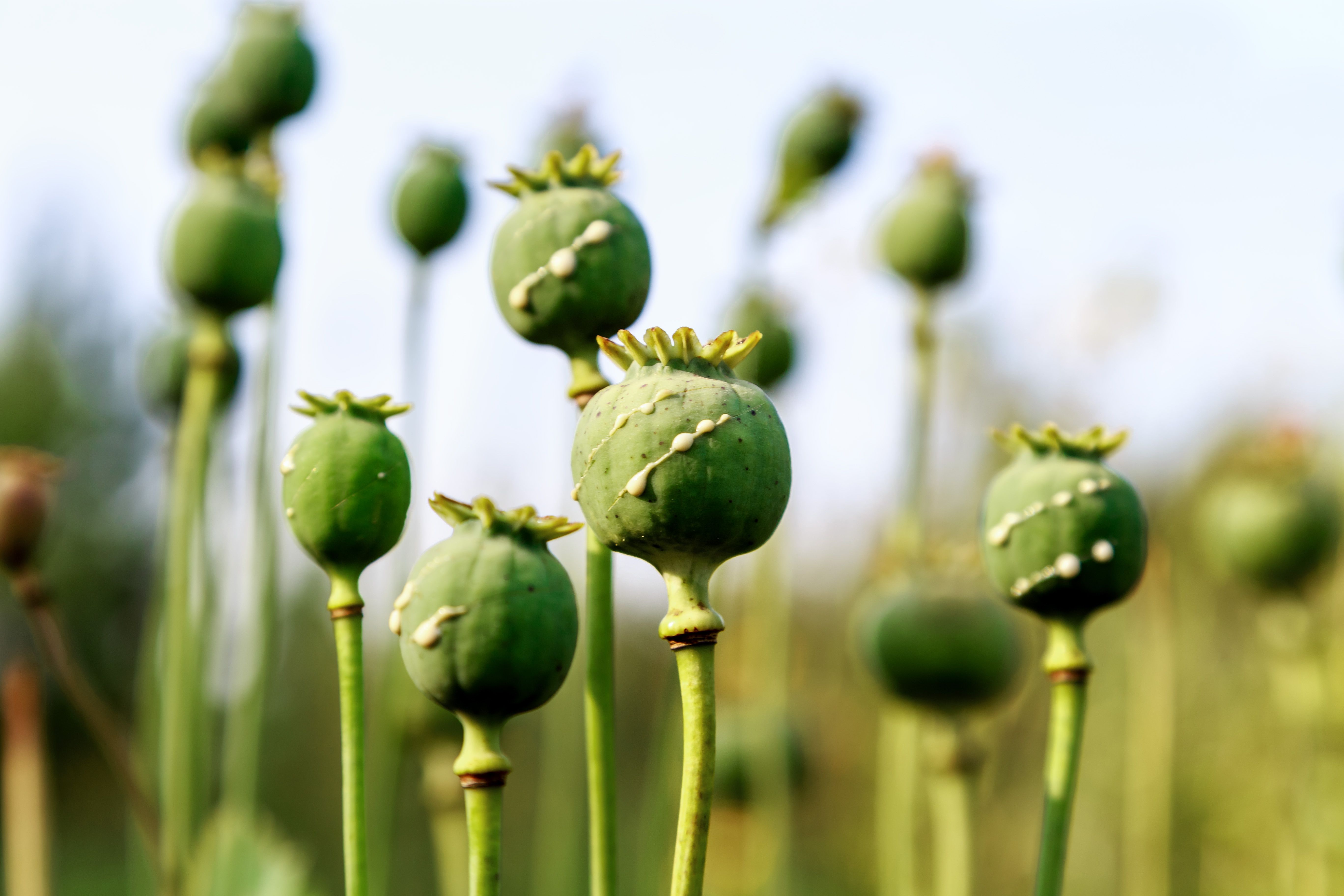
poppy heads with drops of opium milk | Image Credit: © KPad - stock.adobe.com

Opium, which comes from the opium poppy, is the source of many narcotics, including morphine and codeine. Hashish is made from the resin of the cannabis plant. Cannabis is one of the most common psychoactive drugs, due to its psychoactive components: delta-9-tetrahydrocannabinol (THC), cannabinol (CBN), and cannabidiol (CBD).
Opium and hashish are optically dense multi-fluorophore samples and contain fluorophores of unknown concentrations. This makes it difficult for conventional fluorescence measurement tools to analyze these substances. In this study, the researchers used LIF to properly characterize the properties of both opium and hashish.
LIF is often praised for its simplicity, quickness, and portability, making it a practical method of drug identification. In this study, LIF was extensively examined to overcome drawbacks of obtaining specific fluorescence parameters as a novel method of drug analysis. The spectroscopy process was carried out at a single-line excitation of 405 nm over a wide range of concentrations to obtain three distinctive fluorescence parameters (k, α and Cp) based on the modified Beer-Lambert (MBL) formalism.
The typical α value was determined to be 0.30 and 0.15 mL/(cm∙mg) for opium and hashish, respectively. Similarly, typical k is obtained ar 0.390 and 1.25 mL/(cm∙mg), respectively. Furthermore, the concentration at max fluorescence intensity (Cp) was determined for opium and hashish to be 1.8 and 1.3 mg/mL, respectively. These results show that using this method, opium and hashish create their own characteristic fluorescence parameters.
Reference
(1) Shamsi, E.; Parvin, P.; Ahmadinouri, F.; Khazai, S. Laser-induced fluorescence spectroscopy of plant-based drugs: Opium and hashish provoking at 405 nm. Spectrochim. Acta A Mol. Biomol. Spectrosc. 2023, 302, 123055. DOI: https://doi.org/10.1016/j.saa.2023.123055
New Study Reveals Insights into Phenol’s Behavior in Ice
April 16th 2025A new study published in Spectrochimica Acta Part A by Dominik Heger and colleagues at Masaryk University reveals that phenol's photophysical properties change significantly when frozen, potentially enabling its breakdown by sunlight in icy environments.
Tracking Molecular Transport in Chromatographic Particles with Single-Molecule Fluorescence Imaging
May 18th 2012An interview with Justin Cooper, winner of a 2011 FACSS Innovation Award. Part of a new podcast series presented in collaboration with the Federation of Analytical Chemistry and Spectroscopy Societies (FACSS), in connection with SciX 2012 ? the Great Scientific Exchange, the North American conference (39th Annual) of FACSS.
New Fluorescence Model Enhances Aflatoxin Detection in Vegetable Oils
March 12th 2025A research team from Nanjing University of Finance and Economics has developed a new analytical model using fluorescence spectroscopy and neural networks to improve the detection of aflatoxin B1 (AFB1) in vegetable oils. The model effectively restores AFB1’s intrinsic fluorescence by accounting for absorption and scattering interferences from oil matrices, enhancing the accuracy and efficiency for food safety testing.