Optimization of ED-XRF Excitation Configuration Parameters to Determine Trace-Element Concentrations in Organic and Inorganic Sample Matrices
Spectroscopy
This month’s column will describe a novel ED-XRF system, which utilizes a combination of a Bragg polarizer, used simultaneously with a direct excitation source, together with a novel, highly annealed pyrolytic graphite (HAPG) crystal as a band-pass filter. By selection of the optimum configuration, it will allow for high precision of minor and major elements across a wide wavelength range and/or lower detection capability for smaller groups of elements from potassium to manganese. To show the practical benefits of this technology, this study will focus on these performance metrics for the determination of titanium in polymer samples, together with the multielement analysis of high purity graphite using the ashing sample preparation method. In particular, it will be shown that the improved performance for graphite will allow for lower sample weights to be used resulting in significantly shorter ashing times, which is a requirement for high sample workload laboratories and process control applications.
This column installment describes a novel energy-dispersive X-ray fluorescence (ED-XRF) system that uses a combination of a Bragg polarizer, used simultaneously with a direct excitation source, together with a novel, highly annealed pyrolytic graphite (HAPG) crystal as a band-pass filter. By selection of the optimum configuration, this system allows for high analysis precision for minor and major elements across a wide wavelength range and lower detection capability for smaller groups of elements from potassium to manganese. To show the practical benefits of this technology, this study focuses on these performance metrics for the determination of titanium in polymer samples, together with the multielement analysis of high-purity graphite using the ashing sample preparation method. In particular, this installment of “Atomic Perspectives” demonstrates the improved performance for graphite that allows for lower sample weights to be used and results in significantly shorter ashing times, which is a requirement for process control applications and laboratories with a high sample workload.
The improvements in detector and digital signal processing techniques have opened new possibilities for multielement analysis using energy-dispersive X-ray fluorescence (ED-XRF) instruments. The opportunity to process input count rates higher than 1 Mcps are a challenge for the development of new bright excitation optics to further improve elemental sensitivities and detection limits. Excitation of fluorescence lines must be efficient over a large energy range from about 0.5 keV to about 40 keV. The use of Bragg crystals as monochromators in ED-XRF in a selected energy range was first discussed in the early 1980s (1). However, today it is necessary to select crystals with a high integral reflectivity to cover large solid angles of >100 msr (millisteradian) and combine this technology with other excitation optics.
Discussion
Highly annealed pyrolytic graphite (HAPG) is a new mosaic crystal used for X-ray analysis because it has good bending properties, offers a significant increase of integral reflectivity compared to other crystals, and has other useful properties that are required in ED-XRF instrumentation. However, it cannot be used as a monochromator in the first order because of its weak mosaicity at the required bending radius. These properties make HAPG an optimal X-ray band-pass filter in the energy range of <10 keV, if they are placed between the X-ray tube and sample or between the sample and detector. In addition, such a filter can be combined simultaneously with the recognized benefits of a Bragg polarizer or other optics in combination with a binary alloy X-ray tube anode to provide even greater improvement in the sensitivity of the spectrometer in different energy ranges (2). Figure 1 shows the schematic of an XRF excitation system using a Bragg polarizer simultaneously with a direct excitation source.
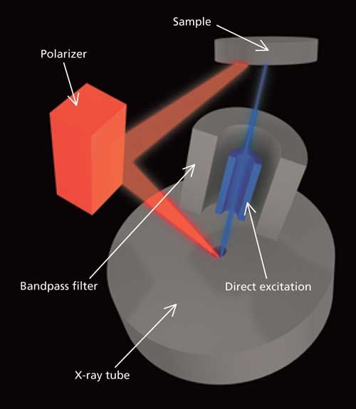
Figure 1: Schematic of an XRF excitation system using a Bragg polarizer simultaneously with a direct excitation source.
Using the HAPG band-pass filter in this configuration allows for optimization of the excitation for different groups of elements. The radiation originating from the tube will be Bragg-reflected at the toroidal shaped crystal in an energy range and will be defined by the mosaic spread, d-spacing of HAPG and the beam guidance. Because of the large solid angle and the intensity of the excitation, radiation will also be increased, resulting in higher intensity of the fluorescence signal combined with lower background. The practical benefits are improved precision for major and minor elements together with an order of magnitude lower detection limits for trace elements. This type of band-pass filter excitation configuration is shown schematically in Figure 2.
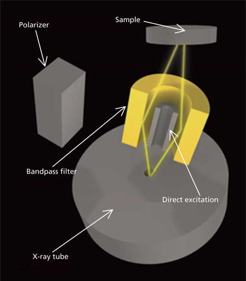
Figure 2: HAPG band-pass filter excitation configuration.
Determination of Titanium in Polymer Samples
One of the most common types of analysis for ED-XRF is the determination of elemental impurities in polymer samples, and in particular trace levels of the transition elements, such as titanium. This is exemplified in Figure 3, which shows spectral measurements from 3.2–5.8 keV for four polymer standards containing Ti at concentration levels of 0, 0.3, 1.0, and 7.7 mg/kg (ppm) at a measurement time of 150 s. It can be clearly seen that the spectral peaks for Ti at about 4.5 keV can all be identified and quantified.
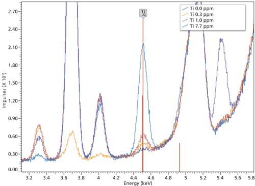
Figure 3: Spectra from measurements of polymer samples containing trace levels of Ti.
To determine the limit of detection and precision for this application, another low standard was analyzed 10 times. The net intensities, normalized to live time and tube current, as well as the analysis results are displayed in Table I.
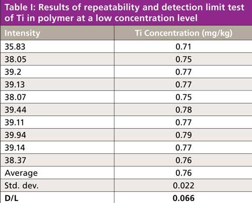
Where traditional ED-XRF instruments can achieve detection limits in the milligrams-per-kilogram (parts-per-million) range for this application, the band-pass-filter excitation approach can achieve a detection limit of about 66 µg/kg (ppb).
Multielement Analysis of Powdered Graphite
Mosaic crystals like HAPG cannot be used for focusing the beam to the sample. The defocusing effects must be eliminated by beam guidance. By using optimized beam guidance, even smaller amounts of sample can be analyzed quantitatively without sacrificing precision. This approach can be used in applications where only small amounts of samples are available, such as the use of high-temperature ashing as a sample preparation technique. With this procedure, the elements are concentrated in the ashed sample. However, typically there is not enough sample to prepare a standard pressed pellet with a diameter of 32 or 40 mm, or to fill a standard XRF sample cup.
One example is the analysis of impurities in high quality graphite with purity better than 99.5% or even 99.9%. To be able to ash enough material to prepare a standard pressed pellet, ~4 g of ash would be required, resulting in a long ashing time and making this procedure unrealistic for a process control application. The alternative is the microwave-assisted ashing procedure, where the sample material is reduced by a factor of 5–10 within 30 min. Using this approach, as little as 120–150 mg of the remaining ash in a 20-mm sample cup allows for an accurate determination of the elemental impurities in the graphite. This approach is exemplified in Figure 4, which shows spectra of a graphite sample with 99.5% purity analyzed directly in a standard 32-mm sample cup compared to the spectra obtained from analyzing the ashed sample in a 20-mm sample cup. It should be emphasized that the determination of volatile elements like S have to be carried out before the ashing procedure, but typically their concentrations are high enough to obtain precise results from the analysis of the powder directly.
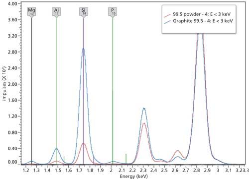
Figure 4: Spectra from the direct measurement of graphite (displayed in red) and after ashing (displayed in blue) of the sample.
There is no question that it’s always going to be challenging to quantify all trace elements with different fluorescent energies up to 40 kV for ashed samples that cannot be prepared as a thin layer, or as a thick pressed pellet. Sherman (3) as well as Shiraiwa and Fujino (4) have developed mathematical algorithms for the computation of count rate values of fluorescent radiation emitted from a standard at a fixed detector position. Additionally, Weber (5) described effects caused by the instrumental and sample geometry that are neglected in the traditional fundamental parameter models for thick samples. As a result, modifications of the traditional fundamental parameter algorithms are therefore necessary to improve the accuracy of the analytical results, and for that reason, the density as well as the thickness of a sample must be known, independently of each other.
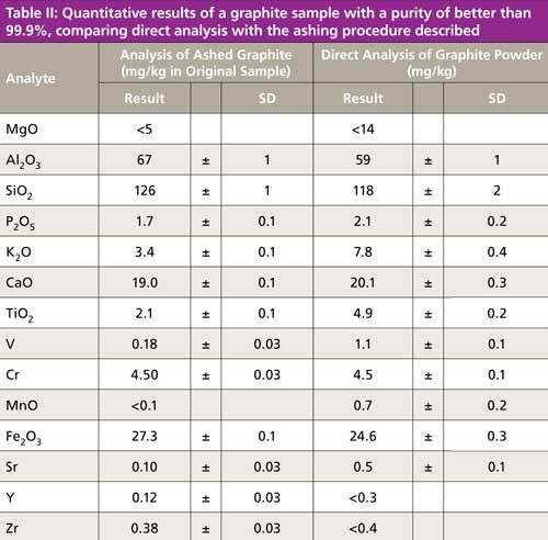
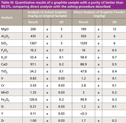
This approach was used to analyze the graphite powder directly in a sample cup with an outer diameter of 32 mm, together with a smaller amount of ashed material in a 20-mm cup. The results for the direct graphite analysis and those determined from the ashed sample, but calculated back to the original substance, are displayed in Tables II and III, respectively. The results obtained demonstrate that there is a good agreement between both sampling methods for elements at concentration levels well above the limit of detection (LOD). However, it can be clearly seen that the standard deviation (SD) of the analytes in the ashed samples are lower, allowing for better precision (see SDs for MgO, Al2O3, and SiO2 as examples) and also better accuracy. In addition, the analysis of the ashed samples allows for the determination of some trace elements, which are not possible by directly analyzing the original material (see results for elements Sr, Y, and Zr as examples). The overall conclusion is that the quantitative analysis of ~150 mg of ashed sample is possible with a standard XRF instrument using the hardware configuration and calculation principles described in this study.
Summary
The improvements in detector and digital signal processing techniques have opened new possibilities for multielement trace-element analysis with stationary and portable ED-XRF instruments. The development of new bright excitation optics improves elemental sensitivities as well as detection limits. The user benefits range from better precision to lower detection limits, which results in improved accuracy. By using optimized beam guidance even smaller amounts of sample can be analyzed quantitatively without sacrificing precision. This capability can be used where only small amounts of sample are available, such as in research and development applications, or when using rapid ashing procedures, which are a requirement for high sample workload laboratories and process control applications.
References
- T.C. Furnas, G.S. Kuntz, and R.E. Furnas, Adv. X-Ray Anal. 25, 59–62 (1981).
- Handbook of X-ray Spectrometry, Second edition (Marcel Dekker Inc., ISBN 0-8247-0600-5, 1982), pp. 618–626.
- J. Sherman, Spectrochim. Acta 7, 283–306 (1955).
- T. Shiraiwa and N. Fujino, Jpn. J. Appl. Phys. 5, 886–899 (1966).
- A. Weber, in X-ray Spectrometry (Wiley Heyden on-line library, 1983), pp. 12,1, 11–18.

Joachim Heckel is the vice president of R&D at Spectro Analytical Instruments in Kleve, Germany, and is responsible for research and development of all products from Spectro.

Dirk Wissmann is the product manager for XRF instruments at Spectro Analytical Instruments and is responsible for the strategic development of the complete product line including application development and product support. Direct correspondence about this article to: SpectroscopyEdit@UBM.com
Applications of Micro X-Ray Fluorescence Spectroscopy in Food and Agricultural Products
January 25th 2025In recent years, advances in X-ray optics and detectors have enabled the commercialization of laboratory μXRF spectrometers with spot sizes of ~3 to 30 μm that are suitable for routine imaging of element localization, which was previously only available with scanning electron microscopy (SEM-EDS). This new technique opens a variety of new μXRF applications in the food and agricultural sciences, which have the potential to provide researchers with valuable data that can enhance food safety, improve product consistency, and refine our understanding of the mechanisms of elemental uptake and homeostasis in agricultural crops. This month’s column takes a more detailed look at some of those application areas.