Potential of Raman Spectroscopy in Recognizing Nucleophosmin Mutant Gene Expression in Leukemia Cells
A new study reveals the potential of Raman spectroscopy in recognizing nucleophosmin (NPM1) mutant gene expression in leukemia cells.
In a recent study published in the journal Applied Spectroscopy, lead authors Yihui Wu and Mingbo Chi from the Chinese Academy of Sciences in Changchun, China, explore the potential of Raman spectroscopy in recognizing the expression of the nucleophosmin (NPM1) mutant gene in leukemia cells (1).
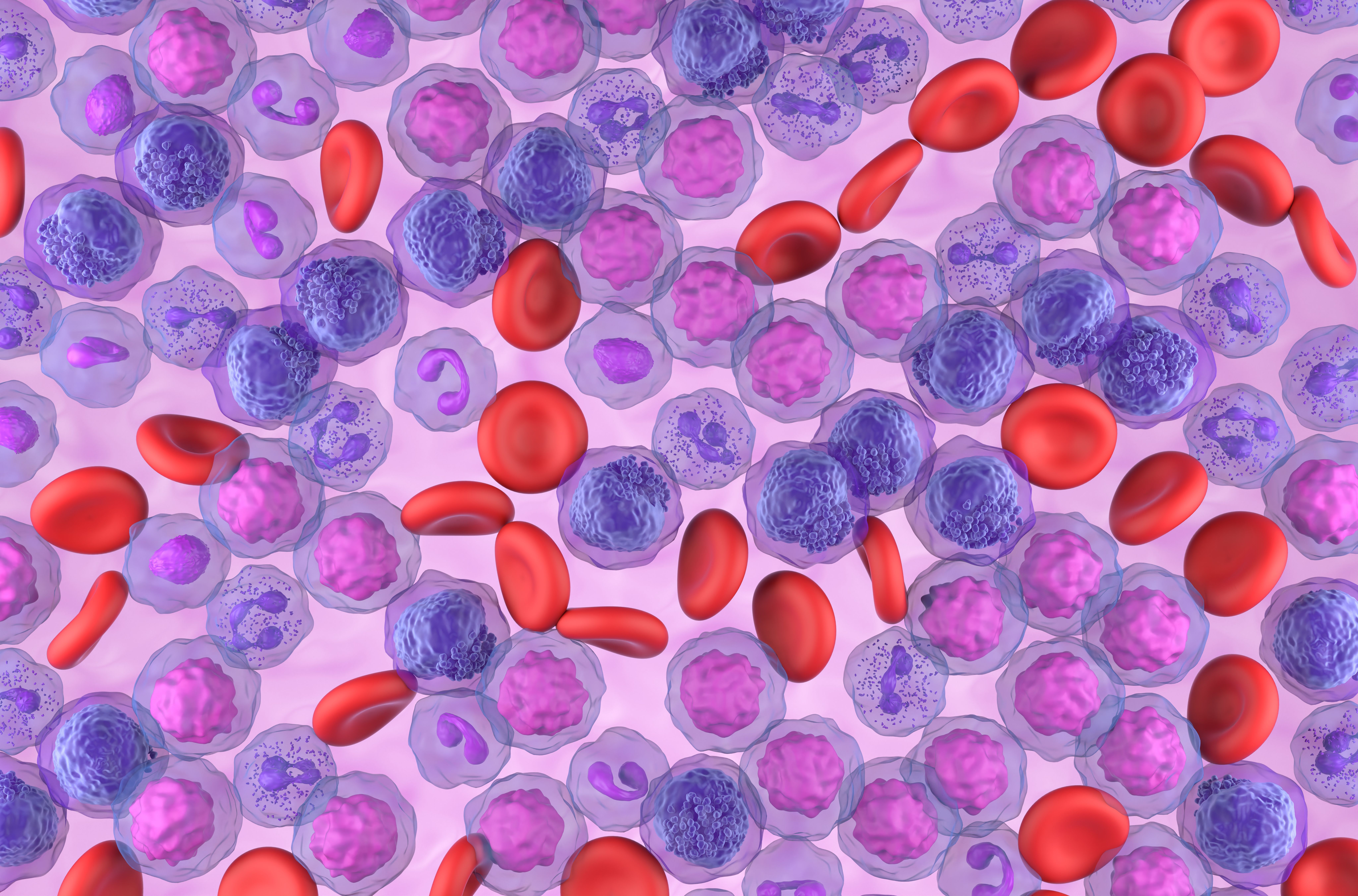
Acute myeloid leukemia (AML) cells field - top view 3d illustration | Image Credit: © LASZLO - stock.adobe.com

Raman spectroscopy is a technique that could be useful in cancer diagnosis applications and biomedical research. This study explores its applicability in detecting the spectral characteristics of acute myeloid leukemia (AML) cells with and without the NPM1 mutation (1). Their study provides insights into the underlying reasons for the observed spectral differences through transcriptomic analysis.
Transcriptomic analysis is a field of study that focuses on analyzing and interpreting the complete set of RNA transcripts, known as the transcriptome, within a cell or a group of cells (1). It involves the identification, quantification, and characterization of all the RNA molecules present in a biological sample, providing insights into gene expression patterns and regulation (1).
Experimental data was collected by culturing and analyzing Raman spectra of two AML cell lines without NPM1 mutation (THP-1 and HL-60) and the OCI-AML3 cell line carrying the NPM1 mutant gene (1). The researchers found distinct intensity differences in multiple peaks corresponding to chondroitin sulfate (CS), nucleic acid, protein, and other molecules between the average Raman spectra of NPM1 mutant and nonmutated cells (1).
To further understand the observed differences, the study involved quantitative analysis of gene expression matrices from the two types of cells (1). Differentially expressed genes were identified, and their roles in the regulation of CS proteoglycan and protein synthesis were analyzed (1). Remarkably, the researchers discovered that the differences expressed through single-cell Raman spectral information aligned with the differences in transcriptional profiles (1).
These findings highlight the potential of Raman spectroscopy as a valuable tool for cancer cell typing, specifically in recognizing the expression of the NPM1 mutant gene in leukemia cells. By providing detailed molecular information without the need for labeling or sample destruction, Raman spectroscopy offers a promising avenue for further advancements in cancer research and diagnosis.
Reference
(1) Li, M.; Wu, Y.; Chi, M.; Wang, Y.; Zhu, M.; Gao, S. Recognition of Nucleophosmin Mutant Gene Expression of Leukemia Cells Using Raman Spectroscopy. Appl. Spectrosc. 2023, ASAP. DOI: 10.1177/00037028231176547
Ancient Meteorite Reveals Space Weathering Secrets Through Cutting-Edge Spectroscopy
Published: May 27th 2025 | Updated: May 27th 2025Researchers in Rome used advanced spectroscopic techniques to probe the mineralogy of the CM2 carbonaceous chondrite NWA 12184. This revealed the effects of space weathering and provided insights into C-type asteroid evolution.
Nanometer-Scale Studies Using Tip Enhanced Raman Spectroscopy
February 8th 2013Volker Deckert, the winner of the 2013 Charles Mann Award, is advancing the use of tip enhanced Raman spectroscopy (TERS) to push the lateral resolution of vibrational spectroscopy well below the Abbe limit, to achieve single-molecule sensitivity. Because the tip can be moved with sub-nanometer precision, structural information with unmatched spatial resolution can be achieved without the need of specific labels.
Multi-Analytical Study Reveals Complex History Behind Ancient Snake Motif in Argentine Rock Art
May 22nd 2025A recent study published in the Journal of Archaeological Science: Reports reveals that a multi-headed snake motif at Argentina's La Candelaria rock shelter was created through multiple painting events over time.

.png&w=3840&q=75)

.png&w=3840&q=75)


.png&w=3840&q=75)


.png&w=3840&q=75)


