Raman-Based Oesophageal Tissue Classification and Modern Machine Learning in the Support of Multi-Center Clinical Diagnostics: An Interview with 2024 Charles Mann Award Winner Nick Stone
A multi-organizational team, believing that a reason for slow adoption is a lack of evidence that data taken on one spectrometer can transfer across to data taken on another spectrometer to provide consistent diagnoses, investigated multi-center transferability using human oesophageal tissue. By using a common protocol, the researchers aimed to minimize the difference in machine learning performance between centers.
While the clinical potential of Raman spectroscopy in a clinical environment has been well established, the technique has yet to become established in routine oncology workflows.
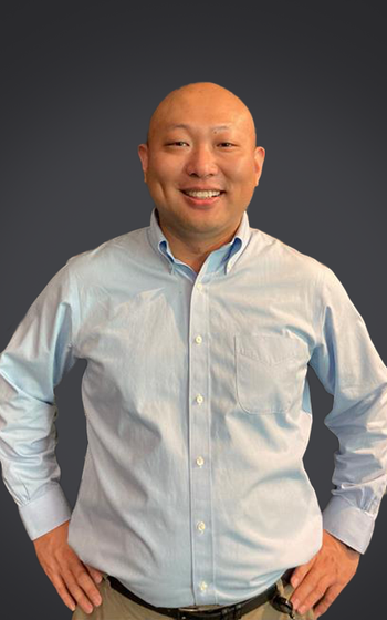
A multi-organizational team, believing that a reason for slow adoption is a lack of evidence that data taken on one spectrometer can transfer across to data taken on another spectrometer to provide consistent diagnoses, investigated multi-center transferability using human oesophageal tissue. By using a common protocol, the researchers aimed to minimize the difference in machine learning performance between centers. A recent paper co-authored by biomedical physicist Nick Stone at the University of Exeter (UK) focused on this investigation and its results. Stone will be receiving the Charles Mann Award in Applied Raman Spectroscopy. The award will be presented at this year’s SciX conference, to be held from October 20 through October 25, in Raleigh, North Carolina. As part of an ongoing series of this year’s SciX conference honorees, Nick spoke to us about this study, its benefits and significance, and the next steps in its development, as well as his thoughts on receiving the Mann Award.
In your paper (1), you state that “the clinical potential of Raman spectroscopy (RS) is well established but has yet to become established in routine oncology workflows.” Why do you believe this to be the case?
We and others have shown that Raman can distinguish between cells, tissues and biofluids from normal to diseased and steps in between (2,3). This is possible using microspectrometers, portable systems, and in vivo probes. However, to date, most studies have been limited to a relatively small number of samples or patients. This is due mostly to the fact that, to obtain fresh tissue samples, one must recruit patients locally, and this means that the numbers available are inherently limited to the patient throughput for that condition, the availability of someone to recruit them, and collect the sample or take the measurement. This is generally limited by the funding available, which is often of the order of a few hundred thousand dollars, pounds or euros, enabling a single postdoctoral scientist to work for 2-3 years on the project.
To enable routine analysis, genuine translation and adoption studies need to be at the scale of multicenter and this involves a great deal of commitment, perseverance, luck, and time delays in seeking investment to achieve this goal and in performing continued studies. These are being done in a few cases around the world, and it should be considered in the context of many medical technologies that are likely to take 15-20 years to translate from the bench to the bedside.
You also state that “oesophageal cancer diagnostics is a setting that could gain much from the clinical translation of RS.” Briefly discuss the reasons why.
Oesophageal cancer is one of the cancers that is both deadly, with around 15% survival at five years from diagnosis, and difficult to diagnose. Patients don’t usually present with symptoms for the disease, such as a difficulty swallowing, until it is too late to provide effective treatments. However, if the disease is detected pre-symptomatically at a non-invasive stage, hugely effective treatments can be provided. Luckily, oesophageal cancers are slow developing and, therefore, if we can use a method (fiber optic Raman) to measure the molecular changes in the cells that line the surface, we can provide an effective method of selecting biopsies initially, but eventually providing a real time decision coupled to treatments in the same procedure.
Equally important is to provide an improved classification of pre-cancerous lesions in tissues that have been removed (biopsies). Pathologists struggle to agree on the classification of these lesions, and this hinders decision making. We have demonstrated that Raman can provide a reproducible method of delivering a classification equivalent to an independent pathologist, enabling a potential support to decision making.
Are there any potential drawbacks in using RS in this space?
The draw backs of using Raman are its relative speed or lack of it. Generally, it takes around a second to take a measurement that can be used for disease classification. This may sound quick, but it is only providing a single measurement of the molecular soup that makes up the sample volume measured, and not an image. This is controlled by the optics and can range from µm3 to cm3; of course, this can also be an advantage, but the abnormal signals must not be diluted too much by the surrounding signals measured. Clinicians are used to seeing images and not spectra. It is important to provide either a likelihood of disease risk or a relative measure of molecule tissue composition that the clinician can interpret.
Briefly state your findings.
We demonstrated in the multicenter study that using three identical spectrometers from the same manufacturer in three centers, that we could get very similar results from measurements of the “same” tissue samples. Note each center was provided with a different section from each patient’s biopsy, so the locations were subtly different, but generally the performance was particularly similar.
Do your findings correlate with what you had hypothesized? Was there anything particularly unexpected that stands out from your perspective?
We did expect to need to provide transfer functions for the instruments to deliver the best results. In this paper, for these samples and well controlled systems, we showed that this was unnecessary, provided care was taken in using temperature stabilized labs and careful calibration routines.
Were there any analytical limitations or challenges you encountered in your work? How did you overcome these challenges?
It can be very hard to link the disease state of a particular cell or cells with the gold standard pathology, particularly when using different tissue sections. Very careful analysis of each section and maps provided for each user to measure the same region on each sample was provided.
As noted, Raman scattering is relatively weak, and measuring maps of large tissue sections is prohibitively slow. The most appropriate method is to target regions of interest selected using another approach, or to only measure samples that a decision can only be accurately made using this approach. Alternatively, in vivo measurements can measure many cells at the same time, which can overcome some of these issues. We have noted in unpublished work that, provided the sample volume is made up of around 10% of cancer cells or more, the disease state can still be identified.
What best practices can you recommend in this type of analysis for both instrument parameters and data analysis?
It is vital to ensure instruments are well calibrated, and the signals generated by the instruments themselves are well understood, including the effect of a changing environment on calibrations and background signals. Use of both a Raman standard and a broadband fluorescence standard enables instruments to be calibrated and monitored. Using the same standard across sites then enables instruments to be appropriately mapped to each other, assuming they are very close in performance characteristics (4,5).
Do you believe this technique could transfer to other cancers? To other disease states?
Yes, there are very few cancers that Raman cannot distinguish; it is also viable in numerous degenerative and infectious diseases. The question will always come down to relative cost and effectiveness, and an acceptance by medical colleagues to grasp the new technologies that could be applied to so many of their problems.
What are the next steps in this research?
We are currently running in vivo trials using out endoscopic Raman in the esophagus, as well as our Smart Raman needle in head and neck cancers and lymph nodes. We expect to build sufficient evidence from these studies to move towards much larger scale multicenter studies and wider adoption of the approach for many other cancers.
What are your thoughts regarding your being named the recipient of the Charles Mann Award in Applied Raman Spectroscopy?
I am delighted and honored to follow in the footsteps of so many pioneering and distinguished scientists in the field.
References
Blake, N., Gaifulina, R., Isabelle, M. et al. System Transferability of Raman-Based Oesophageal Tissue Classification Using Modern Machine Learning to Support Multi-Centre Clinical Diagnostics. BJC Rep. 2024, 2, 52. DOI:
Stone, N.; Kendall, C.; Smith, J.; Crow, P.; Barr H. Raman Spectroscopy for Identification of Epithelial Cancers. Faraday Discuss. 2004, 126, 141–157. DOI:
Kong, K.; Kendall, C.; Stone, N.; Notingher, I. Raman Spectroscopy for Medical Diagnostics--From in-vitro Biofluid Assays to in-vivo Cancer Detection. Adv. Drug Deliv. Rev. 2015, 89, 121–134. DOI:
Butler, H. J.; Ashton, L.; Bird, B.; Cinque, G.; Curtis, K.; Dorney, J.; et al. Using Raman Spectroscopy to Characterize Biological Materials. Nat. Protoc. 2016, 11 (4), 664–687. DOI:
Guo, S.; Beleites, C.; Neugebauer, U.; Abalde-Cela, S.; Afseth, N. K.; Alsamad, F.; et al.Comparability of Raman Spectroscopic Configurations: A Large Scale Cross-Laboratory Study. Anal. Chem. 2020, 92 (24), 15745–15756. DOI:
Newsletter
Get essential updates on the latest spectroscopy technologies, regulatory standards, and best practices—subscribe today to Spectroscopy.


