Raman Spectroscopy Unveils Potential Serum Biomarkers for Bone Marrow Failure Diseases
In a recent study, a research team from China used Raman spectroscopy to uncover serum biomarkers for aplastic anemia and myelodysplastic syndromes.
A recent study conducted by researchers from China uncovered potential serum biomarkers for bone marrow failure (BMF) diseases. In the study, which was published in the journal Spectrochimica Acta Part A: Molecular and Biomolecular Spectroscopy, the researchers used Raman spectroscopy to discover possible serum biomarkers that could be used to help detect BMF diseases such as aplastic anemia (AA) and myelodysplastic syndromes (MDS) (1).
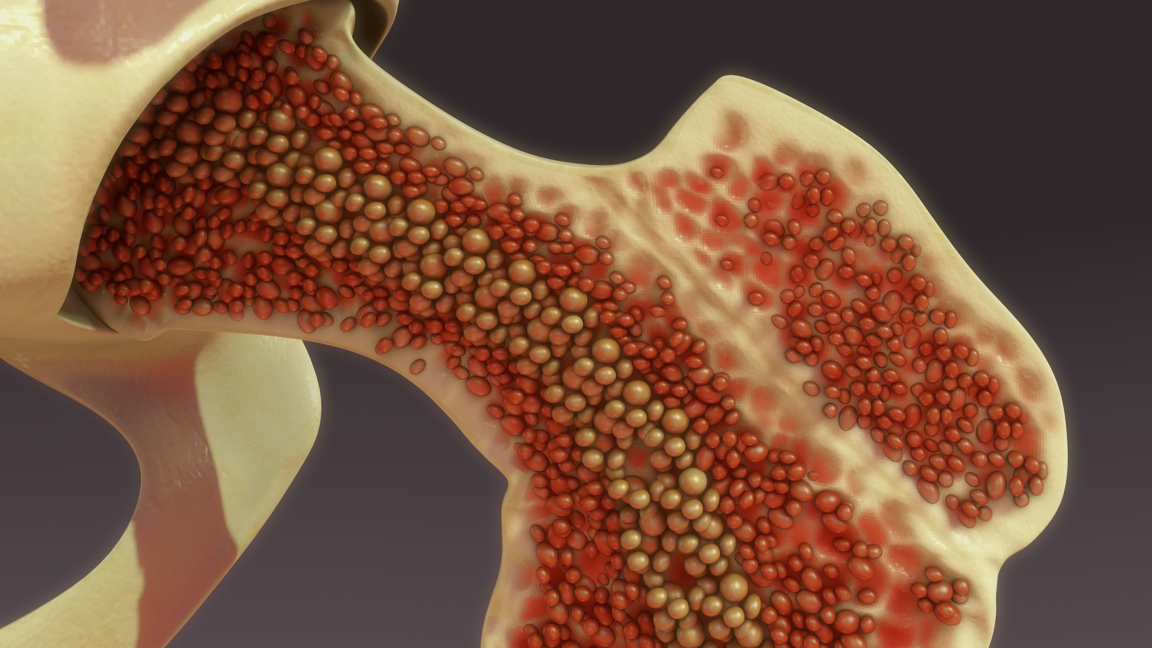
Bone marrow | Image Credit: © 7actionstudio - stock.adobe.com

The study involved analyzing serum samples from 35 AA patients, 25 MDS patients, and 23 control volunteers (1). By utilizing laser Raman spectroscopy coupled with orthogonal partial least squares discrimination analysis (OPLS-DA), the researchers aimed to establish a reliable method for distinguishing between BMF patients and healthy individuals (1).
When compared to the control group, BMF patients exhibited distinct serum spectral patterns. Notably, the intensities of Raman peaks associated with nucleic acids, proteins, phospholipids (cholesterol), and β-carotene decreased significantly, while lipid-related peaks saw an increase (1). Differences were observed between the AA and MDS groups as well, with specific Raman peaks showing significant variations (1).
What makes this study unique is that it is, according to the researchers’, the first time Raman spectroscopy has been applied to analyze sera from BMF patients (1). The analysis revealed specific biomolecular differences that may reflect metabolic changes in these patients (1). These differences could serve as valuable biomarkers for early detection and subtyping of BMF, AA, and MDS, which could have positive effects in the medical field in treating these types of diseases (1).
The study also delved into the association between Raman peaks and serological markers. Notably, glycolipid metabolism-related serological markers were strongly linked to specific Raman peaks, suggesting a potential connection with triglyceride (TG), high-density lipoprotein (HDL), and glucose levels (1). This association opens up new avenues for understanding disease mechanisms and prognosis in BMF patients (1).
However, the study's small sample size warrants further investigation. Because there are not many BMF patients, the sample size for this study was relatively small. As a result, a more comprehensive study would be needed to determine how effective Raman spectroscopy could be as a clinical application for BMF detection and screening (1).
In conclusion, this study showcases the promising potential of Raman spectroscopy as a non-invasive method for detecting and subtyping BMF, AA, and MDS. The researchers found that by using OPLS-DA, AA and MDS subtypes can be distinguished from another (1). As research in this field continues to advance, the hope is that the findings in this study could be used to improve detection of BMF disorders in patients.
Reference
(1) Liang, H.; Kong, X.; Ren, Y.; Wang, H.; Liu, E.; Sun, F.; Zhu, G.; Zhang, Q.; Zhou, Y. Application of serum Raman spectroscopy in rapid and early discrimination of aplastic anemia and myelodysplastic syndrome. Spectrochimica Acta Part A: Mol. Biomol. Spectrosc. 2023, 302, 123008. DOI: 10.1016/j.saa.2023.123008
This article was written with the help of artificial intelligence and has been edited to ensure accuracy and clarity. You can read more about our policy for using AI here.
AI-Powered SERS Spectroscopy Breakthrough Boosts Safety of Medicinal Food Products
April 16th 2025A new deep learning-enhanced spectroscopic platform—SERSome—developed by researchers in China and Finland, identifies medicinal and edible homologs (MEHs) with 98% accuracy. This innovation could revolutionize safety and quality control in the growing MEH market.
New Raman Spectroscopy Method Enhances Real-Time Monitoring Across Fermentation Processes
April 15th 2025Researchers at Delft University of Technology have developed a novel method using single compound spectra to enhance the transferability and accuracy of Raman spectroscopy models for real-time fermentation monitoring.
Nanometer-Scale Studies Using Tip Enhanced Raman Spectroscopy
February 8th 2013Volker Deckert, the winner of the 2013 Charles Mann Award, is advancing the use of tip enhanced Raman spectroscopy (TERS) to push the lateral resolution of vibrational spectroscopy well below the Abbe limit, to achieve single-molecule sensitivity. Because the tip can be moved with sub-nanometer precision, structural information with unmatched spatial resolution can be achieved without the need of specific labels.