RamanFest Highlights a Wide Range of Techniques and Applications
Horiba Scientific?s second annual RamanFest conference was held June 12-13, 2014, in Boston, Massachusetts, in conjunction with Harvard University. The conference took place on the Harvard campus in the Pfizer auditorium. The event chairs were Professor Sunny Xie of Harvard University, Dr. Dan Fu of Harvard University, and Dr. Andrew Whitley of Horiba Scientific. The international, two-day conference featured 16 talks from many well-known scientists as well as 13 posters that were on display both days.
Horiba Scientific’s second annual RamanFest conference was held June 12–13, 2014, in Boston, Massachusetts, in conjunction with Harvard University. The conference took place on the Harvard campus in the Pfizer auditorium. The event chairs were Professor Sunny Xie of Harvard University, Dr. Dan Fu of Harvard University, and Dr. Andrew Whitley of Horiba Scientific. The international, two-day conference featured 16 talks from many well-known scientists as well as 13 posters that were on display both days.
“Last year we decided to start a focused meeting on applied Raman spectroscopy because we were seeing, with the huge growth in Raman spectroscopy, that there was a need to show and teach industry and academia about some of the major advances in the application of this powerful technique,” said Whitley. He went on to say that the first conference, held in Lille, France, in 2013, was a success and the attendees encouraged them to continue the conference in the future. “This year, at Harvard, we really established this conference as a fantastic resource for the applied science community,” he said.
The conference began with an interesting talk by Dr. Fran Adar of Horiba Scientific, who is a long-time Spectroscopy editorial advisory board member and columnist for “Molecular Spectroscopy Workbench.” Adar’s talk was titled “Raman Spectroscopy: The Synergism Between the Instrumentation Evolution and the Emerging Applications.” It was a great start to the conference because it offered a history of Raman instruments and demonstrated how different applications have helped to advance the instrumentation.
The second talk was given by Professor Ping-Heng Tan of the Institute of Semiconductors at the Chinese Academy of Sciences. Tan’s talk was titled “Ultra-Low-Frequency Raman Modes in Two-Dimensional Layered Materials.” He reviewed the recent progress on the rigid-layer vibrations for both shear and layer breathing modes in various two-dimensional layered materials as well as other new research from his group.
After a short coffee break, Dr. Michael J. Pelletier of Pfizer Global Research and Development presented “Low-Wavenumber Stokes and Anti-Stokes Raman Microscopy for Pharmaceutical Tablet Characterization.” Pelletier explained at the start of his talk that it’s important to characterize drugs so that patients receive the correct dosage. His talk focused on the discrimination between polymorphs of an active pharmaceutical ingredient (API) using multivariate analysis from low-wavenumber and fingerprint Raman spectra of a pharmaceutical tablet image pixel.
Next, Professor Lukas Novotny of the Photonics Laboratory at ETH Zürich in Switzerland gave a talk titled “Near-Field Raman Microscopy and Spectroscopy of Nanocarbon Materials.” Novotny explained that he uses a laser-irridated optical antenna to establish a localized optical interaction with a sample surface, and then a hyperspectral image of the sample surface is recorded by raster-scanning the antenna over the sample surface and acquiring a Raman scattering spectrum pixel-by-pixel. His group uses this approach to map out phonons and excitons in nanocarbon materials such as graphene and carbon nanotubes.
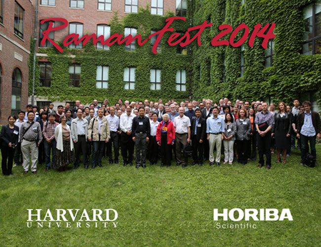
Before breaking for lunch, all of the attendees and speakers gathered for a group photo in the courtyard of the building. A nice lunch was served and everyone was able to mingle and discuss the talks that had already taken place. The consensus in the crowd was that the conference was great and people were excited for the remaining talks of the day.

Figure 1 – Group photo of the attendees and speakers at RamanFest 2014.
After lunch, Professor Christian Pellerin of the Department of Chemistry at the University of Montreal discussed the concept and practical applications of a new procedure proposed by his group to quantify orientation of polymers by Raman spectroscopy based on the most probable orientation distribution function. His talk was titled “Raman Spectroscopy of Individual Electrospun Fibers.”
“Industrial Problem Solving with Raman Spectroscopy” was the next talk presented by Dr. Neil Everall of Intertek-Wilton in the UK. Everall discussed real-world case studies from a laboratory that supports the R&D, production, and technical service activities of a wide range of industries, noting that confocal Raman spectroscopy is often the only technique to solve these particular problems. Some of Everall's examples included Raman imaging of the structure of blends, coatings, and laminates; analysis of defects in composites; mapping of cure in ultraviolet (UV)-cured coatings; and studies of polymer crystallinity on the micrometer scale.
After another brief coffee break, Dr. The-Quyen Nguyen of Northwestern University presented a much-anticipated talk titled “Raman Spectroscopy Goes Out Helping Patients in the Operating Room.” Nguyen discussed a three-dimensional (3D) scanning device, developed by his group, which can measure the entire surface of a resected tumor. The device automatically reconstructs the 3D image of a tumor, scans its entire surface, and evaluates its surgical status in real time using optical spectroscopy. Some tests have been conducted on risk-reducing mastectomy and sarcoma specimens with the outcomes demonstrating a consistent correlation to histopathology results. In the future, this device could make the intraoperative analysis margin achievable in less than 15 min and diagnose the margin status of an excised tumor while the patient is still in the operating room, so if more tissue had to be removed it could be done immediately rather than during a second surgery.
The final talk of the first day was “Supremacy and Variety of Vibrational Spectroscopy for Probing Amyloid Fibrils: From UV Raman to VCD and TERS,” presented by Professor Igor K. Lednev of the University of Albany at the State University of New York. He discussed novel experimental approaches developed by his team and other collaborators that are based on advanced vibrational spectroscopy and were applied to characterizing the structure and dynamics of amyloid fibrils during the last decade. These approaches included deep ultraviolet resonance Raman (DUVRR) spectroscopy, vibrational circular dichroism (VCD), and tip-enhanced Raman spectroscopy (TERS) as well as advanced statistical methods for analyzing spectroscopic data. Lednev also discussed the application of these methods for amyloid fibril characterization.
On Thursday evening a gala dinner was held at the Sheraton Commander Hotel. Several attendees commented on the conference, expressing their appreciation for an event that brought together people from all sides of Raman spectroscopy that might have otherwise never met or heard about each other’s work. The highlight of the evening was a talk given by Professor Mildred “Millie” Dresselhaus of the Massachusetts Institute of Technology (MIT), also affectionately referred to as the “Queen of Carbon” for all of her research work on that element. Dresselhaus’ talk was titled “My Forty-Year Adventure with Raman Spectroscopy and the Future.” She discussed her illustrious career, decade by decade, and offered practical advice for other scientists. One such piece of advice was to save money from grants for “crazy” experiments and to be willing to try out those crazy ideas because you never know where they might lead. Dresselhaus also said that her passion for music helped her get started in science by opening doors with various people who helped her career later on. In honor of her love for music, Horiba presented her with sheet music as a thank you for her great talk.
The second day of the conference began with a talk from Professor Sanford A. Asher of the University of Pittsburgh. Asher’s talk was titled “UV Raman Studies of Protein and Peptide Structure and Folding Studies.” He discussed the details of peptide folding conformation dynamics with laser T-jumps where the water temperature was elevated by a 1.9-µm infrared (IR) nanosecond laser pulse and the ~200-nm UV Raman spectrum was monitored as a function of time. Asher explained that those spectra show the time evolution of conformation. He also discussed the role of salts on stabilizing conformations in solution.
The next talk, titled “SERS and Fluorescence as Analytical Tools to Study Theranostic Nanosystems,” was given by Professor Igor Chourpa of the Université de Tours François Rabelais in France. Chourpa discussed how surface-enhanced Raman scattering (SERS) can be coupled with fluorescence to increase the potential in analytical and diagnostic applications. Some of the topics he covered include nanosystem characterization, the study of drug loading and release in suspension, drug delivery in live cancer cells, and the development of multimodal biomedical imaging concepts. The focus of his talk was the complementary use of SERS and fluorescence based approaches in pluridisciplinary research on novel biocompatible nanosystems developed for theranosis (therapy and diagnosis) of cancers.
Professor Ji-xin Cheng of Purdue University gave a talk titled “Microsecond Time Scale Spectroscopic Imaging for In Vivo Molecular Analysis.” Cheng discussed a novel spectroscopic imaging scheme based on parallel lock-in free detection of spectrally dispersed stimulated Raman scattering signal using a homebuilt tuned amplifier array. The method reduced the spectral acquisition time to 30 µs per pixel, an improvement of three orders of magnitude compared to multiplex coherent anti-Stokes Raman scattering (CARS). Cheng has used this method to monitor molecular penetration into skin tissue in situ and real time. He believes the method has potential for direct visualization of chemistry that occurs at the microsecond time scale as well as highly dynamic environments such as live cells.
Following a brief coffee break, Professor Paul Champion of the physics department at Northeastern University gave a talk titled “Coherent Low-Frequency Vibrational Motion in Proteins and Biomolecules.” Champion said that low frequency modes are prime candidates to serve as biochemical reaction coordinates and discussed their ability to mix with other delocalized low-frequency modes of the protein or binding partners offers a potential control mechanism.
Professor Lawrence D. Ziegler of Boston University gave the next talk, titled “In Vitro Cellular Activity Probed by SERS: Applications for Diagnostics and Forensics.” Ziegler discussed various applications for SERS, including bacterial diagnostics, blood diagnostics, forensics, and cancer cell detection. He also explained that SERS can be used for trace detection and identification of human body fluids such as blood, semen, vaginal fluid, and saliva for ultrasensitive forensic identification at crime scenes.
After lunch, Professor Wei Min of Columbia University presented “Bio-orthogonal Nonlinear Vibrational Imaging.” Min discussed an emerging chemical imaging platform called stimulated Raman scattering (SRS) microscopy, which can enhance the feeble spontaneous Raman transition by virtue of stimulated emission. Min presented information on the emerging biomedical applications such as imaging lipid metabolism, protein synthesis, protein degradation, DNA replication, RNA synthesis, glucose uptake, and drug tracking. This work will be important in studying brain injuries and cancer.
The last talk at the conference was given by Professor Sunny Xie of Harvard University and was titled “Label Free Vibrational Imaging for Medicine.” Xie discussed the recent advances in SRS that have improved the sensitivity, selectivity, robustness, and cost — all of which have opened the door to a wider range of biomedical applications. Xie also did a comparison of SRS to CARS and concluded that SRS was better.
Before the conference wrapped up, Andrew Whitley posed two questions to the audience and speakers in a round table discussion format. The first question was as follows: What are the key factors that need to be addressed to allow Raman and particularly CARS, SRS, TERS, and SERS to be accepted or adopted along side other more mainstream techniques like scanning electron microscopy (SEM), fluorescence, imaging microscopy, FT-IR, and so on? The responses from the group varied: Some said the techniques need to be more useable; others commented that the cost needs to be lowered, that there needs to be a “killer” application, more speed, and that a Raman library needs to be created.
The second question: Are we in the final maturation phase of Raman, CARS, and SRS with not so much real new significant technology progress to come, or if not, what are the technology developments expected? What are the crazy experiments that need to be done — SERS and TERS? SERS-fiber probes for further development? Super resolution Raman or SRS? The audience responses were mixed for this response too: Some people said that Raman and fluorescence should be investigated further, others suggested more research on single-molecule SRS, more UV-Raman, and colored Raman spectra in 1 ns. When asked what the future was for this science, the audience said looking at living cells and cancer detection.
The conference concluded with a tour of Sunny Xie’s laboratory for anyone who was interested. Next year’s 3rd Annual RamanFest is planned for May and will be held in Beijing, China. Updates will be provided in the coming months at www.ramanfest.org.
For more information on Professor Sunny Xie's work click here.
For more information on Horiba's Raman instruments click here.
Real-Time Battery Health Tracking Using Fiber-Optic Sensors
April 9th 2025A new study by researchers from Palo Alto Research Center (PARC, a Xerox Company) and LG Chem Power presents a novel method for real-time battery monitoring using embedded fiber-optic sensors. This approach enhances state-of-charge (SOC) and state-of-health (SOH) estimations, potentially improving the efficiency and lifespan of lithium-ion batteries in electric vehicles (xEVs).