Unveiling the Healing Process: Fluorescence Properties of Wounds on Soybean Seedlings Explored
A research team has developed a nondestructive method to assess the healing ability of plant tissues by analyzing the fluorescence properties of wounds on soybean seedlings.
A recent study published in Spectrochimica Acta Part A: Molecular and Biomolecular Spectroscopy explored plant wound healing by examining the fluorescence properties of wounds on soybean seedlings (1). Excitation emission matrix (EEM) and fluorescence imaging was used in this study (1). What the researchers discovered opens up further exploration of plant regeneration, which can have positive implications for the future of plant-based industries and sustainable agricultural practices (1).
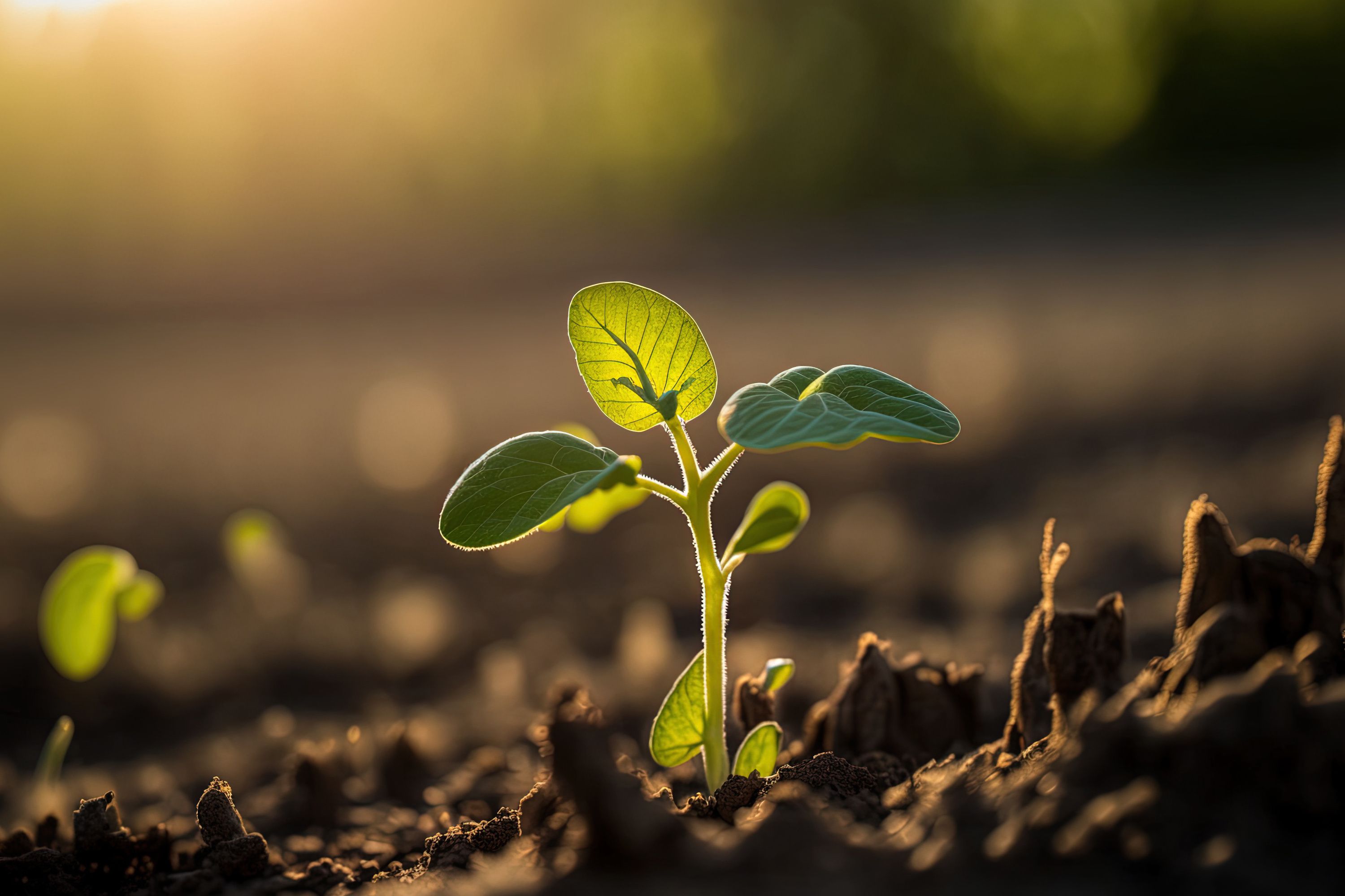
In the field, a thin, vulnerable soybean sprout reaches for the light. Soybean plants growing in rows on a farm. selective attention. Generative AI | Image Credit: © 2ragon - stock.adobe.com

Plants, like humans, are a living organism. Because of this fact, they can heal just like humans to. The researchers sought in this study to learn about plant regeneration processes. To achieve this, the team at Niigata University developed a simple and nondestructive method for evaluating the healing ability of plant tissues (1).
The procedure used is as follows: wounds were created on the stem of soybean seedlings. precisely seven days after sowing. Then, using fluorescence, the research team monitored the wounds over a period of 96 hours (1). By employing excitation emission matrix (EEM) and fluorescence imaging techniques, the researchers captured valuable data that shed light on the healing process at a microscopic level (1).
By using EEM to analyze the wounds, researchers detected the presence of three prominent fluorescence peaks, each exhibiting a decreasing intensity as the healing progressed (1). Moreover, the fluorescence images excited by a wavelength of 365 nm showcased a gradual reduction in the reddish color associated with chlorophyll, indicating a correlation between fluorescence properties and the healing process (1).
A confocal laser microscope was used to study the wounded tissue. This examination uncovered an intriguing phenomenon: the intensity of lignin or suberin-like fluorescence increased with healing time, suggesting that these substances might play a role in blocking the excitation light (1).
These findings open up new avenues for nondestructive assessment of plant wound healing (1). The ability to evaluate the healing process through fluorescence properties offers a valuable tool for plant biologists and agricultural scientists seeking to enhance crop health and productivity. By understanding the intricacies of plant regeneration and response to injuries, researchers can devise targeted strategies to promote healing and resilience in plants (1).
The team's work marks a significant step forward in our comprehension of plant wound healing. It sets the stage for future investigations and paves the way for the development of innovative techniques and tools that will contribute to more sustainable and productive agricultural practices.
Reference
(1) Saito, Y.; Ito, Y.; Tada, T.; Shoda, A.; Shiraiwa, T.; Kondo, N. Characterization of fluorescence properties of wounds on soybean seedlings during healing process using excitation emission matrix and fluorescence imaging. Spectrochimica Acta Part A: Mol. Biomol. Spectrosc. 2023, 298, 122766. DOI:10.1016/j.saa.2023.122766
New Fluorescence Model Enhances Aflatoxin Detection in Vegetable Oils
March 12th 2025A research team from Nanjing University of Finance and Economics has developed a new analytical model using fluorescence spectroscopy and neural networks to improve the detection of aflatoxin B1 (AFB1) in vegetable oils. The model effectively restores AFB1’s intrinsic fluorescence by accounting for absorption and scattering interferences from oil matrices, enhancing the accuracy and efficiency for food safety testing.
Tracking Molecular Transport in Chromatographic Particles with Single-Molecule Fluorescence Imaging
May 18th 2012An interview with Justin Cooper, winner of a 2011 FACSS Innovation Award. Part of a new podcast series presented in collaboration with the Federation of Analytical Chemistry and Spectroscopy Societies (FACSS), in connection with SciX 2012 ? the Great Scientific Exchange, the North American conference (39th Annual) of FACSS.
New Fluorescent Raman Technique Enhances Detection of Microplastics in Seawater
November 19th 2024A novel method using fluorescence labeling and differential Raman spectroscopy claims to offer a more efficient, accurate approach to detect microplastics in seawater. Developed by researchers at the Ocean University of China, this method improves both the speed and precision of microplastic identification, addressing a key environmental issue affecting marine ecosystems.