Using Surface-Enhanced Raman Spectroscopy for the Diagnosis of Pancytopenia-Related Diseases
A recent study examined how surface-enhanced Raman spectroscopy (SERS) is being used to help diagnose pancytopenia-related diseases earlier.
In an effort to improve patient outcomes for pancytopenia-related diseases, researchers recently demonstrated how surface-enhanced Raman spectroscopy (SERS) can help diagnose these diseases earlier, according to a recent study published in Spectrochimica Acta Part A: Molecular and Biomolecular Spectroscopy (1).
Pancytopenia is a severe and complex blood disorder characterized by a significant reduction in red blood cells, white blood cells, and platelets in the peripheral blood (1). The condition can stem from a variety of underlying diseases, many of which carry high mortality rates. Early and accurate identification of the specific cause of pancytopenia is crucial for administering appropriate treatment and improving patient outcomes (1).
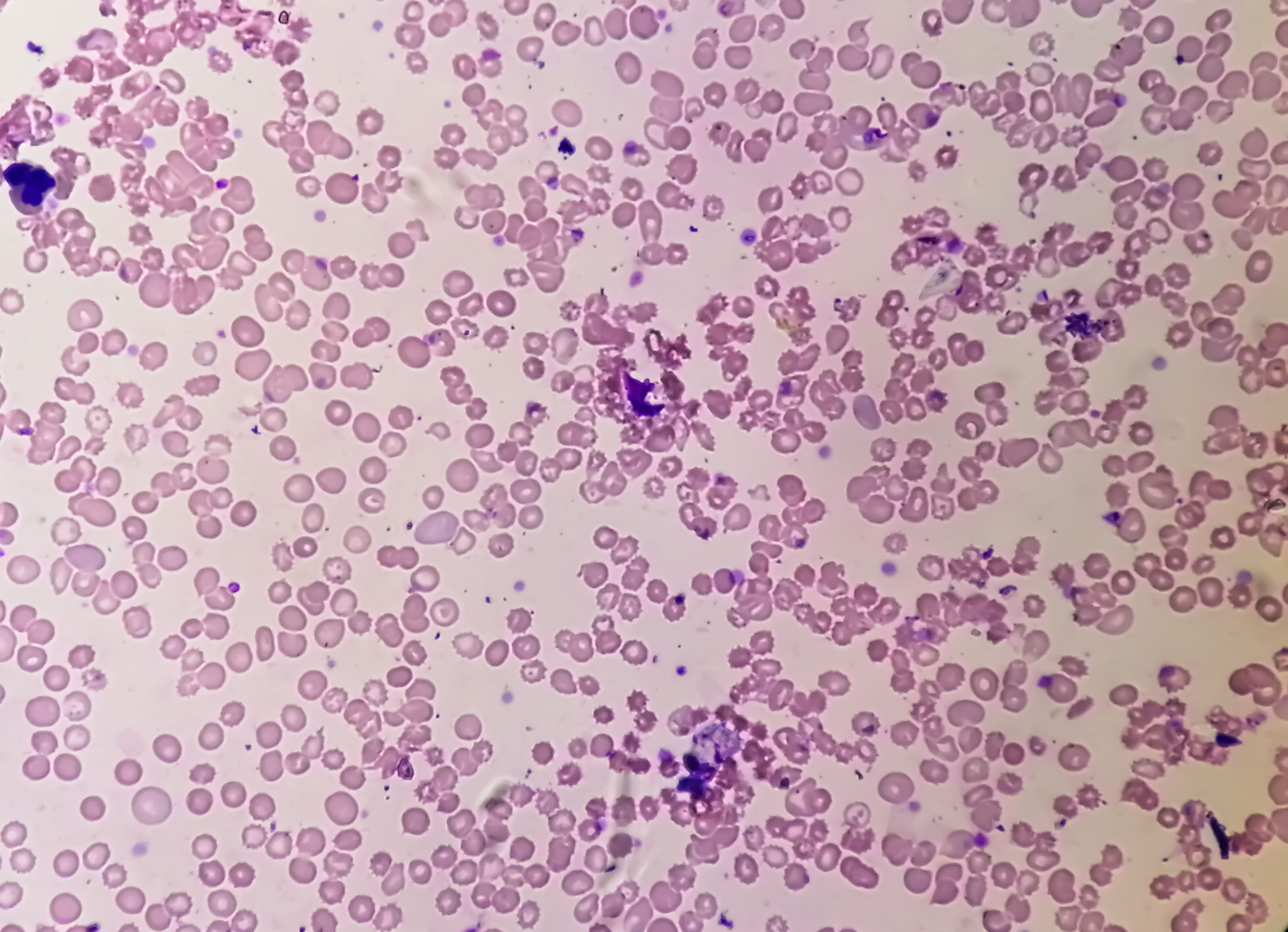
Microscopic view of hematological slide showing Pancytopenia. A condition in which there is a lower number of RBC, WBC and platelets in the blood. | Image Credit: © Saiful52 - stock.adobe.com.

A research team from China, led by Shuo Chen and Guojun Zhang, explored how SERS could analyze serum samples from patients with pancytopenia. SERS is a powerful technique that enhances the Raman scattering signals of molecules adsorbed on rough metal surfaces, making it highly sensitive to molecular changes in biological samples (2). In their study, Chen and Zhang focused on three specific diseases associated with pancytopenia: aplastic anemia (AA), myelodysplastic syndrome (MDS), and spontaneous remission of pancytopenia (SRP) (1). These conditions are similar in that the patient afflicted with them experiences similar initial symptoms, making them difficult to distinguish through conventional clinical examinations alone.
The researchers’ goal of the study, therefore, was to determine whether specific biomolecular changes in the serum could serve as reliable biomarkers for differentiating between AA, MDS, and SRP at an early stage. The SERS spectral analysis revealed significant differences in the serum composition of patients with these diseases (1). Variations in certain amino acids, protein substances, and nucleic acids were identified as potential biomarkers. These findings suggested that SERS could detect subtle molecular changes that were not apparent through traditional diagnostic methods (1).
To further refine their diagnostic approach, the researchers developed a diagnostic model by combining partial least squares analysis and linear discriminant analysis (PLS-LDA) (1). The intention of this statistical model was to process complex SERS data and enhance the accuracy of disease classification, which it was successful in doing so. The model achieved an impressive overall diagnostic accuracy of 86.67%, demonstrating its potential to distinguish between AA, MDS, and SRP even when routine clinical examinations fall short (1).
By enabling early and accurate differentiation of pancytopenia-related diseases, SERS demonstrated its ability to deliver results that can help lead to improved targeted treatments, ultimately improving survival rates for patients (1). The research team also demonstrated in their study how spectroscopic techniques such as SERS can be used in medical diagnostics by playing to the technique’s strengths. In a field where rapid and precise disease identification is paramount, medical diagnostics requires the use of techniques like SERS to identify disease early on.
Chen and Zhang's work represents a significant step forward in the clinical management of pancytopenia. As the SERS technique becomes more refined and accessible, it could be integrated into standard diagnostic protocols, offering a powerful tool for healthcare providers (1). The ability to diagnose these complex conditions accurately and early on could transform patient care and outcomes in hematology (1).
With further research and development, SERS could improve the early diagnosis and treatment of pancytopenia-related diseases, allowing clinicians to provide better care to patients.
References
(1) Chen, Z.; Li, Y.; Zhu, R.; et al. Early Differential Diagnosis of Pancytopenia Related Diseases based on Serum Surface-enhanced Raman Spectroscopy. Spectrochimica Acta Part A: Mol. Biomol. Spectrosc. 2024, 316, 124335. DOI: 10.1016/j.saa.2024.124335
(2) Wetzel, W. An Inside Look at the Latest in Surface-enhanced Raman Spectroscopy. Spectroscopy. Available at: https://www.spectroscopyonline.com/view/an-inside-look-at-the-latest-in-surface-enhanced-raman-spectroscopy (accessed 2024-05-29).
AI-Powered SERS Spectroscopy Breakthrough Boosts Safety of Medicinal Food Products
April 16th 2025A new deep learning-enhanced spectroscopic platform—SERSome—developed by researchers in China and Finland, identifies medicinal and edible homologs (MEHs) with 98% accuracy. This innovation could revolutionize safety and quality control in the growing MEH market.
AI-Driven Raman Spectroscopy Paves the Way for Precision Cancer Immunotherapy
April 15th 2025Researchers are using AI-enabled Raman spectroscopy to enhance the development, administration, and response prediction of cancer immunotherapies. This innovative, label-free method provides detailed insights into tumor-immune microenvironments, aiming to optimize personalized immunotherapy and other treatment strategies and improve patient outcomes.
2 Commerce Drive
Cranbury, NJ 08512
All rights reserved.