Carbon Nanotube Characterization and Quality Control Using Portable Raman: 532-nm Versus 785-nm Laser Excitation
Special Issues
In this paper, we examine the relative performance of 532 and 785 nm portable Raman systems, as well as demonstrate an automated analytical methodology applicable for carbon nanotube (CNT) characterization and quality control applications. Both 532 and 785 nm Raman spectra were used to directly analyze and compare important CNT structural parameters and properties including CNT diameters, diameter distributions, CNT structural quality (% of defects), CNT types, and other properties. The data indicate advantages in a number of areas for using 532 versus 785 nm excitation for CNT Raman measurements.
Recent growth in commercial carbon nanotube (CNT) production resulted in a strong demand for analytical techniques capable of rapid characterization of these nanomaterials. The latest developments in portable Raman spectroscopy made this technique one of the most viable options for the task. This paper compares the relative performance of 532- and 785-nm portable Raman systems, as well as demonstrates an automated analytical methodology suitable for CNT characterization and quality control applications. Both 532- and 785-nm Raman spectra were used to directly analyze and compare important CNT structural parameters and properties including CNT diameters, diameter distributions, CNT structural quality (% of defects), CNT types, and other properties. The data indicate advantages in a number of areas for using 532- versus 785-nm excitation for CNT Raman measurements.
Carbon nanotubes (CNTs) are a relatively new class of nanomaterials first discovered by Iijima (1). The unique electrical and mechanical properties of CNTs make them one of the most attractive building blocks for multiple nanomaterials designed for various applications. The typical applications include but are not limited to nanoelectronics and nanomechanics, ultrastrong yarns, interconnects, batteries and capacitors, and aerospace and defense (2,3). As a result, the global CNTs and CNT-based product market is projected to reach 5.64 billion US dollars by 2020, with an annual growth rate (CAGR) of 20.1% (4).
Manufacturing, modification, functionalization, and customization of the CNT-based materials require bulk quantities of well-defined CNTs, with a high degree of structural purity. Since tight control of the CNT sizes, chirality, and structural quality still remains one of the major challenges during CNT fabrication (3), analytical techniques capable of thorough CNT characterization have become a critical part of the manufacturing process.
Several techniques are currently used for CNT characterization, including electron microscopy, Raman spectroscopy, thermogravimetric analysis (TGA), and optical absorption spectroscopy. However, Raman spectroscopy has become increasingly attractive for CNT characterization because of its intrinsic ability to detect even small changes in CNT structure, its low analysis cost, little or no sample preparation required, and dramatically improved analysis efficiency due to recent engineering breakthroughs in Raman instrumentation.
CNT Raman spectra can provide information about the CNT diameter, chirality, structural quality, conductor or semiconductor character, crystallinity, and degree of functionalization (5–8). A brief introduction to major Raman features of carbon nanotubes is given below.
The radial breathing mode (RBM) band represents radial expansion–contraction of a nanotube. This is a very important band because it is a marker of the presence of single-wall carbon nanotubes (absent in the spectra of essentially all other graphite-like materials). The RBM band frequency can also be directly related to CNT diameter (7,8).
The G-band is a major Raman band in graphite-like materials representing a tangential shear mode of carbon atoms. In CNTs, this mode has two major contributions, G+ and G- (7). The shape of the G-band and its frequency profile can be used to estimate whether the CNT is “metallic” or semi-conducting; or roughly estimate the nanotube diameter.
Because of Raman selection rules, the D-band is a forbidden Raman mode in ideal nanotubes and requires a defect to show up in the Raman spectra (7). Thus, the D/G band ratio can directly serve as a measure of CNT structural quality.
The G′-band is the first overtone of the D-band. However, unlike the D-band, it does not require a defect to show up in Raman spectra. The G′-band can be used, in particular, to probe CNT electronic structure and independently verify CNT chirality (7).
In this article, the relative performance of 532- and 785-nm portable Raman spectrometers was compared for CNT characterization and quality control applications. In addition, an analytical methodology is presented that enables the calculation of CNT structural parameters directly from measured Raman spectra.
Instrumentation
Carbon nanotube samples were examined using the RamTest-NANO and Mobile µ-Raman analyzers with Raman technology (both from BioTools, Inc.). The RamTest-NANO analyzer is a handheld unit with 532-nm laser excitation, ~120–4000 cm-1 spectral range, 4–6 cm-1 spectral resolution, and a maximum laser power of 30 mW. The Mobile-µ-Raman analyzer is a portable unit with 785-nm excitation and variable laser power within a 0–100 mW range. A high resolution (HR) configuration of the Mobile µ-Raman analyzer was used in this study, with ~200–2000 cm-1 spectral range and ~4 cm-1 resolution to match the resolution of the RamTest unit.
The RamTest unit was run in automated mode (requiring no prior knowledge of Raman spectroscopy), where all measurement parameters are automatically adjusted by the instrument to optimize signal-to-noise ratio and minimize fluorescence, with the remaining fluorescence background (if any) automatically subtracted.
The Mobile µ-Raman analyzer was run in a mode where all measurement parameters were manually set before the measurement. The laser power was set to 30 mW to match that of the RamTest unit.
Data Analysis
BioTools’ Ram-NANO software add-on is capable of calculating the following CNT parameters or properties: structural quality (percent of structural defects), CNT diameter distribution, type of nanotube, and several other parameters. This software add-on is directly applicable to both RamTest-NANO and Mobile µ-Raman data to enable automated CNT Raman data processing, and a PDF or Excel report generation.
Technical Results
532- and 785-nm Raman Spectra of Carbon Nanotubes
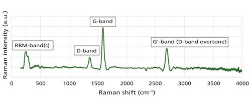
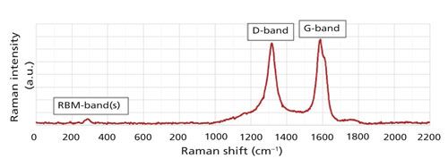
Figures 1 and 2 show 532- and 785-nm Raman spectra of CNTs recorded at identical laser powers of 30 mW at both wavelengths, respectively. There are four major Raman bands observed for the CNT samples measured during this study: RBM bands around 190–320 cm-1, D-band at ~1320–1370 cm-1, G-band at ~1580 cm-1, and G′-band at ~2700 cm-1 (out of instrument spectral range for the 785-nm Raman spectrum shown in Figure 2). The meaning of these CNT Raman bands has been extensively described in the literature (5–8).

Figure 1: 532-nm Raman spectrum of carbon nanotubes measured at 30-mW laser power. This spectrum contains all of the important CNT Raman bands, which can be used for CNT characterization (see text for detailed discussion).

Figure 2: 785-nm Raman spectrum of carbon nanotubes measured at 30-mW laser power. Low RBM/G-band intensity ratio combined with high D/G band intensity ratio may indicate that the 785-nm laser partially destroys CNTs at the laser power density used in this study (see the text for detailed discussion).
There are three major observations from Figures 1 and 2. First, the 532-nm Raman spectrum shows a much greater RBM/G band intensity ratio than the 785-nm Raman spectrum. Second, the D/G band intensity ratio is much smaller for the 532-nm Raman spectrum than that for the 785-nm Raman spectrum. Third, the G-band of the 532-nm Raman is fairly symmetric, whereas the 785-nm G-band shows a distinct high-frequency shoulder.
Since the RBM band is indicative of CNT presence and the D-band is a measure of fraction of structural defects in CNTs, these observations may indicate that a 785-nm laser partially destroys CNTs, at least at the laser power density used in this study. The increased heating or destruction of the CNTs by 785-nm light (versus 532-nm light at an identical power of 30 mW) can likely be explained by the increased absorption of the 785-nm light by these particular CNT samples. A further decrease of laser power helps to reduce the 785-nm laser-induced CNT damage (data not shown). However, it also results in reduction of the 785-nm Raman signal and, therefore, the necessity to increase measurement time.
Thus, the preliminary conclusion is that both 532- and 785-nm Raman can be used for CNT characterization. However, care must be taken to ensure that relatively delicate CNT samples are not being overheated or even destroyed during analysis.
Homogeneous Linewidth of the RBM Raman Band of an Isolated CNT
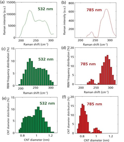
The RBM band profile of CNT samples measured in this study is fairly broad and spans the ~190–320 cm-1 spectral region (Figures 3a and 3b). Since the RBM frequency is related to CNT diameter (7,8), the observed RBM frequency span is likely to result from a subset of CNTs with different diameters. Thus, if we knew the homogeneous linewidth of the CNT RBM band, we could directly estimate the CNT diameter population distributions directly from the RBM band profile.

Fortunately, Raman spectra of isolated single-wall CNTs were reported in the literature (8), and their RBM bandwidths are in the range from 9 to 14 cm-1 full width at half height (FWHH). These RBM bandwidths are wider than the resolution of both the 532- and 785-nm portable Raman instruments used in this study (~4 cm-1). Thus, for the sake of simplicity, we can take a “rough average” of the reported values, and estimate the RBM band homogeneous linewidth of an individual CNT with a particular diameter, Γ, as ~6 cm-1 half-width at half-height (HWHH).
Deconvolution of RBM-Band Frequency
Using the ~6 cm-1 homogeneous linewidth of a single isolated CNT with a particular diameter, and assuming that Raman cross-sections of all CNT conformations are independent of their frequencies, we can estimate the population distribution of each CNT conformation directly from the RBM band profiles using a method similar to that reported by Asher and colleagues (9). We assume that an inhomogeneously broadened RBM band profile, RBM(ν) is a sum of N Lorentzians with identical intensities and homogeneous linewidths Γ = 6 cm-1, occurring at different central frequencies ν = νi0.
[1]

where Pi is a probability for a band to occur at frequency νi0.
The resulted histograms of frequency populations, Pi, are shown in Figures 3c and 3d for 532- and 785-nm Raman RBM bands, respectively.
CNT Diameter Distributions Obtained Directly from 532- and 785-nm Raman Spectra
CNT diameter can be estimated from RBM band frequency using the following relationship (8):
[2]

where νRBM is the RBM band frequency in cm-1 and dCNT is the CNT diameter in nm.
Using equation 2, we can convert the RBM frequency population distributions into CNT diameter population distributions, as shown in Figures 3e and 3f. These diameter distributions reflect the percentage of CNTs with their diameters falling into a specified diameter interval and, thus, can be directly utilized for quality control purposes.
Figure 3 demonstrates that RBM band profiles and the resulting CNT diameter distributions obtained from the 532-nm Raman spectra differ from those obtained from the 785-nm Raman spectra. This is despite the fact that both 532- and 785-nm Raman measurements were taken on the identical CNT samples, and using the same laser power of 30 mW on both 532- and 785-nm instruments.
Theoretically, the difference in diameter distributions obtained from the 532- and 785-nm Raman spectra could have been explained by selective resonance enhancement of different subsets of CNTs by 532- and 785-nm light, because of the different resonance Raman conditions (5). However, as it was already discussed above, comparison of 532- and 785-nm relative Raman intensity ratios for RBM, D, and G bands indicates that, most likely, the 785-nm laser at 30-mW power partially destroys the CNTs (in contrast to 532-nm laser used at the same power). If this conclusion is correct, then Figure 3 may indicate that the 785-nm laser selectively destroys CNTs with higher diameters.
Conclusions
Both 532- and 785-nm portable Raman instruments can be used for CNT characterization and quality control applications. Specifically, CNT diameters, diameter distributions, and structural quality (percent of structural defects), other important parameters, and multiple controls plots can be directly obtained or generated from 532- and 785-nm Raman spectra using the automated analytical methodology demonstrated in this paper.
The differences observed between 532- and 785-nm Raman spectra of identical CNT samples can likely be explained by partial destruction of CNT samples by the 785-nm laser, at the laser power used in this study. Alternatively, these differences can be explained by selective resonance Raman enhancement of different subsets of CNTs by 532- and 785-nm lasers, respectively (5).
Even though multiple Raman excitations are still preferable for complete characterization of CNTs, the following techno-economic benefits are identified for 532-nm as opposed to 785-nm portable Raman, if, for any reason, an end-user decides to use only one Raman wavelength for CNT analysis:
- A 532-nm laser at 30-mW power does not readily destroy CNTs examined here, whereas the 785-nm laser must only be used at a significantly lower power. This can likely be explained by increased absorption of the 785-nm light by these particular CNT samples.
- The Raman signal at 532-nm excitation is approximately fivefold stronger than that at 785 nm, per unit laser power. This is because Raman signal is ~(1/λEX)4, where λEX = 532 nm, 785 nm, and so forth (10). Thus, 532-nm portable Raman units are fundamentally capable of approximately five times faster analysis or improved analysis quality. In addition, in the case of especially delicate samples, the 532-nm Raman analyzers can be used at up to approximately fivefold lower laser power than the 785-nm based units, respectively, without compromising spectral analysis quality.
- The 532-nm Raman technique provides a superior combination of spectral resolution and spectral range (compared to the 785-nm Raman technique) to enable superior CNT structural characterization. This is because the use of 532-nm light provides an engineering advantage in space-limited portable instruments.
- Quantum efficiencies of best in class charge-coupled devices (CCD) detectors and optics are greater at 532 nm (compared to 785 nm) to even further increase the observed Raman signal in favor of 532-nm excitation.
- The 532-nm Raman analyzer has a superior performance not only for the analysis of CNTs “as is,” but also for the analysis of CNT suspensions in aqueous solutions, because of the reduced absorption of 532-nm light by water versus that of 785-nm light.
References
- S. Iijima, Nature354, 56–58 (1991).
- M.F.L. De Volder et al., Science339, 535–539 (2013).
- A. Barreiro et al., Carbon45, 55–61 (2007).
- Data obtained from www.marketsandmarkets.com.
- R. Saito and H. Kataura, in Carbon Nanotubes: Synthesis, Structure, Properties, and Applications, M.S. Dresselhaus, G. Dresselhaus, and Ph. Avouris, Eds. (Springer-Verlag, 2001), pp. 213–246.
- A.G. Souza Filho et al., Nanotechnology14(10), 2043–2061 (2003).
- M.S. Dresselhaus et al., Carbon40, 2043–2061 (2002).
- F. Hennrich et al., J. Phys. Chem. B109, 10567–10573 (2005).
- S.A. Asher et al., J. Am. Chem. Soc. 126, 8433–8440 (2004).
- Handbook of Vibrational Spectroscopy, J.M. Chalmers and P.R. Griffiths, Eds. (John Wiley & Sons, Chichester, 2002).
Aleksandr V. Mikhonin and Rina K. Dukor are with BioTools Inc., in Jupiter, Florida. Laurence A. Nafie is with BioTools Inc., and the Department of Chemistry at Syracuse University in Syracuse, New York. Direct correspondence to: alex@btools.com

AI-Powered SERS Spectroscopy Breakthrough Boosts Safety of Medicinal Food Products
April 16th 2025A new deep learning-enhanced spectroscopic platform—SERSome—developed by researchers in China and Finland, identifies medicinal and edible homologs (MEHs) with 98% accuracy. This innovation could revolutionize safety and quality control in the growing MEH market.
New Raman Spectroscopy Method Enhances Real-Time Monitoring Across Fermentation Processes
April 15th 2025Researchers at Delft University of Technology have developed a novel method using single compound spectra to enhance the transferability and accuracy of Raman spectroscopy models for real-time fermentation monitoring.
Nanometer-Scale Studies Using Tip Enhanced Raman Spectroscopy
February 8th 2013Volker Deckert, the winner of the 2013 Charles Mann Award, is advancing the use of tip enhanced Raman spectroscopy (TERS) to push the lateral resolution of vibrational spectroscopy well below the Abbe limit, to achieve single-molecule sensitivity. Because the tip can be moved with sub-nanometer precision, structural information with unmatched spatial resolution can be achieved without the need of specific labels.