Discriminating Paints with Different Clay Additives in Forensic Analysis of Automotive Coatings by FT-IR and Raman Spectroscopy
An investigation of the different kinds of clay used as paint additives with the goal of discriminating the paints
The analysis of automotive coatings is important for forensic scientists in the investigation of hit-and-run accidents. However, many kinds of paint are similar in structure and cannot be discriminated easily. In this study, Fourier-transform infrared (FT-IR) and Raman microscopy were employed to investigate different kinds of clay as the additives in paints to help discriminate the paints. The IR and Raman spectra were measured and tentatively interpreted. The indicative peaks distinguishing kaolin and bentonite were summarized in the IR spectrum. For some other kinds of clay that could not be discriminated from kaolin in the IR spectrum, several peaks in the Raman spectrum of kaolin were found to be weak but characteristic as indicators of kaolin. The method was applied and verified in a complex paint analysis case successfully. The data in this study can help forensic scientists identify paints accurately, particularly in cases with interference in the spectrum (<3000 cm-1).
Pigments and resins, as important ingredients in paints, are widely used in the classification and identification of paints and in the investigation of hit-and-run accidents (1–12). However, few studies focus on the classification of paints with additives. Clays are now widely used as additives in paints, which can enhance the coating's mechanical strength and resistance against degradation. They exist in almost all kinds of paints including amino resin paints, alkyd resin paints, polyurethane paints, and acrylic resin paints. The ability to discriminate these kinds of clay will be of great help in paint discrimination.
Infrared (IR) spectroscopy, scanning electron microscopy (SEM), X-ray fluorescence (XRF), and pyrolysis gas chromatography–mass spectrometry (GC–MS) are useful techniques frequently used in paint identification (13–15). Among all of these techniques, IR spectroscopy has been proven to be effective and accurate for paint analysis since the 1970s, requiring only a small quantity of sample to achieve rapid analysis and high-quality spectra (1–12). The Raman spectrum usually has different peak positions compared to the Fourier-transform infrared (FT-IR) spectrum for the same substance, so Raman spectroscopy can yield extra peak information. Raman spectroscopy covers some shortcomings of FT-IR spectroscopy and has proven to be a powerful tool in polymer characterization. Optical observation for different clays with these two methods was performed in this study to investigate their spectral characteristics. The spectroscopic properties of clay minerals have been investigated in many previous studies (16,17). However, to the best of our knowledge, few studies have examined the spectroscopic properties (particularly in Raman) of clays in paints. In addition, how to use the weak but indicative peaks in spectra to discriminate the paints is not clear and needs to be solved.
Experiments
Kaolin, bentonite, and some other clay samples were obtained from the National Technical Committee on Plastic Products of Standardization Administration of China. Calcium carbonate (CaCO3), titanium dioxide (TiO2), polystyrene, and acrylic resin paint samples were collected in cases. A Nicolet iN10 attenuated total reflectance (ATR)-FT-IR system (Thermo Fisher Scientific) with a mercury–cadmium–telluride (MCT) detector and Omnic Picta software (Thermo Fisher Scientific) was employed for IR observation. Spectra were collected in high-resolution (8-cm-1) mode. A Spectrum GX 2000 system from PerkinElmer with a diamond anvil cell, a deuterated triglycine sulfate (DTGS) detector, and Spectra 5.01 software was employed to verify the IR results from the iN10 system. The background was subtracted for every measurement. Triplicate tests were performed at different sites for each sample.
A Renishaw inVia confocal Raman microscopy system with two lasers emitting at 532 nm and 633 nm, respectively, and a charge-coupled device (CCD) detector was employed to collect the Raman spectrum. The 532-nm laser was chosen for emission in 50–100% power. Crystal silicon with a fixed peak position at 520 cm-1 was used to calibrate the Raman shifts before measurement. The spectra (100–4000 cm-1) were collected in WIRE 3 software (Renishaw) in extensive mode. Spectra were obtained by three accumulations to enhance the signal-to-noise ratio if necessary. Multiple measurements (n = 3) were made at different regions of the same sample to avoid variation.
Results and Discussion
IR Spectroscopy
Figure 1 shows the IR spectra of kaolin and bentonite obtained using the PerkinElmer Spectrum GX 2000 system. Peaks of kaolin and bentonite appeared at similar positions. The main IR peaks of kaolin were at 3696 cm-1, 3670 cm-1, 3655 cm-1, 3622 cm-1, 1117 cm-1, 1038 cm-1, 1013 cm-1, 938 cm-1, 915-1, 797 cm-1, 756 cm-1, 694 cm-1, 542 cm-1, 472 cm-1, and 432 cm-1. The main peaks of bentonite were at 3625 cm-1, 1638 cm-1, 1034 cm-1, 916 cm-1, 799 cm-1, 695 cm-1, 530 cm-1, and 469 cm-1. Four peaks in the region greater than 3000 cm-1 (3696 cm-1, 3670 cm-1, 3655 cm-1, and 3622 cm-1) and the peak at 938 cm-1 help to identify kaolin, while the peaks at 3625 cm-1 and 1638 cm-1 help to identify bentonite. In addition, peaks at 542 cm-1 and 472 cm-1 in kaolin shifted slightly to 530 cm-1 and 469 cm-1 for bentonite. These changes in peak position also will help discriminate these two clays.

Figure 1: Comparison of kaolin (top) and bentonite (bottom).
Kaolin (Al4 [Si4O10][OH]6) and bentonite ([Na,Ca]0.33 [Al,Mg]2 [Si4O10][OH]2 -nH2O), two kinds of clay minerals with mass production, were used in paint to extend titanium dioxide and thus modify gloss levels of coatings. Kaolin is a layered silicate mineral, with one tetrahedral sheet linked through oxygen atoms to one octahedral sheet of alumina octahedral. There are various types of bentonite, and they are named after the respective dominant element, such as potassium, sodium, calcium, and aluminum. Feng revealed that different types of bentonite had similar IR spectra (18). According to the component and the structure, IR spectra of kaolin and bentonite were tentatively interpreted as follows.

Figure 2: IR spectrum of acrylic resin paint containing TiO2, bentonite, and polystyrene.
For kaolin, 3696 cm-1, 3670 cm-1, 3655 cm-1, and 3622 cm-1, OH stretching; 1117 cm-1 and 1038 cm-1, Si-O stretching; 938 cm-1 and 915 cm-1, OH bending; 797 cm-1, Si-O-Mg; 542 cm-1, Si-O-Al; 472 cm-1, Si-O-Mg. The intensity of peaks at 3696 cm-1, 3670 cm-1, 3655 cm-1, and 3622 cm-1 indicated the crystallinity of kaolin. For bentonite, 3625 cm-1, OH stretching; 1638 cm-1, OH bending; 530 cm-1, Si-O-Al; 469 cm-1, Si-O-Mg. Comparison of IR spectra showed that peaks of kaolin at 3696 cm-1, 3670 cm-1, 3655 cm-1, 3622 cm-1, 938 cm-1, 915 cm-1, and those of bentonite at 3625 cm-1, 1638 cm-1, and 916 cm-1 were characteristic. These peaks can be used to discriminate the two clay minerals.
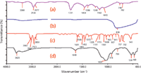
Figure 3: IR spectra of (a) acrylic resin paint, (b) TiO2, (c) polystyrene, and (d) bentonite.
Case Studies
Figure 2 shows the IR spectrum of acrylic resin paint containing TiO2, clay, and polystyrene in a hit-and-run case. Figure 3 shows the IR spectra of acrylic resin paint, TiO2, clay, and polystyrene used to interpret the spectrum of Figure 2. Figure 4 shows the IR spectra of acrylic resin paint containing TiO2, CaCO3, bentonite, and polystyrene in a case. Figure 5 shows the IR spectra of acrylic resin paint, TiO2, CaCO3, clay, and polystyrene to interpret the spectrum of Figure 4.

Figure 4: IR spectrum of acrylic resin paint containing TiO2, polystyrene, kaolin, and CaCO2.
The spectra in both Figure 2 and Figure 4 are acrylic resin paint containing TiO2, clay mineral, and polystyrene, so the two spectra were similar and hard to discriminate. The peak positions of the above substances were as follows: acrylic resin paint: 2875 cm-1, 1732 cm-1, 1453 cm-1, 1394 cm-1, 1162 cm-1, 759 cm-1, and 701 cm-1; TiO2: 800 cm-1 and ~500 cm-1; polystyrene: 3027 cm-1, 1493 cm-1, 1453 cm-1, 1330 cm-1, 1028 cm-1, 843 cm-1, 759 cm-1, and 701 cm-1; clay minerals: 3625 cm-1, 1120 cm-1, 1028 cm-1, 535 cm-1, and 473 cm-1. The assignments of the functional groups in the spectrum are listed in Table I. Without the information from clay minerals and CaCO3 in Figure 4, it would not be easy to discriminate between the two samples and avoid a wrong certificate of authenticity.
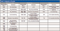
Table I: Assignment of IR peaks
In Figure 2, only one peak (3625 cm-1) was found above 3000 cm-1. Weak absorbance appeared at 1648 cm-1 and only one peak (916 cm-1) appeared in the 900–1000 cm-1 region. Si-O-Al and Si-O-Mg vibration appeared at 530 cm-1 and 469 cm-1, respectively. In Figure 4, there were four peaks in the region greater than 3000 cm-1 (3696 cm-1, 3670 cm-1, 3655 cm-1, and 3622 cm-1) and two peaks in the 900–1000 cm-1 region (938 cm-1 and 915 cm-1), which is not the same as in Figure 2. Si-O-Al and Si-O-Mg vibration appeared at 546 cm-1 and 473 cm-1, respectively. Bentonite and kaolin were identified from the two spectra separately; therefore, the two samples were discriminated.
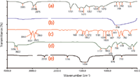
Figure 5: IR spectra of (a) acrylic resin paint, (b) TiO2, (c) polystyrene, (d) kaolin, and (e) CaCO3.
Raman Spectroscopy
There are many kinds of clay minerals with different components and crystallinity. Some minerals cannot be easily discriminated by IR spectroscopy. Furthermore, peaks of many inorganic compounds (including clay minerals in this study) often appeared in the region less than 500 cm-1 and cannot be analyzed with IR spectroscopy. Raman spectroscopy offers a new and powerful tool in solving this problem. In this study, Raman spectroscopy was employed for future determination of clay minerals and paints.
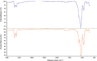
Figure 6: IR spectra of GBW03101a (top, clay) and GBW 03121 (bottom, kaolin).
Figure 6 shows the IR spectra of two China National Standard Materials: GBW03101a (one kind of clay minerals) and GBW 03121 (kaolin). Indicative peaks above 3000 cm-1, 900–1000 cm-1, and 1640 cm-1 were too weak to employ as qualitative peaks even with an MCT detector. For Raman analysis, both samples were found to have serious fluorescence interference. Sample bleaching for 60 min and 3–9 accumulations were used in sample preparation to obtain better spectra. Signals in the 1000–3000 cm-1 region of Raman shifts still were affected by fluorescence interference, which will not be discussed further in this study. Figure 7 shows the Raman spectra of GBW03101a (clay) and GBW 03121 (kaolin) in the 130–1000 cm-1 region. Figure 8 shows the Raman spectra of GBW03101a (clay) and GBW 03121 (kaolin) in the 3000–4000 cm-1 region. Significant differences in Raman spectra of the two minerals were observed in Figures 7 and 8. Peaks of kaolin appeared at 144 cm-1, 196 cm-1, 240 cm-1, 274 cm-1, 334 cm-1, 393 cm-1, 428 cm-1, 464 cm-1, 508 cm-1, 636 cm-1, 706 cm-1, 749 cm-1, 790 cm-1, 912 cm-1, 934 cm-1, 3240 cm-1, 3623 cm-1, 3654 cm-1, 3667 cm-1, and 3698 cm-1. Peaks of GBW03101a appeared at 140 cm-1, 198 cm-1, 278 cm-1, 459 cm-1, 603 cm-1, 616 cm-1, 687 cm-1, 708 cm-1, 779 cm-1, 930 cm-1, 3240 cm-1, and 3800 cm-1. The two samples were discriminated clearly in Raman spectra.

Figure 7: Raman spectra of GBW03101a (top, clay) and GBW 03121 (bottom, kaolin) (130â1000 cm-1).
Conclusion
In this study, FT-IR and Raman microscopy were employed to investigate different kinds of clays to discriminate the paints in hit-and-run cases. The IR and Raman spectra were tentatively interpreted. The indicative peaks distinguishing kaolin (3696 cm-1, 3670 cm-1, 3655 cm-1, 3622 cm-1, 938 cm-1, and 915 cm-1) and bentonite (3625 cm-1, 1638 cm-1, and 916 cm-1) were summarized in IR spectra. Raman spectroscopy was used for some kinds of clay that could not be discriminated from kaolin in IR spectrum. Kaolin peaks in the region greater than 3000 cm-1 and at 934 cm-1, 636 cm-1, and 274 cm-1 in its Raman spectrum were found to be characteristic and can be used as indicators. The method was applied and successfully verified in a complex case. Three kinds of clay minerals were investigated in this study to set up a database with adequate spectra. Spectra of more kinds of clay minerals and the combined spectra of clays and paints will be involved in future studies.

Figure 8: Raman spectra of GBW03101a (top, clay) and GBW 03121(bottom, kaolin) (3000â4000 cm-1).
Yanming Cai and Rongguang Shi are with the Agro-Environment Monitoring Center, Ministry of Agriculture in China.
Jungang Lv, Jimin Feng, Yong Liu, Zhaohong Wang, and Meng Zhao are with the Procuratoral Technology and Information Research Center at the Supreme People's Procuratorate in Beijing, China.
Please direct correspondence to: sdtaljg@163.com.
References
(1) P.G. Rodgers, R. Cameron, N.S. Cartwright, W.H. Clark, J.S. Deak, and E.W.W. Norman, Can. Soc. Forensic. Sci. J. 9, 1–14 (1976).
(2) E.M. Suzuki, J. Forensic. Sci. 41, 376–392 (1996).
(3) ASTM E1610-02 Standard Guide for Forensic Paint Analysis and Comparison (2008).
(4) Scientific Working Group on Materials Analysis (SWGMAT). Forensic Paint Analysis and Comparison Guidelines (2000).
(5) S. Ryland, G. Bishea, L. Brun-Conti, M. Eyring, B. Flanagan, T. Jergovich, D. MacDougall ,and E. Suzuki, J. Forensic. Sci. 46, 31–45 (2001).
(6) F.T. Tweed, R. Cameron, J.S. Deak, and P.G. Rodgers, Forensic. Sci. 4, 211–218 (1974).
(7) B.D. Thorburn and K.P. Doolan, Anal. Chim. Acta. 539, 145–155 (2005).
(8) J. Zieba-Palus, J. Molecular. Struc. 511, 327–335 (1999).
(9) K. Flynn, R. O'Leary, C. Lennard, C. Roux, and B.J. Reedy, J. Forensic. Sci. 50, 832-841 (2005).
(10) G.P. Voskertchian, J. Forensic. Sci. 40, 823–825 (1995).
(11) S.E.J. Bell, L.A. Fido, S.J. Speers, W.J. Armstrong, and S. Spratt, Appl. Spectrosc. 59, 1333–1339 (2005).
(12) S.E.J. Bell, L.A. Fido, S.J. Speers, W.J. Armstrong, and S. Spratt, Appl. Spectrosc. 59, 340–1346 (2005).
(13) T.L. Beam and W.V. Willis, J. Forensic. Sci. 35, 1055–1063 (1990).
(14) J. Zieba-Palus and R. Borusiewicz, J. Mol. Struct. 792, 286–292 (2006).
(15) B.K. Kochanowski and S.L. Morgan, J. Chromatogr. Sci. 38, 100–108 (2000).
(16) B. Tyagi, C.D. Chudasama, and R.V. Jasra, Spectrochim. Acta. A. 64, 273–278 (2006).
(17) J. Madejová, H. Pálková, and P. Komadel, IR Spectroscopy of Clay Minerals and Clay Nanocomposites Spectroscopic Properties of Inorganic and Organometallic Compounds (RSC Publishing, 2010).
(18) J.M. Feng, Application of Infrared Spectrometry in The Trace Evidence (Chemistry industry Press, 2009) (in Chinese).

The State of Forensic Science: Previewing an Upcoming AAFS Video Series
March 10th 2025Here, we provide a preview of our upcoming multi-day video series that will focus on recapping the American Academy of Forensic Sciences Conference, as well as documenting the current state of the forensic science industry.
Previewing the American Academy of Forensic Sciences Conference
February 14th 2025This year, the American Academy of Forensic Sciences Conference is taking place from February 17–22, 2025. We highlight the importance of spectroscopy in this field and why we’re covering the conference this year.
Raman Spectroscopy Takes a Leap Forward in Forensic Drug Detection
January 29th 2025Researchers have demonstrated the potential of deep ultraviolet Raman spectroscopy (DUVRS) as a rapid, nondestructive, and sensitive tool for detecting antihistamines like cetirizine in oral fluid samples, paving the way for broader forensic applications.