Electron Ionization: More Ins and Outs
October 2006. Columnist Ken Busch continues his discussion of the El source and how source design and operational details directly affect source performance.

The last column closed with an in and out calculation challenge. To reiterate: We consider an EI source sensitivity specified as 1 × 10⁻⁷ C/µg of methyl stearate introduced into the source (this is a reasonable number for instruments of moderate performance). Detail-oriented readers immediately ask about the time associated with that sample introduction. Is it 1 s? 10 s? 100 s? These times represent the period over which the ion current produced is integrated. For methyl stearate and EI, the molecular ion of the compound at m/z 298 usually is specified. Given the molecular weight and Avogadro's number, the actual fraction of neutral sample molecules converted into molecular ions can be calculated. Let's complete the calculation. Methyl stearate of molecular formula CH3 (CH2)16COOCH3 has a molecular mass of 298.51. Avogadro's number is 6.02 × 10²³ . A microgram of methyl stearate, therefore, is 2 × 10¹⁵ molecules. The elementary charge has a value of 1.602176 × 10⁻¹⁹ C; this is the charge, in coulombs, of a singly charged ion. The sensitivity of 1 × 10⁻⁷ C, therefore, represents 6.24 × 10¹¹ ions. Setting up the ratio yields the ratio of ions to molecules as 3.1 × 10⁻⁴ . In words (with rounding), in an optimized EI source, one of every 3200 molecules in is represented by an ion at the detector. This is not yet an out value, because the sensitivity (as mentioned above) convolutes mass analyzer transmission with ionization efficiency. An estimate of mass analyzer transmission is about 10%, which leads to an ionization efficiency for the optimized EI source in which one molecule of about every 300 is ionized. A time of 1 s for production of this ion current is concordant with gas flows into and pumping speeds out of an EI source used for GC–MS. We'll save gas dynamics in relationship to ion sources for a future column and concentrate in the remaining space here on charged-particle physics. A less-than-1% ionization efficiency is not, on its face, impressive. This fact, perhaps, explains why sensitivity is most often specified for an instrument rather than for a source alone. The sensitivity value so quoted is improved by the high gain of the electron multiplier usually used as the detector. It is claimed that other ionization sources demonstrate a higher specific ionization efficiency than the generic EI source, which is constrained by its fundamental dependence upon the interaction of a gaseous molecule with an electron, and the sometimes difficult-to-control flow of molecules (gas dynamics) into and out of the source. A subtext to such arguments, reserved for a later time, is whether ionization sources operate as mass- or concentration-dependent devices.

Kenneth L. Busch
In our previous EI description, we listed some suggested methods through which the chances for that interaction between the gas-phase sample molecule and the electron might be increased — a more concentrated sample gas density, a more intense electron beam, or a longer path length over which the molecule–electron interaction might take place. We described the use of a source magnet that caused electrons to follow a spiral path in the course and effectively increase the path length. The use of such a source magnet was first described in a paper by Walker Bleakney of the University of Minnesota (Minneapolis, Minnesota) in 1929 (1). This was Bleakney's first scientific publication, and part of his doctoral work under Professor John T. Tate. The usual field strengths of 100–500 G are achieved with small, permanent magnets, usually fitted to the outside of the source housing itself.
For his doctoral research work, Bleakney measured ionization potentials using MS. Upon his graduation, he moved to Princeton University (Princeton, New Jersey), and he became immersed in a research career in which he studied shock-tube measurements. Al Nier, a self-described "gadgeteer," began his own scientific career at the University of Minnesota about five years later (2), and concentrated on the use of MS for the determination of isotopic abundances. (Nier was also a doctoral student of John Tate, and his early papers, as with Bleakney, were published without the authorship of the research advisor.) In such determinations, the EI source must operate in a constant and reproducible fashion, and the inherent discrimination against ions of different masses must be delineated. Nier, with the aid of a superb departmental machinist, devised a stable EI source and a new sector-based mass analyzer, and applied the new instrument to the study of isotopes. These instruments, including their EI sources, are described in Nier's seminal 1940 and 1947 publications (3,4), part of his doctoral dissertation work. Nier spent most of his research career at Minnesota, and his pioneering contributions to the development of sector mass analyzers have been reviewed in a recent historical special feature (5). His contributions to EI source design are equally noteworthy, supplemented by the work of many others, notably Mark Inghram. The Nier–Inghram source is widely used with a Knudsen cell for generation of transient reactive species, and Inghram made his own contributions to the field of dating through the study of isotopes.
The Nier EI source has been referred to as a "three-electrode source." Ions are formed within the source volume, held at an appropriate voltage relative to the electron-emitting filament; the source volume is the first electrode. The ions are promptly pushed out of the source by the voltage applied to the repeller plate (the second electrode). The third electrode is the source slit, which establishes the accelerating voltage difference between the source volume and the mass analyzer, resulting in extraction of the ions into the mass analyzer of the sector instrument. The EI source that Nier joined to the sector mass analyzer in his 1940 instrument is shown in Figure 1. Notice the use of glass insulators (hand-blown). Nier and other "gadgeteers" eventually sought to increase source efficiency, sometimes by increasing sample pressure in the source (termed a closed source as opposed to the open design used, for instance, in ionization gauges used to measure pressure). The electron entrance to the source volume (an in) could be restricted to a slit (to change the shape of the ion image formed within the source volume), or could (in later designs of the EI source) become a circular aperture to reduce the energy spread of the ionizing electrons. Concomitantly, for the same reasons, the ions' exit aperture (an out) could be modified in size and distance from the source slit, which also had the effect of decreasing the penetration of the high-magnitude acceleration voltage into the confines of the ion volume. The Nier ion sources are known by their high ion output relative to their compact size.

Figure 1
These first- and second-generation EI sources were studied carefully with respect to their ability to accurately determine ionization energies and appearance energies, along with isotopic abundances. At the same time, however, versions of these same sources were being fitted onto the first analytical mass spectrometers (the CEC series) used for organic analysis, in which the requisite characteristics were noticeably different, and included sample clearance times, resistance to contamination, and ease of cleaning and reassembly. While the early rules for mass spectral interpretation for organic analysis were being established by the "chemists," many of Nier's physicist colleagues determined "repeller curves," which were plots of ion output from the source as a function of the repeller plate voltage (6). Not all of the ions in an EI source are formed in the same place with the same energy. The collection of ions represent an "object" characterized by space and energy of individual constituents (time is left out for now). As in a physically real object that we perceive through light optics, the collection of ions is an image for which our source should provide an accurate representation, without discrimination. For a source in which gas dynamics creates a more-or-less homogeneous distribution of molecules, the image is constrained by, and can be the same as, the physical volume intersected by the ionizing electron beam. "Imaging" ion sources have always focused our attention on delineation of the space and energy characteristics of the ion image to a higher dimension and a higher resolution.
For EI sources used for isotopic abundance measurements, lack of (or known) discrimination based upon mass was especially important. Stated succinctly, each different ion mass produces a slightly different ion image in the source. Each different physical iteration of an EI source, and each change in the voltages applied to the electrodes within it, changed the image. Settling on the standard EI source design and a calibration scheme introduced by Nier, the EI source could be used to make measurements with an extraordinary degree of accuracy.
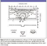
Empirical determination of repeller curves, or ion optics, is both tedious and somewhat unsatisfying, because the underlying rules of physics are known. Scientists match theory with experiment, and so directed studies of the ion optical properties of EI sources were first accomplished using the techniques of ray tracing, followed by calculations using infinitely thin electrodes, and then more realistic calculations that allowed for thick electrodes of various shapes, as in the actual EI source. Predictably, the power of the computer was brought to bear on the problem. Werner (7) modeled an EI source of the basic Nier type, factoring in such subtleties as the effect of the source magnet (no significant effect found), the penetration of the magnetic field of the sector into the source (no effect found), and the space charge (an effect that varied in proportion with the electron beam current and the number of ions produced). A particularly instructive result of such a computation is illustrated in Figure 2, adapted from Werner's original paper. The bottom part of the figure highlights (for a given set of operational conditions) the ionization region, the ion extraction region, and an overlap of the two, which represents the ion image.

Figure 2
We have concentrated here on the EI source modeled after the design of Nier, which has persevered for over 60 years in its basic form since its introduction in Nier's 1940 mass spectrometer. Specialized EI sources for specialized applications abound, and there are other eponymous EI sources such as the Brink ion source, which has itself been further modified (8). The modifications are designed to change the most basic parameters of the source operation — the ins of samples in a molecular beam, and the outs of ions generated from beam components as compared to background components. The patent literature, too, is replete with new sources and variants of existing sources. The patent claims are examined using the concept of ins and outs. Accurate measurements of ion abundances are still the focus in isotope ratio MS, and this applications group in particular has continued to advance the optimization of the design of the EI source.
For use in combined GC–MS instruments, the optimized EI source design is found at the balance point between physical characteristics [of size, cost, operational stability, response time] and ionization efficiency, which affects the overall sensitivity of the instrument. Once the basics of the source ins and outs and their underlying physical phenomena are understood, the genuine advances and the insights of the past and current generations of "gadgeteers" can be accurately assessed.
Kenneth L. Busch has overtightened more filament posts, cracked more spacer ceramics, and misaligned more repeller plates on various EI sources than he will ever admit in any public forum. He once designed an EI source for operation in a preparative mass spectrometer to be placed on the lunar surface, where the molecular losses due to pumping disappear from the balance of ins and outs. Reach the author at WyvernAssoc@yahoo.com
References
(1) W. Bleakney, Phys. Rev. 34, 157–160 (1929).
(2) John H. Reynolds, Biographical Memoirs, Volume 74 (National Academy of Sciences Press, Washington, D.C., 1998).
(3) A.O. Nier, Rev. Sci. Instrum. 11, 212–216 (1940).
(4) A.O. Nier, Rev. Sci. Instrum. 18, 398–411 (1947).
(5) J. De Later and M.D. Kurz, J. Mass Spectrom. 41, 847–854 (2006).
(6) T.D. Mark and A.W. Castleman, Jr., J. Phys. E Sci. Instrum. 13, 1121–1124 (1980).
(7) H.W. Werner, J. Phys. E Sci. Instrum. 7, 115–121 (1974).
(8) A. Amirav, A. Fialkov, and A. Gordin, Rev. Sci. Instrum. 73(8), 2872–2876 (2003).

Newsletter
Get essential updates on the latest spectroscopy technologies, regulatory standards, and best practices—subscribe today to Spectroscopy.
The Rising Role of Near-Infrared Spectroscopy in Biofuel Innovation
July 25th 2025A new bibliometric study published in Infrared Physics & Technology highlights the growing global impact of near-infrared (NIR) spectroscopy in biofuel research, revealing key trends, contributors, and future directions for advancing sustainable energy solutions.
Best of the Week: The Emerging Leader in Molecular Spectroscopy, Big Pharma’s Manufacturing Shift
July 25th 2025Top articles published this week include a feature article about big pharma’s investments in U.S.-based manufacturing, an article about the 2025 Emerging Leader in Molecular Spectroscopy Lingyan Shi, and some news items detailing the winners of the Coblentz Society’s student awards.