An Archaeometric Investigation into the Former Cataract House Hotel via Elemental Analysis
Over the course of the 19th century, the former Cataract House Hotel of Niagara Falls, New York, became one of the largest hotels in the region while also serving as an important “station” on the Underground Railroad. A park now occupies the area covering its demolished ruins. Since 2017, archaeological excavations of the site have taken place, led by the Anthropology Department at SUNY Buffalo. Although much is known about the overall design of the Cataract House Hotel, a clearer understanding of its construction phases, as well as its role in the Underground Railroad, could be determined from spectroscopic analysis in tandem with ongoing archaeological investigations. In 2022, in situ data collection was performed on plaster walls at the excavation site using a portable X-ray fluorescence (pXRF) instrument. These elemental data were used in conjunction with archaeological information to form conclusions regarding different construction phases of the hotel. Samples of plaster walls were also collected for further ex situ analyses with pXRF and portable laser-induced breakdown spectroscopy (pLIBS) in a laboratory setting. Future work will include data collection and analysis by additional spectroscopic methods of other artifacts collected at the site, such as pigment samples removed from an unearthed stone step.
Archaeometry, the combination of archaeological knowledge and quantitative measurements, offers empirical information regarding material origin, time of use or construction, and artifact conservation (1,2). Spectroscopic techniques have been shown to be valuable for characterizing archaeological materials and may provide chemical data that can be used to determine the relative ages of those features (2). In particular, elemental composition has been useful for gauging similarities among man-made materials because the makeup of building materials often can be associated with a specific time period, raw material sources, or both (2). Two elemental analysis techniques that are well suited for the analysis of precious archaeological samples are X-ray fluorescence (XRF) and laser-induced breakdown spectroscopy (LIBS). In XRF analysis, a sample is bombarded by X-rays, and the inner electrons of elements in the sample are removed, allowing outer electrons to fill their empty spaces and causing energy to be released in the form of a fluorescent X-ray photons. The energy of the photon released during this process is characteristic of the element being interrogated. In LIBS, a high-energy laser is fired at a sample, a portion of which becomes ablated. At the same time, the energy density yields a high-temperature plasma, which atomizes and excites ablated material. As the elements decay to a lower energy state, light is emitted at wavelengths characteristic of the elements in the sample.
These methods are appropriate for this task because they are minimally or non-destructive in the case of portable LIBS (pLIBS) and portable XRF (pXRF), respectively. Furthermore, both are available as handheld instruments and provide spectral information in a matter of seconds. Although each technique has its unique drawbacks, when they are used in conjunction, many of these limitations are minimized. For instance, XRF provides qualitative and quantitative information for larger elements, whereas low-Z elements, such as carbon and lithium, are not detected. Meanwhile, LIBS is capable of measuring a wider range of elements, including light elements. The nature of the LIBS method also allows for faster data collection in much more focused areas; for LIBS instruments with automated two-dimensional (2D) elemental mapping modes, the technique can easily be applied for evaluating the homogeneity of a sample (3). However, the LIBS spectra are much more data-rich and with higher background than XRF spectra and require more analysis before the elements present can be identified and quantified. The LIBS method is also quasi-destructive. Depending on the intensity of the laser, the number of pulses used to generate a spectrum, and the type of material being ablated, LIBS leaves small, sometimes visible craters on the surface of a sample. The X-rays emitted in XRF leave no marks on any sample during data collection.
The development and proliferation of handheld chemical analysis instruments, such as XRF, LIBS, Raman spectroscopy, and infrared (IR) spectroscopy, has greatly expanded archaeometric capabilities: samples can be rapidly analyzed in situ where access can be sharply limited (4). Obtaining this information in the field allows conclusions to be made during an excavation and inform the direction of a project, such as taking additional samples or deciding where next to dig. This work describes the use of two spectroscopic techniques, XRF and LIBS, to analyze architectural features uncovered during the recent archaeological excavation of the Cataract House, which was once a spacious, luxury hotel that served as an important node on the Underground Railroad (5).
Historical Background
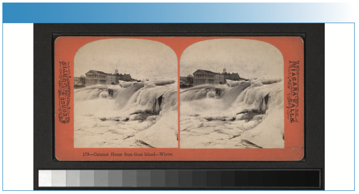
The Cataract House, located in Niagara Falls, New York, was built in 1825 and named for the nearby cataracts, or waterfalls. It began as a simple, three-story building, but by the mid-1840s, the Cataract House had undergone multiple expansions to become one of the largest hotels in the area (Figure 1) (6). The hotel drew tourists from across the United States, including slave-holding families from the southern United States, who were often accompanied by several enslaved persons (5).
FIGURE 1: Photograph of the Cataract House viewed from Goat Island in the Niagara River circa 1800s.
In the early 1840s, the Cataract House became the first hotel in Niagara Falls to employ African-Americans. Many of those hired were from southern states and may have been former slaves themselves (5). The hotel waitstaff made use of their positions as employees of the Cataract House and their proximity to Canada, where slavery had been abolished in 1834, to aid enslaved visitors in escaping to freedom (5). Their efforts resulted in several successful escapes, including in the cases of Cecelia Reynolds, Nancy Berry, and Patrick Sneed (5,7). Other less successful rescue attempts resulted in some negative press around the hotel, but the Underground Railroad “station” continued to operate out of the hotel through the end of the American Civil War in 1865 (5,7).
Between the 1890s and 1920s, the practice of hiring African-American waiters in American hotels and restaurants declined significantly, and the composition of the waitstaff at the Cataract House reflected this trend. The Cataract House continued operating as a hotel until 1945, when a fire destroyed the building (8,9). The ruined hotel was razed in 1946, with rubble from above-ground structures being used to fill in basements. Plans to rebuild were never realized and the site was covered. Today, Heritage Park occupies much of the former hotel’s location (8,9).
Recently, there has been renewed interest in the site, particularly with regard to the Underground Railroad activity. In 2015, preliminary work began to establish an excavation of the hotel (8,9). Digging started in 2017, and subsequent excavations were carried out in 2018 (7) and resumed in 2022. In 2017 and 2018, excavations took place along the former eastern wall of the hotel. In 2017, test units (TUs), which are one square meter trenches dug at areas of cultural interest and carefully documented, were dug, making up two larger trenches. Trench 1 contained TUs 1 and 2, Trench 2 contained TUs 3 and 4, and TU 5 was dug in a separate location (Figure 2). Both excavated trenches contained foundation-wall segments from the Cataract House, referred to as features, which are indicated in Figure 2. Feature 1 (Figure 3a), along the eastern side of Trench 1, is a semi-intact wall of an unknown substrate covered in plaster. Feature 2 (Figure 3b), on the northern side of Trench 2, is a semi-intact brick wall partially covered by a layer of plaster approximately 25 mm thick. In 2018, two additional trenches, Trench 3 containing TUs 6 and 7, and Trench 4 containing TUs 8 and 9, were dug and TU 10 was dug in a separate location (see Figure 2). These trenches lay to the east of the Cataract House’s easternmost wall. The spaces between these trenches and TU 5 were excavated, connecting them and turning TUs 5–9 into one large trench (8,9).
FIGURE 2: Field map of Test Unit (TU) 1–10 locations relative to Heritage Park landmarks. Each numbered TU is one square meter.
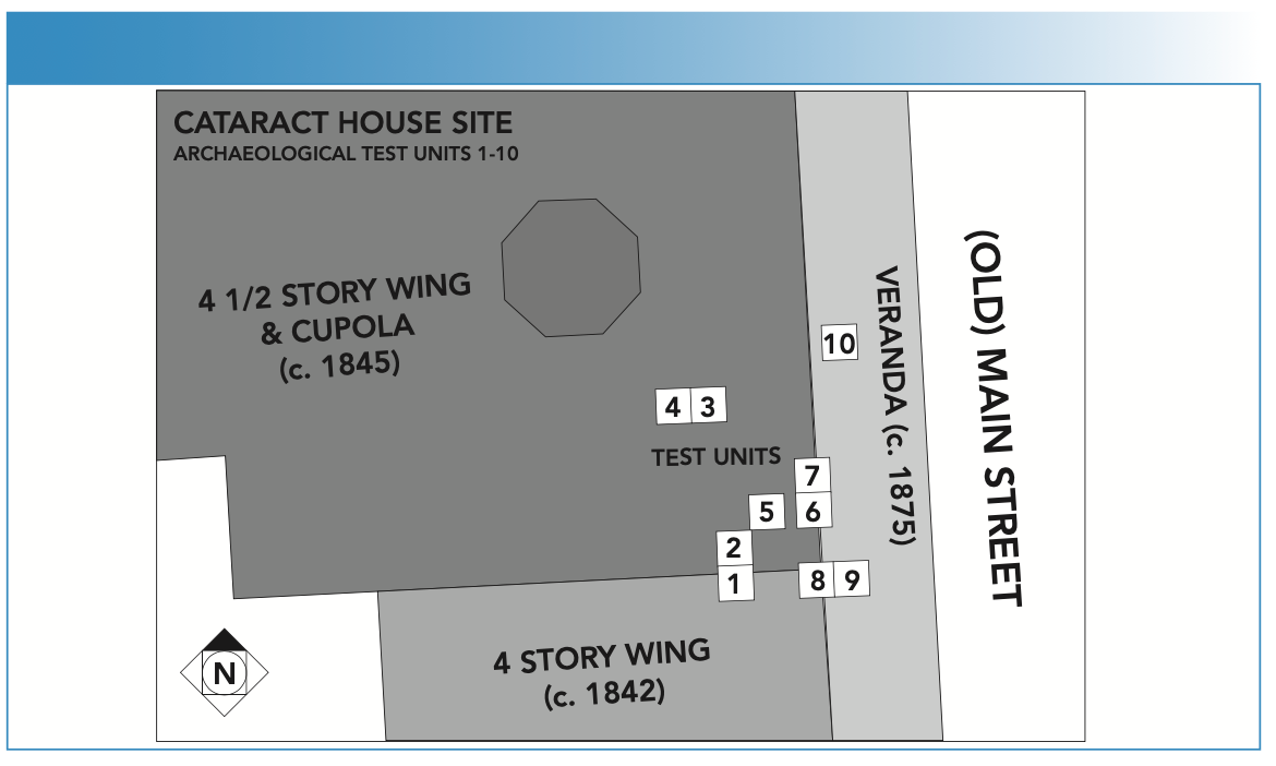
FIGURE 3: Photographs of Cataract House excavation sites of (a) Feature 1; (b) Feature 2; and (c) Feature 6 in May 2022.

In 2022, the focus of the excavations shifted toward the Cataract House’s kitchen, which is believed to have been the hub of local Underground Railroad activity. A new trench with four TUs (TUs 11–14) was dug several yards to the west of the original TUs. Site archaeologists used floor plans of the hotel to guide placement of the trench, approximating the location of the kitchen. Uncovered in this trench was Feature 6 (Figure 3c), an entryway flanked by plaster-covered wall remnants and lined on one side with wooden trim. To investigate the relative ages of these features and verify the accuracy of the hotel’s floor plan, atomic spectroscopic methods of analysis were employed.
Materials and Methods
In Situ Data Collection
In Summer 2022, a handheld XRF spectrometer was brought to the ongoing Cataract House excavation with the intention of determining whether construction features in separate trenches were of similar elemental composition and, therefore, likely built as part of the same expansion or modification. Because of the simplicity of the spectra produced and the possibility that the features being analyzed may be present in small quantities or considered precious, XRF was used for in situ data collection. Spectra were obtained with a Bruker Tracer III-V+ and fitted with a rhodium X-ray tube and a yellow filter with 305-μm aluminum and 25-μm titanium. The X-ray tube was operated at 40 keV of excitation energy and 3 μA of current. Each interrogate spot was analyzed with the head of the XRF instrument flush with the plaster surface for 120 s. Duplicate spectra were collected for select locations that were easily accessible based on geometric placement of the feature to be analyzed and instrument dimensions. The spectra were taken at points on the flattest parts of the plaster and were at least 15 cm apart from other points. A total of 39 spectra were obtained onsite from Features 1 and 6.
Ex Situ Data Collection
Excavation time constraints limited the number of sample spots and spectra obtained in situ. However, plaster wall samples were removed from Features 1, 2, and 6, and they were further analyzed in the laboratory. There were six samples collected from Feature 1, 11 samples collected from Feature 2, and seven samples collected from Feature 6. Additional pXRF data was collected with the same handheld XRF instrument and settings used for in situ analysis. For the laboratory measurements, the XRF instrument was mounted to a stand pointing upward and samples were placed on top of the device so that they were flush with its head. Replicate XRF spectra were obtained at every analyzed location.
To verify and support XRF results, pLIBS spectra were collected in the lab using the same set of samples from Features 1, 2, and 6. The pLIBS instrument used (the Z-300 LIBS Analyzer from SciAps) contained three charge-coupled device (CCD) spectrometers that together cover a wavelength range of 190–950 nm. The instrument utilizes an Nd:YAG laser at a wavelength of 1064 nm and a beam diameter of 100 μm. Spectra were obtained with a laser repetition rate of 50 Hz. Two cleaning shots and four data-collection shots were used to generate each spectrum. Additionally, argon gas (high purity grade from SciAps) was used to purge the sample area to increase the spectral intensity of most elemental lines; argon purge is an integrated feature of the instrument used for this study that delivers a few milliliters of gas per analysis. The samples were placed flush with the head of the instrument during analysis. Replicate analyses with LIBS were performed in certain locations to verify consistency in intensity and elements detected. These two instruments were used to collect data sets from the same set of samples from Features 1, 2, and 6. A total of 116 XRF and 60 LIBS spectra were collected in the laboratory, including replicate spectra.
Results and Discussion
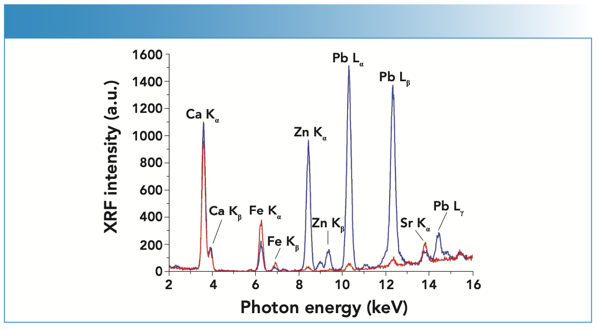
Spectra taken in situ with pXRF were visually distinguishable by the intensity of the peaks. For example, spectra taken from the left side of Feature 1 had much more intense lead peaks than those taken from the right side. To demonstrate, a plot of overlayed spectra from both sides of Feature 1 is shown in Figure 4. For a more definitive conclusion regarding the elemental compositions of the plasters from the site, principal component analysis (PCA) was performed with Unscrambler (version 10.1 from AspenTech) on three data sets extracted from the Cataract House plaster sampling frame to compare the data from the different features; each is discussed below. Prior to principal component analysis (PCA), a subset of data was evaluated using a Shapiro-Wilkes test and was found to be normal.
FIGURE 4: Overlay of two XRF spectra collected in situ from different parts of Feature 1 showing dramatic differences in lead and zinc content.
XRF Data Extracted In Situ from Features 1 and 6
The XRF spectra from the in-field analysis was used for PCA analysis. This fully cross-validated PCA was performed at the site during the 2022 excavation season so that results could be quickly relayed to archaeologists. This initial PCA was produced using the XRF spectral intensity data that was mean centered for each analysis. Feature 1 was found to contain two different elemental compositions that were distinct from the single composition across Feature 6. Both features contained calcium, iron, zinc, lead, and strontium, though in different abundances. Feature 1 was found to be composed of two distinct regions, marked by different levels of lead, according to XRF spectra collected from various points on the feature (Figure 4). The left side of Feature 1 displayed higher amounts of lead than the right side of Feature 1 or any part of Feature 6. A thin, grayish coating—possibly paint—on the left side of Feature 1 may explain the higher lead levels. Lead was commonly used in paint formulations, with its use in the United States peaking in the 19th century. Because Feature 1 was a continuous piece of plaster, the higher lead composition on the left side is not indicative of a second construction event. Feature 6 has more strontium than Feature 1 and it is believed that Feature 6 was built earlier, a hypothesis supported by the floor plans and records available. At the site, the XRF analysis data corroborated the theory that Features 1 and 6 were built during separate construction events.
XRF Data Extracted In Situ Combined with XRF Data Extracted Ex Situ
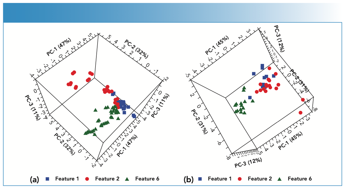
Fully cross-validated PCA with XRF results verified that the in situ and ex situ data could be combined while also comparing the elemental compositions of Features 1, 2, and 6. The score plot from this PCA can be seen in Figure 5a. Integrated peak areas from the XRF spectra were used to generate this PCA and data were normalized to one, mean centered, and divided by the standard deviation of the data set. Elements included in the analysis included calcium, iron, nickel, lead, strontium, and zinc. Spectra taken from the high-lead portion of Feature 1 were excluded from this PCA because the high lead levels were found to be from paint (see above) and not part of the plaster composition. Sodium peaks, which are often inflated by soil contamination, were similarly excluded from the PCA. The explained variance of PCs 1 through 3 was 90.1%. The majority variance in PC1 was because of contributions from nickel, iron, and calcium. Meanwhile, zinc and lead contributed the most to variance in PC2 and iron and calcium to variance in PC3. This combined in situ and ex situ data analysis supports the earlier finding that Features 1 and 6 are made of different plasters. The XRF data collected ex situ also included Feature 2, which was not analyzed at the site. According to PCA results, Feature 2 has a third distinct plaster composition, containing more zinc than the other two features.
FIGURE 5: Three-dimensional PCA score plots for (a) in situ and ex situ XRF; and (b) ex situ LIBS from analyses of wall samples from Features 1 (blue squares), 2 (red circles), and 6 (green triangles).
LIBS Data Extracted Ex Situ from Features 1, 2, and 6
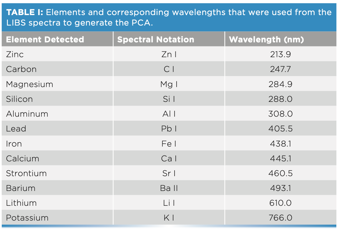
The fully cross-validated PCA from the laboratory-based LIBS analysis corroborated the XRF results and further distinguished the features with a larger subset of elements detected by LIBS, which included nickel, carbon, magnesium, silicon, aluminum, lead, iron, calcium, strontium, barium, lithium, and potassium. Prior to PCA analysis, the data were normalized to one, mean centered, and divided by the standard deviation of the data set. This PCA was performed using integrated peaks at selected wavelengths, which are listed in Table I, with the score plot shown in Figure 5b. The explained variance across PCs 1–3 was 87.0%, where aluminum, strontium, silicon, carbon, and magnesium contributed the most to the variance in PC1. Variances for PC2 and PC3 was because of mainly barium, calcium, and potassium, respectively. As anticipated, analysis of these data revealed additional elements that could not be detected by XRF, providing information that may link the plasters to specific time periods. For example, carbon is identified in all three plasters, but almost twice as much carbon is present in Features 1 and 2 as in Feature 6. Calcium carbonate (CaCO3), or lime, was commonly used in North American plasters before the 20th century (10). These LIBS results also showed that Feature 6, in addition to high amounts of strontium, contained more lithium than either Feature 1 or 2 and revealed a higher degree of heterogeneity in Feature 2’s plaster than in Features 1 and 6, with Feature 2 being speckled with small, high-barium areas. However, the separation between Features 1 and 2 can be seen more clearly in the score plot of the XRF data. Overall, the LIBS and XRF results both demonstrated that Features 1 and 2 share many elemental components and are more similar to each other than either is to Feature 6, suggesting that Features 1 and 2 were built at approximately the same time.
Conclusion
Combining archaeological knowledge with empirical evidence established using analytical techniques, we have been able to distinguish features at the Cataract House excavation from one another and relate them to specific time periods. Through the application of handheld instruments for collecting and analyzing data in situ, archaeological endeavors can be supported by rapidly supplying information during an ongoing investigation. Such information may inform the direction of an excavation, enhancing or modifying conclusions based solely on a feature’s appearance and surroundings. Future work will focus on analysis of other, non-plaster artifacts recovered from the site, allowing archaeologists to confidently develop a clearer picture of the Cataract House during its time as a “station” on the Underground Railroad.
References
(1) R.M. Ehrenreich, J. Archaeol. Method Theory 2(1), 1–6 (1995). DOI: 10.1007/BF02228433
(2) M.K. Donais, S. Wojtas, A. Desmond, B. Duncan, and D.B. George, Appl. Spectrosc. 66(9), 1005–1012 (2012). DOI: 10.1366/12-06616
(3) R.S. Harmon, D. Khashchevskaya, M. Morency, L.A. Owen, M. Jennings, J.R. Knott, et al, Molecules 26(17), 5200 (2021). DOI: 10.3390/molecules26175200
(4) M.K. Donais and P. Vandenabeele, in Portable Spectroscopy and Spectrometry 2: Applications, R. Crocombe, P. O’Leary, and B. Kammrath, Eds. (Wiley & Sons Ltd., West Sussex, United Kingdom, 2021), pp. 523–544.
(5) J. Wellman, “Niagara Falls Underground Railroad Heritage Area Management Plan, Appendix C: Survey of Sites Relating to the Underground Railroad, Abolitionism, and African American Life in Niagara Falls and Surrounding Area, 1820–1880”, for Companies and Niagara Falls Underground Railroad Heritage Area Commission, April 2012.
(6) F.C. Pierce, Descendants of John Whitney, Who Came from London, England, to Watertown, Massachusetts, in 1635 (Chicago, IL, 1895).
(7) K. Whalen, R. Austin, N. Montague, and D.J. Perrelli, Reports of the Archaeological Survey 50(31) (2018).
(8) Niagara Falls Gazette, Famed Cataract House Turned Into Mass of Charred Ruins. (accessed October, 1945).
(9) Niagara Falls Gazette, Historic Hotel to be Rebuilt (accessed October, 1945).
(10) R.F. Austin, “Analysis of Wall Plasters at the Cataract House Site, City of Niagara Falls, Niagara County, New York”, State University of New York at Buffalo, 2022.
Magdalena Jackson and Jacob T. Shelley are with the Department of Chemistry and Chemical Biology at Rensselaer Polytechnic Institute, in Troy, New York. Douglas Perrelli is with the Department of Anthropology, at the State University of New York at Buffalo, in Buffalo, New York. Mary Kate Donais is with the Chemistry Department at St. Anselm College, in Manchester, New Hampshire. Direct correspondence to: MDonais@Anselm.edu. ●

LIBS Illuminates the Hidden Health Risks of Indoor Welding and Soldering
April 23rd 2025A new dual-spectroscopy approach reveals real-time pollution threats in indoor workspaces. Chinese researchers have pioneered the use of laser-induced breakdown spectroscopy (LIBS) and aerosol mass spectrometry to uncover and monitor harmful heavy metal and dust emissions from soldering and welding in real-time. These complementary tools offer a fast, accurate means to evaluate air quality threats in industrial and indoor environments—where people spend most of their time.
Laser Ablation Molecular Isotopic Spectrometry: A New Dimension of LIBS
July 5th 2012Part of a new podcast series presented in collaboration with the Federation of Analytical Chemistry and Spectroscopy Societies (FACSS), in connection with SciX 2012 — the Great Scientific Exchange, the North American conference (39th Annual) of FACSS.
AI Shakes Up Spectroscopy as New Tools Reveal the Secret Life of Molecules
April 14th 2025A leading-edge review led by researchers at Oak Ridge National Laboratory and MIT explores how artificial intelligence is revolutionizing the study of molecular vibrations and phonon dynamics. From infrared and Raman spectroscopy to neutron and X-ray scattering, AI is transforming how scientists interpret vibrational spectra and predict material behaviors.
Advancing Corrosion Resistance in Additively Manufactured Titanium Alloys Through Heat Treatment
April 7th 2025Researchers have demonstrated that heat treatment significantly enhances the corrosion resistance of additively manufactured TC4 titanium alloy by transforming its microstructure, offering valuable insights for aerospace applications.