Analytical Biology: An Emerging Discipline for the Future
The field of analytical chemistry is well established. It’s time to develop the field of analytical biology—an emerging discipline that blends various research fields to provide a holistic view of biological phenomena.
Interdisciplinary research at the intersection of physics, biology, and chemistry is attracting increasing interest and generating many innovative ideas. One example is the application of Raman spectroscopy to biological and medical research. Although the Raman effect was applied to chemical identification shortly after its discovery, it took nearly six decades for the first Raman spectrum of a live cell to be measured, because of the inherently low efficiency of Raman scattering and the complexity of biological samples (1,2). The later advances in laser technologies, such as solid-state ultrashort-pulse lasers (3,4), high-sensitivity detectors (5), and high-quality optical filters (6) have brought Raman spectroscopy into the realm of cell biology.
Raman spectroscopy offers unique capabilities, such as label-free and comprehensive detection of biological molecules in cells and tissues. Unlike infrared spectroscopy, Raman spectroscopy does not strongly interact with water and can provide molecular information under physiological conditions without damaging samples. Raman spectroscopy is used for a broad range of measurements ranging from general Raman spectral analysis to high-resolution Raman imaging with subcellular sensitivity. An example can be seen in Figure 1, which shows high-resolution Raman images of hepatocytes with and without the treatment of the drug rifampicin (7). In particular, high-speed Raman spectroscopic imaging, coupled with microscopy, has gained significant interest in the life sciences (8–10). Raman microscopy faithfully translates molecular information from biological samples, such as DNA, proteins, carotenoids, and lipids—and their intracellular balance—into spectra that can provide information about cells, tissues, and species. This comprehensive analysis of intracellular molecules can be employed to detect cell apoptosis and differentiation and to diagnose diseases such as cancer that have not been fully explained using traditional biological methods. This capability can pave the way for new approaches in cell and tissue analysis in medical diagnostics, regenerative medicine, and drug development.
FIGURE 1: Raman images of different cellular components in hepatocytes (a) without and (b) with drug (rifampicin) treatment.

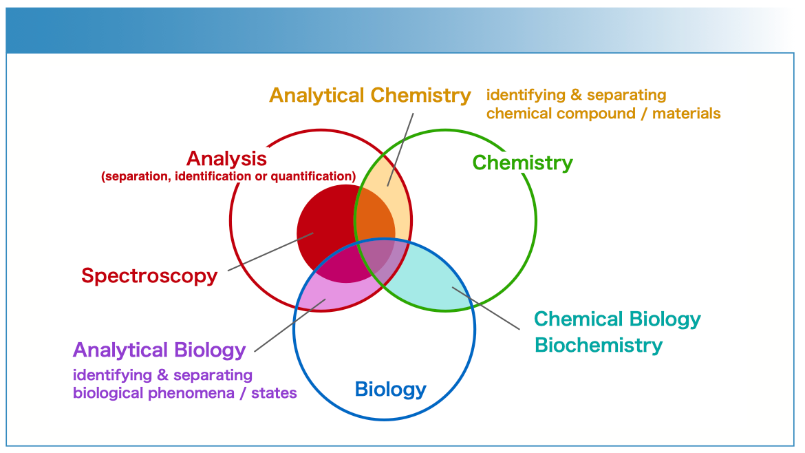
In many applications, Raman microscopy is used to distinguish biological phenomena at the levels of biomolecules, cells, tissues, and even organisms. This approach is somewhat akin to analytical chemistry, which aims to analyze chemicals (that is to separate, identify, or quantify the chemistry of a sample in sufficient detail in order to gain a better understanding of its nature, structure, or function), but with this use of Raman spectroscopy and microscopy one aims to also analyze biological phenomena. From this perspective, these studies are shaping a new discipline that might be referred to as analytical biology. Although this term has rarely been used as a research field, its roots can be traced back to a 1950 book titled Analytical Biology by G. Sommerhoff (11) and a 1966 Nature article by I.J. Good (12). However, these early works focused more on mathematical and theoretical biology rather than the analysis of biological materials or phenomena. From our viewpoint, as shown in Figure 2, the field analytical biology would stand at the intersection of analysis and biology, indicating a research field that includes studies, instruments, and methods aimed at analyzing biological substances and phenomena.
FIGURE 2: The overlap of related disciplines: analytical biology stands between analysis and biology, just as analytical chemistry stands between analysis and chemistry, and as chemical biology stands between chemistry and biology.

The strategy in analytical biology is different from typical biological methods that are mainly based on reductionism. Raman microscopy offers a holistic and unbiased approach to analyzing multiple biological molecules simultaneously, allowing for non-targeted analysis. This phenotypic and high-content strategy can complement target-oriented approaches. The concept of analytical biology also aligns with other state-of-the-art and emerging techniques, particularly omics approaches such as genomics, transcriptomics, proteomics, and metabolomics. With this strategy it becomes possible to first obtain an overarching view of a given biological phenomenon and then proceed to elucidate the underlying mechanisms using target-oriented methodologies. This approach can be combined with chemical biology, which includes studies using multimodal analysis with targeted methods, such as those using Raman tags and probes (13). The relationship between these approaches can be seen in Figure 2.
Because biological phenomena are not solely associated with a single protein, holistic approaches like omics techniques have garnered interest for their potential to provide a comprehensive view of biological phenomena instead of focusing on a single or limited number of biomolecules. Unbiased and phenotypic screening has been found to be valuable for gaining a comprehensive understanding of complex phenomena such as multifaceted and multilevel hepatotoxicity. Analytical biology represents an emerging discipline that blends various research fields to provide a holistic view of biological phenomena. By incorporating spectroscopy and other cutting-edge techniques, analytical biology allows scientists to explore new aspects of biology and uncover deeper insights into the mechanisms behind complex biological processes. As this field continues to evolve, it promises to enrich our understanding of the intricacies of life and open new avenues for discovery in medicine, drug development, and related areas.
We came up with this idea of analytical biology when we encountered the difficulties in choosing a category of research on submitting our papers to journals. Throughout history, there are numerous examples of new technologies paving the way for new research fields. We are excited by the prospect that studies in advanced spectroscopy may stand at the forefront of this exciting and emerging new field of analytical biology and all that it promises.
Menglu Li is an Associate Investigator at the Shenzhen Medical Academy of Research and Translation. Katsumasa Fujita is a Professor of Applied Physics at Osaka University. Direct correspondence to: fujita@ap.eng.osaka-u.ac.jp ●

AI-Powered SERS Spectroscopy Breakthrough Boosts Safety of Medicinal Food Products
April 16th 2025A new deep learning-enhanced spectroscopic platform—SERSome—developed by researchers in China and Finland, identifies medicinal and edible homologs (MEHs) with 98% accuracy. This innovation could revolutionize safety and quality control in the growing MEH market.
New Raman Spectroscopy Method Enhances Real-Time Monitoring Across Fermentation Processes
April 15th 2025Researchers at Delft University of Technology have developed a novel method using single compound spectra to enhance the transferability and accuracy of Raman spectroscopy models for real-time fermentation monitoring.
Nanometer-Scale Studies Using Tip Enhanced Raman Spectroscopy
February 8th 2013Volker Deckert, the winner of the 2013 Charles Mann Award, is advancing the use of tip enhanced Raman spectroscopy (TERS) to push the lateral resolution of vibrational spectroscopy well below the Abbe limit, to achieve single-molecule sensitivity. Because the tip can be moved with sub-nanometer precision, structural information with unmatched spatial resolution can be achieved without the need of specific labels.