The C=O Bond, Part IV: Acid Anhydrides
Spectroscopy
Acid anhydrides are unique in that they have two carbonyl groups in them. The intensity and position of their IR peaks can be used to determine which of the four types of anhydride exist in a sample.
There are a number of carboxylic acid derivatives that, like their parent functional group, are easy to detect by infrared (IR) spectroscopy. Acid anhydrides are one of these. These molecules are unique in that they have two carbonyl groups in them, and the intensity and position of their IR peaks can be used to determine which of the four types of anhydride exist in a sample. Other diagnostic peaks for acid anhydrides are also discussed.
So far in our investigation of the infrared (IR) spectroscopy of the carbonyl or C=O functional group, we have looked at ketones, aldehydes, and carboxylic acids. One thing they had in common was a single C=O bond and hence a single C=O stretching peak. There exists a functional group called acid anhydrides that contain two carbonyl groups. They are made by reacting two carboxylic acid molecules to generate the anhydride and a molecule of water as shown in Figure 1.

Figure 1: The reaction of two carboxylic acid molecules to give an acid anhydride and water.
The "R" moieties represent carbon atoms. The term anhydride means "no water" and is appropriate because of the water molecule that is split off during this reaction. Anhydrides are named after the carboxylic acids they are made from. For example, two molecules of acetic acid react to give acetic anhydride. Note that the anhydride functional group consists of two carbonyl groups connected by a single oxygen atom. As a result of this unique structure, the IR spectra of acid anhydrides contain not one but two C=O stretching peaks, and a C-O stretch. The anhydride group is somewhat reactive, so these molecules are often used as polymeric crosslinkers or as chemical intermediates.
The nature of the R-groups attached to the anhydride framework determine the type of anhydride. If the two attached carbons are saturated so is the anhydride, whereas if one or both of the anhydrides is unsaturated then the anhydride is said to be unsaturated.
Unlike some functional groups, it seems the most common anhydrides are cyclic. Examples of the structures of two common cyclic anhydrides, succinic anhydride and phthalic anhydride, are shown in Figure 2.
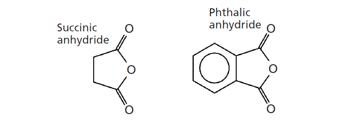
Figure 2: The structures of succinic anhydride, a saturated cyclic anhydride, and phthalic anhydride, an unsaturated cyclic anhydride.
In addition to whether an anhydride is saturated or unsaturated, whether its substituents are in a ring or not also matters. Thus, it takes two adjectives to describe an anhydride and there are four types: noncyclic saturated, noncyclic unsaturated, cyclic saturated, and cyclic unsaturated. Succinic anhydride is a saturated cyclic anhydride, since there are methylene groups attached to the carbonyls in the anhydride functionality. On the other hand, phthalic anhydride is a cyclic unsaturated anhydride because there are aromatic carbons attached to the carbonyls in the anhydride functionality. An example of a noncyclic saturated anhydride would be acetic anhydride, where the two R-groups attached to the anhydride carbonyls are CH3 groups. Fortunately, we can distinguish between the four distinct types of anhydrides using IR spectroscopy as we will see below.
The Infrared Spectroscopy of Acid Anhydrides
The two carbonyl groups in acid anhydrides give rise to two carbonyl stretching peaks. The vibrations involved are a symmetric C=O stretch where the two carbonyl groups stretch in phase with each other, and an asymmetric C=O stretch where the two carbonyl groups stretch out of phase with each other as shown in Figure 3.

Figure 3: The symmetric (left) and asymmetric (right) carbonyl stretches of the acid anhydride functional group.
Noncyclic Anhydrides
The IR spectrum of a noncyclic saturated anhydride, valeric anhydride, is shown in Figure 4.
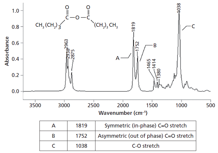
Figure 4: The IR spectrum of valeric anhydride (C10H18O3).
The double carbonyl stretching peaks for valeric anhydride fall at 1819 and 1752 cm-1 (going forward assume all peak positions are in cm-1) and are labeled A and B in Figure 4. The first unusual thing about this pair of peaks is that they are at higher wavenumbers than the carbonyl stretches. The second unusual thing is that the symmetric stretch is at a higher wavenumber than the asymmetric stretch we have seen so far. In general, and for reasons too lengthy to go into here (2), for functional groups that have asymmetric and symmetric stretches the asymmetric stretch is typically higher in wavenumber. In general, for noncyclic saturated anhydrides the symmetric C=O stretch falls at 1820 ± 5 whereas the asymmetric stretch falls at 1750 ± 5. For noncyclic unsaturated acid anhydrides these peaks fall at 1775 ± 5 and 1720 ± 5, respectively. Note that based on their C=O stretching peak positions and the narrowness of the range where the peaks fall, it is easy to distinguish between saturated and unsaturated noncyclic anhydrides.
Anhydrides of course have a C-O stretch, and based on previous columns (3,4) we expect this bond to have a stretching peak between 1300 and 1000. Valeric anhydride follows this rule with its most intense peak being a C-O stretch at 1038, labeled C in Figure 4. In general, for noncyclic anhydrides this peak falls from 1060 to 1035 and is in the same range whether the anhydride is saturated or unsaturated.
Note in Figure 4 that the high-wavenumber C=O stretch is more intense than the low-wavenumber C=O stretch. This intensity ratio is typical of all noncyclic anhydrides and can be used to distinguish the noncyclic from cyclic forms. Table I summarizes the useful group wavenumbers for noncyclic acid anhydrides. In Table I, note that for both types of noncyclic anhydride the higher-wavenumber symmetric C=O is more intense.

A double carbonyl peak with one or both features at a relatively high wavenumber for a C=O stretch is a strong indication of an anhydride being present in a sample. If the higher-wavenumber peak is stronger it is noncyclic. Then use the positions of the two C=O stretches and Table I to determine whether the noncyclic anhydride is saturated or unsaturated.
Cyclic Anhydrides
The IR spectrum of an unsaturated cyclic anhydride, maleic anhydride, is shown in Figure 5. This anhydride is unsaturated because there is a C=C bond containing unsaturated carbons in the ring attached directly to the carbonyl carbons of the two C=O groups.
The symmetric carbonyl stretching peak for maleic anhydride is at 1853, while the asymmetric stretch is at 1780. For unsaturated cyclic anhydrides these two peaks typically fall from 1860 to 1840 and 1780 to 1760, respectively. For saturated cyclic anhydrides, such as succinic anhydride whose structure is seen in Figure 2, these peaks fall at 1870–1845 and 1800–1775.
Note in Figure 5 that the high-wavenumber symmetric C=O stretching peak is weaker than the lower-wavenumber asymmetric C=O stretching peak. This pattern is opposite that for noncyclic anhydrides as seen above. Thus, the peak intensity ratio of the two anhydride C=O stretching peaks can be used to determine whether an anhydride is noncyclic or cyclic. For noncyclic anhydrides the higher-wavenumber symmetric C=O stretch is more intense, while for cyclic anhydrides the lower-wavenumber asymmetric stretch is more intense. Again, a double carbonyl stretch with one or both peaks at a relatively high wavenumber is a strong indication of an anhydride. If the lower-wavenumber peak is more intense it is cyclic. Then use the position of the two C=O stretches and Table II to determine whether the anhydride is cyclic saturated or cyclic unsaturated.
Like noncyclic anhydrides, cyclic anhydrides have a C-O bond and hence a C-O stretch. It falls anywhere from 1300 to 1000. In the spectrum of maleic anhydride in Figure 5 this peak falls at 1058 and is labeled C. Additionally, cyclic anhydrides have a strong C-C stretching peak from 960 to 880, in Figure 5 this peak falls at 894.
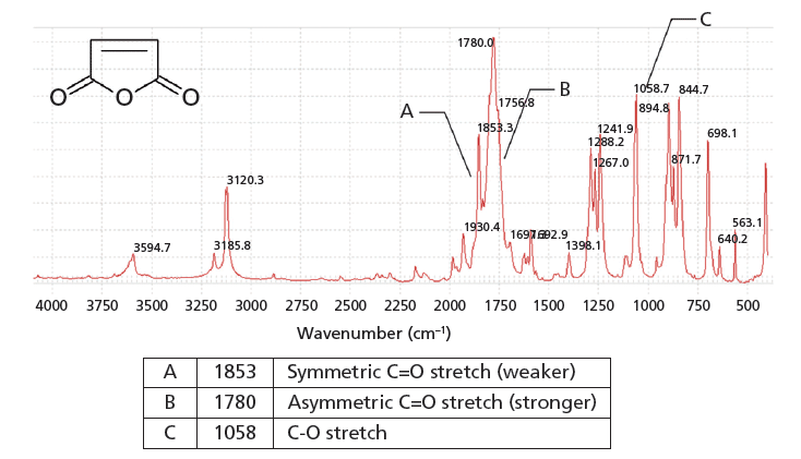
Figure 5: The IR spectrum of maleic anhydride (C4H2O3).
Table II lists the important group wavenumbers for cyclic acid anhydrides. Note that it states in Table II that for both types of cyclic anhydride the lower-wavenumber C=O stretch is more intense.
In summary there are four kinds of acid anhydride: saturated noncyclic, unsaturated noncyclic, saturated cyclic, and unsaturated cyclic. A double carbonyl stretch, with one or more of the peaks at relatively high wavenumber for a C=O stretch is a strong indication of an acid anhydride. Next, look at the intensity ratio of the two carbonyl stretching peaks. If the higher-wavenumber peak is more intense the anhydride is noncyclic. If the lower-wavenumber peak is more intense the anhydride is cyclic. Once this is known, then use the peak positions of these peaks and either Table I (noncyclic) or Table II (cyclic) to determine whether the anhydride is saturated or unsaturated. In this way, you can distinguish the four types of acid anhydrides from each other using IR spectroscopy.

Conclusion
Acid anhydrides are made by reacting two carboxylic acid molecules with each other. One of the products is water, hence the name anhydride. Acid anhydrides come in four varieties: saturated noncyclic, unsaturated noncyclic, saturated cyclic, and unsaturated cyclic. Their most significant spectral feature is a double carbonyl stretch because of the symmetric and asymmetric stretching of the two carbonyl groups, one of which sometimes occurs above 1800. For noncyclic anhydrides the higher-wavenumber C=O stretching peak is more intense. For cyclic anhydrides the lower-wavenumber C=O stretching peak is more intense. The positions of these two peaks can then be used to determine whether the anhydride is saturated or unsaturated.
References
(1) B.C. Smith, Spectroscopy 33(1), 14–20 (2018).
(2) B.C. Smith, Infrared Spectral Interpretation: A Systematic Approach (CRC Press, Boca Raton, Florida, 1999).
(3) B.C. Smith, Spectroscopy 32(1), 14–21 (2017).
(4) B.C. Smith, Spectroscopy 32(9), 31–36 (2017).
(5) B.C. Smith, Spectroscopy 30(4), 18–23 (2015).
(6) B.C. Smith, Spectroscopy 30(9), 40–45 (2015).
Brian C. Smith, PhD

Brian C. Smith, PhD, has more than three decades of experience as an infrared spectroscopist. He has published numerous peer reviewed papers and has written three books on the subject: Fundamentals of FTIR and Infrared Spectral Interpretation, both published by CRC Press, and Quantitative Spectroscopy: Theory and Practice published by Elsevier. As a spectroscopic trainer, he has helped thousands of people around the world improve their infrared analyses. He earned his PhD in physical chemistry from Dartmouth College. He can be reached at: SpectroscopyEdit@UBM.com

AI Shakes Up Spectroscopy as New Tools Reveal the Secret Life of Molecules
April 14th 2025A leading-edge review led by researchers at Oak Ridge National Laboratory and MIT explores how artificial intelligence is revolutionizing the study of molecular vibrations and phonon dynamics. From infrared and Raman spectroscopy to neutron and X-ray scattering, AI is transforming how scientists interpret vibrational spectra and predict material behaviors.
Real-Time Battery Health Tracking Using Fiber-Optic Sensors
April 9th 2025A new study by researchers from Palo Alto Research Center (PARC, a Xerox Company) and LG Chem Power presents a novel method for real-time battery monitoring using embedded fiber-optic sensors. This approach enhances state-of-charge (SOC) and state-of-health (SOH) estimations, potentially improving the efficiency and lifespan of lithium-ion batteries in electric vehicles (xEVs).
New Study Provides Insights into Chiral Smectic Phases
March 31st 2025Researchers from the Institute of Nuclear Physics Polish Academy of Sciences have unveiled new insights into the molecular arrangement of the 7HH6 compound’s smectic phases using X-ray diffraction (XRD) and infrared (IR) spectroscopy.