Fourier Transform Infrared Microspectroscopy Reveals Insights into Ovarian Cancerous Tissues in Paraffin and Deparaffinized Samples
The research highlights the significance of tissue preparation methods and demonstrates the feasibility of accurate classification, providing valuable insights into the biomolecular composition of ovarian cancer tissues.
Ovarian cancer is a highly lethal disease often diagnosed at an advanced stage, making effective treatment challenging. However, a new study conducted by Patryk Stec at the University of Science and Technology in Krakow, Poland, and published in the journal Spectrochimica Acta Part A: Molecular and Biomolecular Spectroscopy, explores the potential of Fourier transform infrared microspectroscopy to aid in the diagnosis of ovarian cancer (1). The research investigates the analysis of ovarian cancerous tissues in paraffin-embedded and deparaffinized samples using infrared microspectroscopy, shedding light on the impact of tissue preparation methods on biomolecular composition and the feasibility of accurate classification.
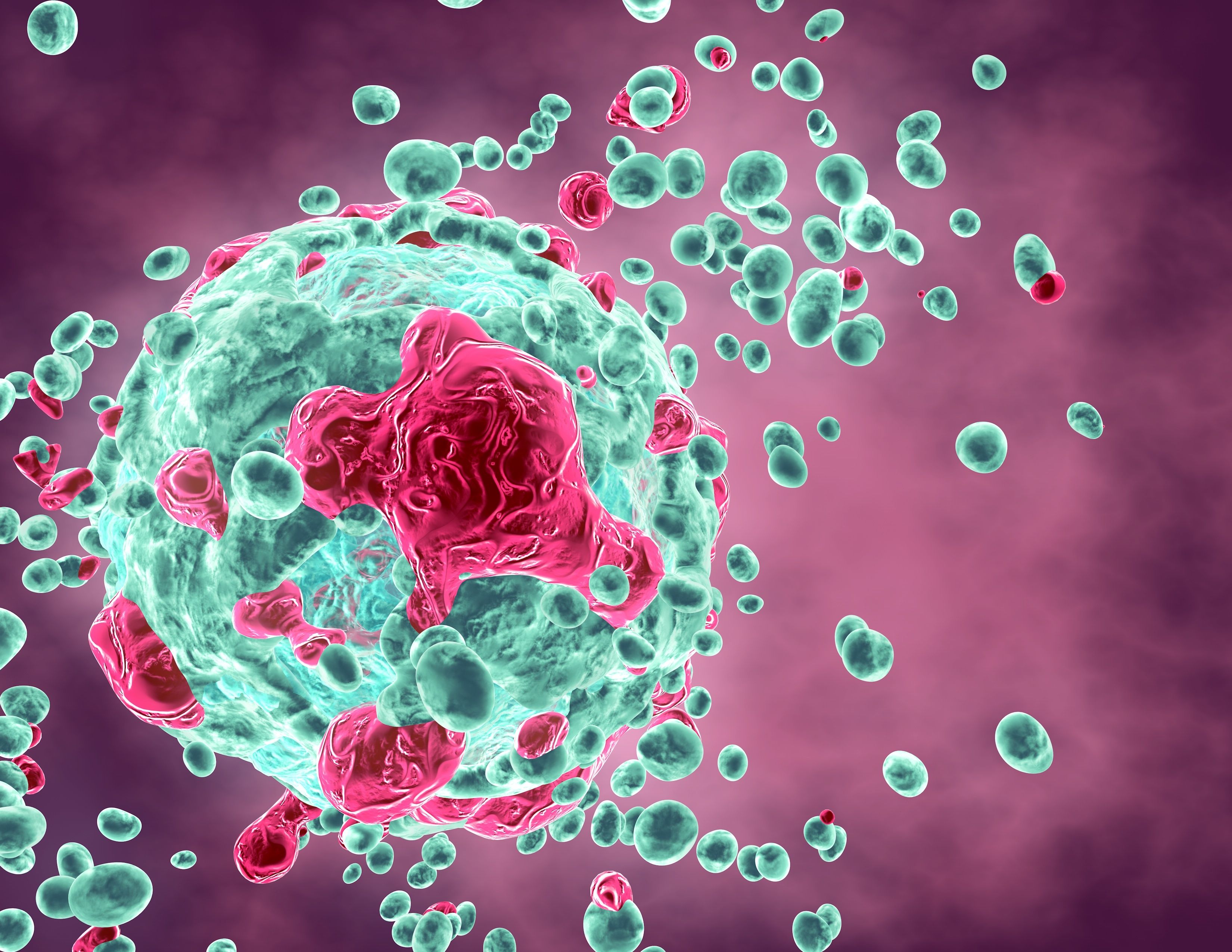
Cancerous tissue destroying human cell, tumor concept illustration. | Image Credit: © picture-waterfall - stock.adobe.com

Infrared microspectroscopy provides a valuable tool for examining the biomolecular composition of tissues, offering insights into the molecular changes associated with diseases such as cancer. However, the presence of paraffin in archival paraffin-embedded tissue samples can hinder accurate analysis due to its strong absorption of infrared radiation. To overcome this challenge, the researchers employed a deparaffinization process to remove the paraffin prior to analysis.
The study involved analyzing archival paraffin-embedded preparations of ovarian tissues, including tumor and control samples. Fourier transform infrared (FT-IR) measurements were performed on samples both in paraffin and after deparaffinization. The researchers calculated ratios of integrated peaks and massifs within the obtained spectra for different types of tissues, including diseased and healthy controls.
Statistical analysis revealed significant differences in the calculated ratios between various types of tissues. Moreover, the researchers employed random forest models, demonstrating that both paraffin-embedded and deparaffinized samples contained sufficient information for reliable tissue classification. Further analysis identified the most important features for distinguishing between different sample types.
The study highlighted that the deparaffinization process induces changes in the biomolecular composition of the analyzed tissues. Despite these alterations, the classification of tissues based on FT-IR measurements remained feasible. Importantly, the research contributes to enhancing the understanding of ovarian cancer and underscores the potential of infrared microspectroscopy as a valuable tool for its diagnosis and classification.
These findings pave the way for future advancements in the application of FT-IR microspectroscopy in the analysis of cancerous tissues, offering hope for improved diagnostic accuracy and personalized treatment strategies for ovarian cancer patients.
Reference
(1) Stec, P.; Dudala, J.; Wandzilak, A.; Wrobel, P.; Chmura, L.; Szczerbowska-Boruchowska, M. Fourier transform infrared microspectroscopy analysis of ovarian cancerous tissues in paraffin and deparaffinized tissue samples. Spectrochimica Acta Part A: Mol. Biomol. Spectrosc. 2023, 297, 122717. DOI: 10.1016/j.saa.2023.122717
New Study Reveals Insights into Phenol’s Behavior in Ice
April 16th 2025A new study published in Spectrochimica Acta Part A by Dominik Heger and colleagues at Masaryk University reveals that phenol's photophysical properties change significantly when frozen, potentially enabling its breakdown by sunlight in icy environments.
AI Shakes Up Spectroscopy as New Tools Reveal the Secret Life of Molecules
April 14th 2025A leading-edge review led by researchers at Oak Ridge National Laboratory and MIT explores how artificial intelligence is revolutionizing the study of molecular vibrations and phonon dynamics. From infrared and Raman spectroscopy to neutron and X-ray scattering, AI is transforming how scientists interpret vibrational spectra and predict material behaviors.
Real-Time Battery Health Tracking Using Fiber-Optic Sensors
April 9th 2025A new study by researchers from Palo Alto Research Center (PARC, a Xerox Company) and LG Chem Power presents a novel method for real-time battery monitoring using embedded fiber-optic sensors. This approach enhances state-of-charge (SOC) and state-of-health (SOH) estimations, potentially improving the efficiency and lifespan of lithium-ion batteries in electric vehicles (xEVs).