Frontiers of NIR Imaging
After a brief introduction to the technique, this review will discuss the state-of-the-art NIR imaging instrumentation and its applications, including applications of an ordinary NIR imaging system to solvent diffusion into a polymer. Other imaging applications discussed include the use of a portable NIR imaging system in the pharmaceutical industry, high-speed and wide-area monitoring of polymers, and imaging for fish embryos development research. Also discussed is an imaging-type two-dimensional Fourier spectroscopy (ITFS) system and its application to medaka (Japanese rice fish, Oryzias latipes) egg development. Finally, general perspectives on NIR imaging will be provided.
Near-infrared (NIR) imaging has developed significantly for the last two decades (1–5). It enables the obtaining of information about the spatial distribution of chemical components, even for thick materials. It is an in situ and non-destructive analysis method that allows non-contact analysis and also analysis using optical fibers. Moreover, by using NIR imaging, it is possible to probe an aqueous or a solvent dispersion much more easily than IR imaging. NIR imaging has been applied to various fields, such as investigations of component and crystallinity distributions in polymers (6–11), quality evaluation of foods (12–16), measurements of pharmaceutical tablets (17–20), explorations of diffusion process of solvents in drugs and polymers (6,18), blend process monitoring (17), and studies of the distributions of biological components and water species in eggs (21–26).
In this review, we report recent progress of NIR imaging, demonstrating six examples of the applications of NIR imaging from our studies:
1) Time-dependent NIR imaging study of the diffusion process of butanol (OD) into polyamide 11 (6).
2) Blend process monitoring (17).
3) Investigation of inhomogeneity of a pharmaceutical tablet during a grinding process (19).
4) NIR imaging of changes in hydrogen bonds of water during the process of medaka embryonic development (26).
5) A high speed and wide-area monitoring NIR imaging system with a novel NIR camera (Compovision) (24).
6) Non-staining blood flow imaging using optical interface due to Doppler shift in developing fish egg embryos (25).
Temperature-Dependent NIR Imaging Study of the Diffusion Process of Butanol (OD) into Polyamide 11
Unger and colleagues carried out a time-resolved NIR imaging study of the diffusion process of butanol (OD) into polyamide 11 (PA11) to see if there are any significant differences of the diffusion rate below and above the glass transition temperature (37 °C) of PA11 (6). The mobile NH protons of PA11 are subjected to hydrogen-deuterium (H/D) exchange with the diffusant butanol (OD), which is directly linked to the diffusion of butanol (OD) into PA11. Therefore, NIR imaging can be used to probe the diffusion process as a function of time. NIR spectra in the 5050–4750 cm-1 region of PA11 film before and after deuteration for 5 h 35 min at 50 °C are shown in Figure 1 (6). It is noted in Figure 1 that the intensity decrease in the 4965 cm-1 band derived from the combination of ν(NH) and amide I is much less than that in the 4875 cm-1 band, due to the combination of ν(NH) and amide II and because the former band is increasingly superimposed by the evolving second overtone band of ND group with progressing deuteration. Therefore, we used the 4875 cm-1 band to monitor time-dependent deuteration progress. To develop NIR contour plots, the 4875 cm-1 band was integrated into the 4935–4800 cm-1 region with a baseline from 5030 to 4800 cm-1.
FIGURE 1: NIR spectra of PA 11 before (- - -) and after 5 h 35 min (—) deuteration with butanol (OD) at 50 °C. Reproduced from reference (6) with permission.
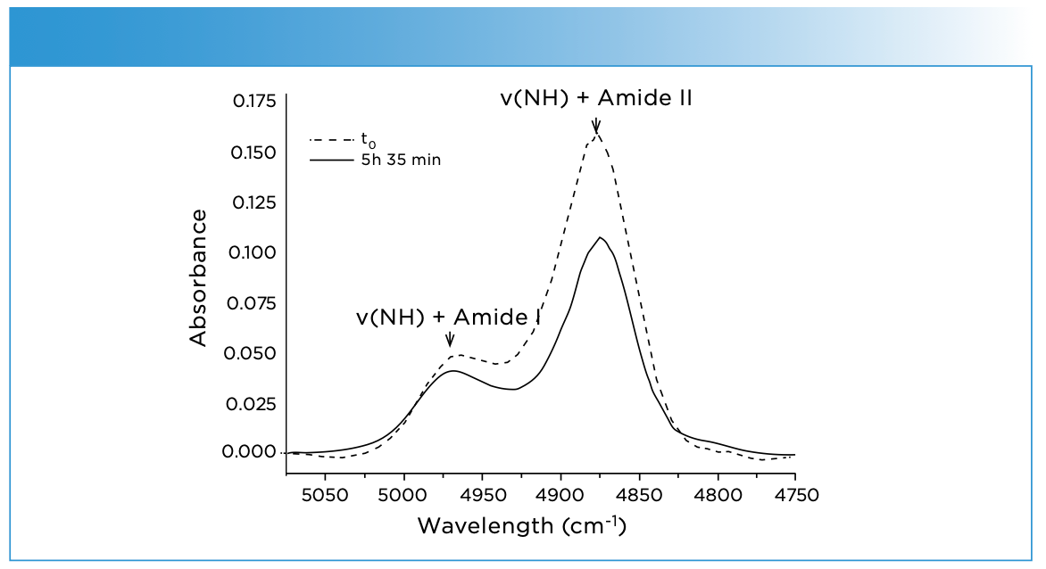
Figure 2 shows NIR images constructed by the integrated 4875 cm-1 band for (a) the deuteration times t0; (b) 10 h 15 min at 25 °C; (c) t0; and (d) 1 h 45 min at 50 °C (6). Note that the blue color on the left side of each image indicates the butanol (OD), while the light pink part on the right side represents the PA11 film. At the starting point (t = 0 min) no butanol (OD) diffused into the polymer film. Thus, the PA11 part of the image was homogeneously lightly pink colored (Figure 2b)). After 10 h 15 min at 25 °C, the deuteration front had moved into the PA11 area, and the 4875 cm-1 band decreased with the NH/ND exchange, yielding a yellow color (Figure 2b). In contrast to the deuteration progress at 25 °C, at 50 °C the front of the NH/ND exchange already reached approximately the same position at about 1 h 45 min (Figure 2d). It was found from Figure 2 reveals that a significant difference in the diffusion rate occurs below and above the glass transition temperature of PA11 (6).
FIGURE 2: NIR images based on the integrated 4875 cm-1 absorbance for (a) the deuteration times t0 and (b) 10 h 15 min at 25 °C, (c) t0 and 9d) 1 h 45 min at 50 °C. Reproduced from reference (6) with permission.

Unger and colleagues also investigated the type of diffusion from the NIR imaging data (6). A case-II diffusion type was found for 25 °C, whereas at 50 °C a Fickian-type diffusion was observed. Based on the assumption that the diffusion of the butanol (OD) and the NH/ND exchange occur simultaneously, diffusion coefficients of 1.00 × 10⁻⁹ and 2.92 × 10⁻⁹ cm2/s were derived for the experiments at 25 and 50 °C, respectively (6).
Blend Process Monitoring
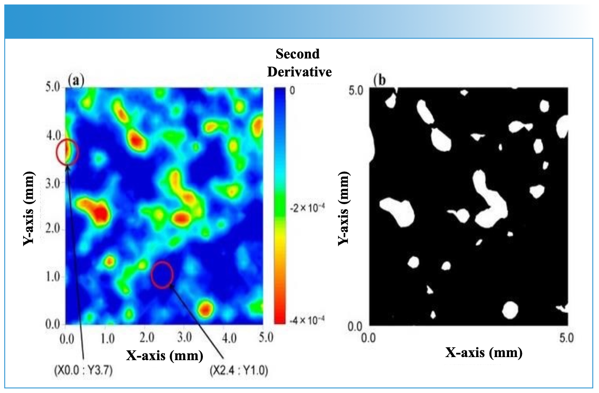
Figure 3 depicts a NIR spectrometry-based process monitoring set up for a blending process (17). To determine an accurate end point of the blending process, NIR spectra in the 950–1700 nm region were collected inline using the setup shown in Figure 3. NIR images of the sample during blending were developed using the D-NIRs system (Yokogawa [17]) atline. Figures 4a and 4b display the second-derivative image and binary image obtained during the blending period. The estimated concentration of ascorbic acid corresponded to actual concentration, showing that blending time was sufficient to provide a homogeneous blend (17).
FIGURE 3: A photograph of the NIR spectroscopy-based process monitoring setup for a blend process (a) P-NIRs, Yokogawa, see reference (17), for the inline blending process monitoring. (b) A vessel type blending machine. Reproduced from reference (17) with permission.
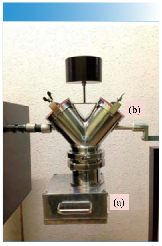
FIGURE 4: (a) An image of the mixing sample developed by second derivative at 1458 nm for 15 min after the start of the blending. (b) A binary image of the sample at 15 min after the start of the blending obtained by thresholding. Note that the mapping was performed under spatial resolution of 0.1 mm. Threshold was defined by standard deviation of second-derivative spectra. Reproduced from reference (17) with the permission.
Investigation of Inhomogeneity of a Pharmaceutical Tablet During a Grinding Process
NIR imaging was used to investigate inhomogeneity of pharmaceutical tablets containing two ingredients, a soluble active ingredient, pentoxifylline (PTX), and an insoluble excipient, palmitic acid (19). Figures 5a and 5b display a series of concentration profiles of PTX and palmitic acid, respectively, for each tablet ground for 0, 2, and 45 min. SMCR was applied to the NIR spectra of tablets in the 7600–4500 cm-1 region, and concentration profiles and pure component spectra were obtained. These concentration profiles can be used to estimate the distribution of PTX and palmitic acid within the tablets.
FIGURE 5: Concentration profiles of (a) PTX and (b) palmitic acid for each tablet ground for 0, 2, and 45 min. Reproduced from reference (19) with permission.
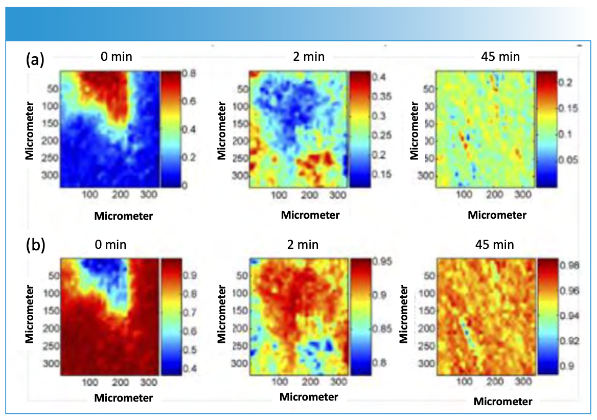
It was found from this imaging study that the homogeneous distribution of chemical ingredients in the samples strongly depends on the grinding time, and that this mixing process plays a central role in quantitative control. In addition, pure component spectra obtained by SMCR showed a sequential change of specific NIR peak intensities with the increase of the grinding time (19). The spectral changes are concerned with structure changes related to its crystallinity during the grinding process. Therefore, it was found from this study that NIR imaging combined with SMCR is a powerful tool to explore chemical or physical mechanisms induced by the manufacturing process of pharmaceutical products (19).
NIR Imaging of Changes in Hydrogen Bonding of Water During the Process of Medaka Embryonic Development
Ishigaki and associates investigated how metabolic activity by embryonic development affects the hydrogen bonding state of water molecules in medaka eggs (26). Very complex biochemical changes, such as DNA expression and associated translation into proteins, occur in the egg during embryonic development Instead of following these complex changes one by one, we evaluated their impact on the water that encompasses them, and verified whether it is possible to distinguish which embryos develop normally.
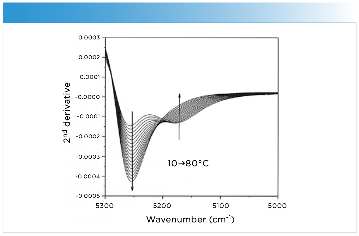
In this study, Ishigaki compared four types of medaka embryos, the first being embryos on the first day of fertilization with activated embryonic development. This was followed by three types of embryos with inactive embryonic development—embryos cultured at low temperatures to stop embryo development; embryos frozen and thawed with liquid nitrogen; and unfertilized eggs. (26). For analyzing the water in the egg by NIR spectroscopy, NIR spectra in the 10,000–4000 cm-1 region of pure water were measured over a temperature range of 10 to 80 °C at 5 °C intervals, with results shown in Figure 6. Two broad bands at around 6900 and 5200 cm-1 are due to the combination of OH stretching and antistretching modes and that of OH bending and OH antistretching modes, respectively. Figure 7 shows the second derivative spectra in the 5300–5000 cm-1 region. It is noted that the band at around 5200 cm-1 consists of two bands. The relative intensity changes of the two bands at 5250 and 5170 cm-1 indicate the variation of hydrogen bonds in water. The component whose second derivative intensity becomes stronger with increasing temperature is due to “water with weak hydrogen bonding” (WHB), while that whose second derivative intensity becomes weaker arises from “water with strong hydrogen boning” (SHB). When principal component analysis (PCA) was performed on the NIR spectra obtained from the yolk portion of the medaka embryo, the component representing the contribution of WHB of principal component 2 (PC2) was relatively higher in the embryo 1 than in the embryos 2–4 (26).
FIGURE 6: NIR spectra of pure water measured over a temperature range of 10–80 oC, at 5 oC intervals.
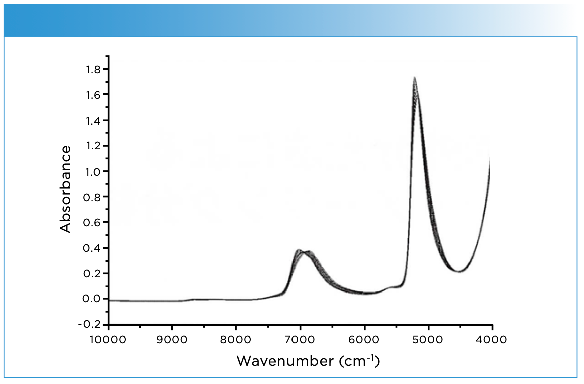
FIGURE 7: Second derivative of the spectra shown in Figure 6.
Therefore, to narrow down the candidates for factors that cause changes in the hydrogen bond state of water molecules, changes in protein concentration, ion concentration, temperature, and secondary structural changes of proteins were applied to water as perturbations. The results suggest that secondary structural changes (α helices → β sheets) of proteins may cause changes in the hydrogen bonding state of water molecules in similar extent to that detected in medaka embryos. Raman spectroscopy then confirmed the results, indicating that the secondary structure of the proteins was changed in actual medaka embryos.
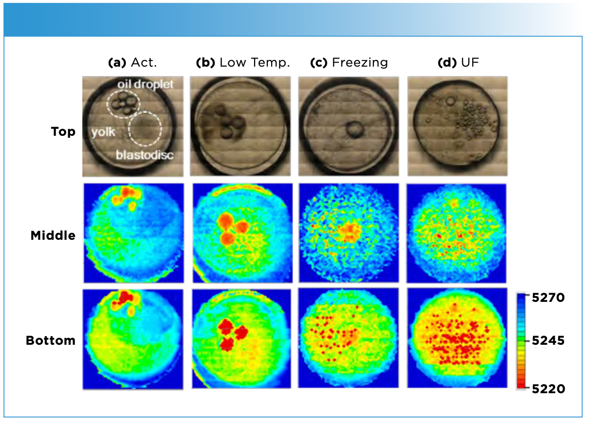
Figure 8 (middle) displays NIR imaging of water in the four kinds of embryos constructed by the intensity ratio of I5250/I5170 (IWHB/ISHB). It was found from the images that the proportion of WHB water in the yolk portion was high in embryos with activated embryonic development. Figure 8 (bottom) exhibits NIR images of water in the embryos generated using the shift of 5250 cm-1 band (26). Note that the images in the bottom of Figure 8 are similar to those in the middle of the same figure. This means that shift image also worked well (26).
Figure 8: (Top) Visible images of medaka embryos. (a) Embryo on the first day of fertilization in which embryonic development was activated, and three types of embryos in which embryo development was inactive [(b) embryo that has been cultured at low temperatures and stopped developing, (c) embryo frozen and thawed by liquid nitrogen, (d) unfertilized egg]. (Middle) NIR images of water in the four kinds of embryos constructed by the intensity ratio of I5250/I5170 (IWHB/ISHB). The dark and light parts indicate a high proportion of water with strong hydrogen bonds (SHB) and water with weak hydrogen bonds (WHB), respectively. (Bottom) NIR images of water in the embryos generated using the shift of 5250 cm-1 band.
We were able to grasp embryonic development not from a microscopic perspective such as DNA, proteins, and lipids, but from a macroscopic perspective of water in the egg, which encompasses all of them, and in a living state. Another feature of this study is that the primary use of near-infrared spectroscopy and Raman spectroscopy has allowed us to clearly show the relationship between structural changes in proteins in vivo and the hydrogen bonding state of water molecules.
A High Speed and Wide-Area Monitoring NIR Imaging System with a Novel NIR Camera (Compovision)
Sumitomo Electric Industries developed a high-speed and wide-area monitoring NIR imaging system with a novel NIR camera (Compovision) (27). These characteristic features of the camera are due to a newly developed InGaAs detector. The detector is equipped with InGaAs/GaAsSb type-II quantum wells, laminated on an indium phosphide substrate. Thus, one can measure an NIR spectrum in the 1000–2350 nm region of a 150 mm × 200 mm2 area (approximately 100,000 pixels) within 2–5 s.
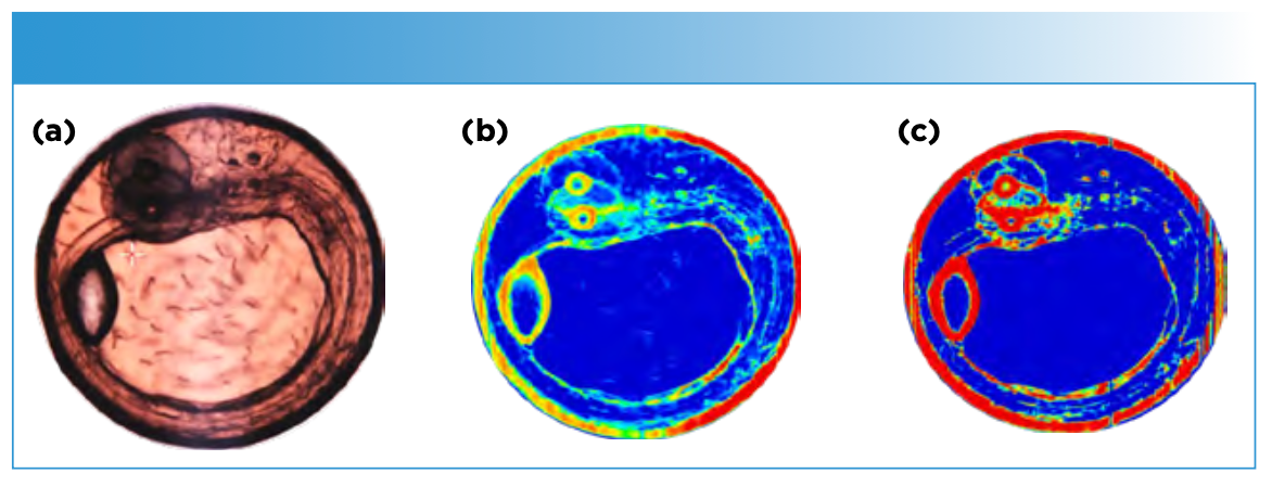
Figure 9 displays (a) an optical image from the NIR camera of eight-day medaka egg, and NIR images developed by bands (b) at 6191 cm-1 due to lipids and (c) at 6659 cm-1 arising from proteins. NIR imaging allows visualization of important organs such as otilishes and eyes. The image in Figure 9b is an image of lipids, while that in Figure 9c is that of proteins. Both are clearly different from each other. It is of note that these images were obtained in only two seconds.
Figure 9: (a) An optical image from Compovision of eight-day medaka egg, and NIR images developed by bands at (b) 6191 cm-1, and (c) 6659 cm-1.
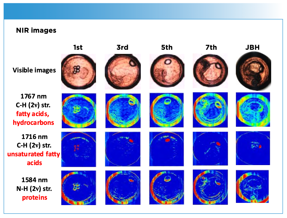
Using the NIR camera, Ishigaki and colleagues demonstrate the high-speed in vivo NIR imaging of the growth of fertilized fish eggs at the molecular level (24). Figure 10 exhibits visible and NIR images of medaka eggs at 1st, 3rd, 5th, and 7th days after fertilization and just before hatching. The structure of the membrane phospholipid bilayer, distribution of unsaturated fatty acids in oil droplets and egg membranes could be studied (24).
Figure 10: Visible and NIR images of medaka eggs at 1st, 3rd, 5th, and 7th days after fertilization and just before hatching (JBH).
Non-Staining Blood Flow Imaging Using Optical Interface due to Doppler Shift in Developing Fish Egg Embryos
Ishigaki and associates explored non-staining blood flow imaging using an optical interface due to the Doppler shift in developing fish egg embryos (25]) In this study an imaging-type two-dimensional Fourier spectroscopy (ITFS) system developed by Ishimaru of Kagawa University and AOI Electronics Co., Ltd. was used. The characteristic feature of the system is that it has a partial movable mirror, which gives optical path differences to the object light, and an interferogram can be obtained by continuously changing the spatial phase difference. Since interference only occurs in the rays that come from the same point in the system, interference coming from outside the focal plane is not observed in the alternate current (AC) component but detected only in the direct current (DC) component. Therefore, the system can obtain confocal 3D imaging data in principle.
In the reflectance mode for a medaka fish egg on the 5th day after fertilization, an interferogram was obtained from the yolk and heart parts. Figure 11a and 11b show spectroscopic light intensity in the 1000–2500 nm and 2000–15000 nm spectral regions, respectively, extracted from the interferogram (25). In the spectra, the strong peak was detected at 3768 nm, and two medium peaks were also observed at 1884 and 1256 nm. The peak at 3768 nm originates from the fundamental mode of the heartbeat, whereas the two peaks at 1884 and 1256 nm are ascribed to the first and second overtones of the fundamental mode, respectively. By plotting the intensities of (a) detected light and (b) absorbance at 1256 and 1884 nm in two dimensions, non-staining blood flow images were successfully accomplished as shown in Figure 12.
Figure 11: Spectra in the (a) 1000–2500 nm and (b) 2000–15,000 nm regions obtained by Fourier transformation of the data. Reproduced from reference (25) with permission.
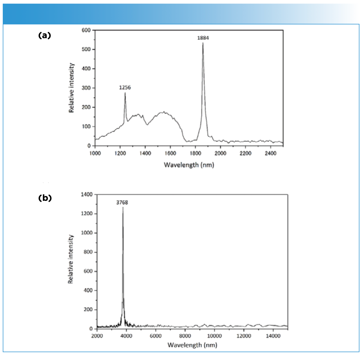
Figure 12: Blood flow images of a medaka fish egg on the 5th day after fertilization obtained by plotting intensities of (a) detected light from the sample and (b) absorbance at 1260 and 1860 nm. Reproduced from reference (25) with permission.

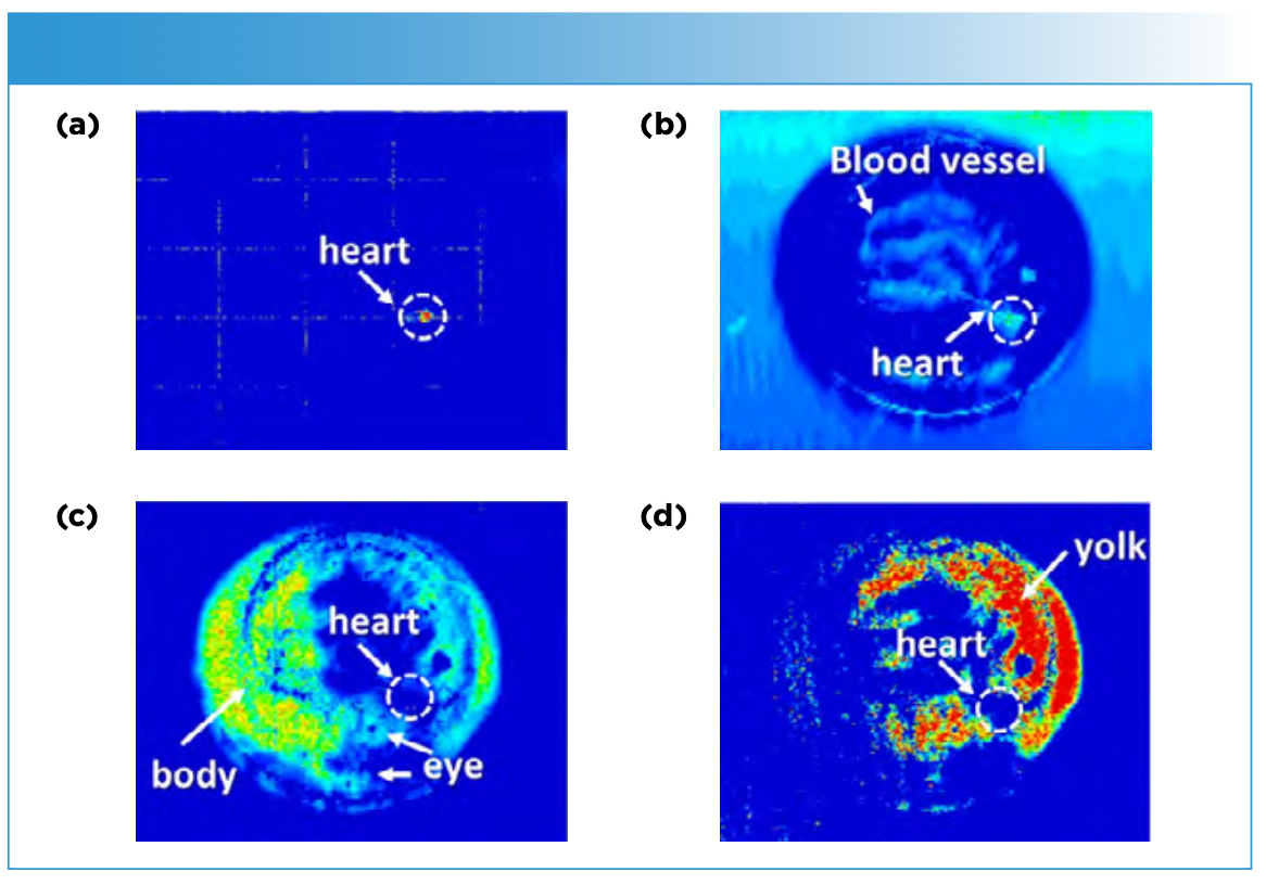
The measurement was carried out also using transmission mode. Two peaks derived from the heart beat were observed at 1880 and 2260 nm in FT spectra. Moreover, absorbance peaks due to molecular vibrations were observed at 1940 and 2360 nm originating from the antisymmetric O-H stretching and O-H bending modes of water, and C-H stretching and bending modes of hydrocarbons and/or aliphatic compounds, respectively (25). Figure 13 displays NIR images developed by plotting the intensities of (a) detected light at 2260 nm and absorbance at (b) 1880 nm, (c) 1940 nm, and (d) 2360 nm. Figure 13a and 13b depict the position of the heart and that of the blood vessels spread within embryo in addition to the heart. Figure 13c and 13d reveal the detailed structure of embryo and yolk, respectively. In this way, molecular vibration and heart beat signals can be simultaneously obtained in vivo using optical interference caused by optical path differences and light frequency shifts.
Figure 13: NIR images obtained by plotting intensities of (a) detected light at 2260 nm and absorbance at (b) 1880 nm, (c) 1940 nm, and (d) 2360 nm. Reproduced from reference (25) with permission.
Perspectives
Imaging-visualization technology is extremely important for various purposes, such as investigations in materials science, nanoscience, pharmaceutical, biomedical, agriculture, foods, and polymers. NIR imaging has been used extensively in various fields. As perspectives expand, we can expect the following six main developments: 3D imaging; more rapid imaging; portable imaging; development of combined instruments (for example, NIR imaging instrument sand blending machines); new analysis methods, such as machine learning; and development of new research fields.
Further innovation of NIR imaging instruments is particularly important. Moreover, NIR imaging should be used more widely. One should recognize more the importance of NIR imaging in observing solvent diffusion by NIR imaging.
References
(1) Ozaki, Y.; Huck, C.; Tsuchikawa, S.; Engelsen, S. B.Near-Infrared Spectroscopy: Theory, Spectral Analysis, Instrumentation, and Applications;Springer, 2000.
(2) Sasic, S.; Ozaki, Y. Raman, Infrared, and Near-Infrared Chemical Imaging;Wiley, 2010..
(3) Salzer, R.; Siesler, H. W.Infrared and Raman Imaging, 2nd ed.; Wiley-VCH, 2014.
(4) Amigo, J. M.; Benson, V. Hyperspectral Imaging; Elsevier, 2019.
(5) Ishikawa, D.; Shinzawa, H.; Genkawa, T.; Kazarian, S. G.; Ozaki, Y. Recent Progress of Near-Infrared (NIR) Imaging—Developments of Novel Instruments and Their Practical Applications for Practical Situations. Anal. Sci. 2014, 30, 143–150. DOI: 10.2116/analsci.30.143
(6) Unger, M.; Ozaki, Y.; Siesler, H. W. Variable-Temperature Fourier Transform Near-Infrared (FT-NIR) Imaging Spectroscopy of the Diffusion Process of Butanol(OD) into Polyamide 11. Appl. Spectrosc. 2011, 65, 1051. DOI: 10.1366/11-06309
(7) Shinzawa, H.; Nishida, M.; Tsuge, A.; Ishikawa, D.; Ozaki, Y.; Morita, S.; Kanematsu, W. Thermal Behavior of Poly(lactic acid)-nanocomposite Studied by Near-Infrared Imaging Based on Roundtrip Temperature Scan.Appl. Spectrosc. 2014, 68, 371–378. DOI: 10.1366/13-07176
(8) Shinzawa, H.; Mizukado, J. Near-Infrared (NIR) Disrelation Mapping Analysis for Poly(lactic) Acid Nanocomposite.Spectrochim. Acta A 2017, 181, 1–6. DOI: 10.1016/j.saa.2017.03.026
(9) Ishikawa, D.; Nishii, T.; Mizuno, F.; Kazarian, S. G.; Ozaki, Y. Potential of a Newly Developed High Speed Near-Infrared (NIR) Camera (Compovision) in Polymer Industrial Analyses – Monitoring of Crystallinity and Crystal Evolution of Polylactic Acid (PLA) and Concentration of PLA in PLA/ Poly-(R)-3-hydroxybutyrate (PHB) Blends. Appl. Spectrosc. 2013, 67, 1411–1416. DOI: 10.1366/13-07103
(10) Muroga, S.; Hikima, Y.; Ohshima, M. Visualization of Hydrolysis in Polylactide Using Near-Infrared Hyperspectral Imaging and Chemometrics. J. Appl. Polym. Sci. 2018, 135, 45898. DOI: 10.1002/app.45898
(11) Ishikawa, D.; Furukawa, D.; Tseng, T. W.; Kummetha, R. R.; Motomura, A.; Igarashi, Y.; Kazarian, S. G.; Ozaki, Y. High-Speed Monitoring of Crystallinity Change in Poly Lactic Acid during Photodegradable Process by Using a Newly Developed Wide Area NIR Camera (Compovision).Anal. Bioanal. Chem. 2015, 407, 397–403. DOI: 10.1007/s00216-014-8211-z
(12) Vejarano, R.; Siche, R.; Tesfaye, W. Evaluation of Biological Contaminants in Foods by Hyperspectral Imaging: A Review. Int. J.Food Prop. 2017, 20, 1264–1297. DOI: 10.1080/10942912.2017.1338729
(13) Roberts, J.; Power, A.; Chapman, J.; Chandra, S.; Cozzolino, D. A Short Update on the Advantages, Applications and Limitations of Hyperspectral and Chemical Imaging in Food Authentication. Appl. Sci. 2018, 8,505. DOI: 10.3390/app8040505
(14) Gowen, A. A.; O’Donnell, C. P.; Cullen, P. J.; Downey, D.; Frias, J. M. Hyperspectral Imaging – An Emerging Process Analytical Tool for Food Quality and Safety Control. Trends Food Sci. Tech. 2007, 18, 590–598. DOI: 10.1016/j.tifs.2007.06.001
(15) Baiano, A. Applications of Hyperspectral Imaging for Quality Assessment of Liquid Based and Semi-Liquid Food Products: A Review. J. Food Eng. 2017, 214, 10–15. DOI: 10.1016/j.jfoodeng.2017.06.012
(16) Liu, Y.; Pu, H.; Sun, D. W. Hyperspectral Imaging Technique for Evaluating Food Quality and Safety During Various Processes: A Review of Recent Applications. Trends Food Sci. Tech. 2017, 69, 25–35. DOI: 10.1016/j.tifs.2017.08.013
(17) Murayama, K.; Ishikawa, D.; Genkawa, T.; Sugino, H.; Komiyama, M.; Ozaki, Y. Image Monitoring of Pharmaceutical Blending Process and the Determination of an End Point by Using a Portable Near-Infrared Imaging Device Based on a Polychromator-Type Near-Infrared Spectrometer with a High-Speed and High-Resolution Photo Diode Array, Molecules 2015, 20, 4007–4019. DOI: 10.3390/molecules20034007
(18) Ishikawa, D.; Murayama, K.; Awa, K.; Genkawa, T.; Komiyama, M.; Kazarian, S. G.; Ozaki, Y. Application of a Newly Developed Portable NIR Imaging Device to Dissolution Process Monitoring of Tablets. Anal. Bioanal. Chem. 2013, 9401–9409. DOI: 10.1007/s00216-013-7355-6
(19) Awa, K.; Okumura, T.; Shinzawa, H.; Otsuka, M.; Ozaki, Y. Self-Modeling Curve Resolution (SMCR) Analysis of Near-Infrared (NIR) Imaging Data of Pharmaceutical Tablets. Anal. Chim. Acta 2008, 619, 81–86. DOI: 10.1016/j.aca.2008.02.033
(20) Ishikawa, D.; Murayama, K.; Genkawa, T.; Kitagawa, Y.; Ozaki, Y. An Identification Method for Defective Tablets by Distribution Analysis of NIR Imaging. J. Spectral Imaging 2019, 8, a15. DOI: 10.1255/jsi.2019.a15
(21) Ishigaki, M.; Kawasaki, S.; Ishikawa, D.; Ozaki, Y. Near-Infrared Spectroscopy and Imaging Studies of Fertilized Fish Eggs: In Vivo Monitoring of Egg Growth at the Molecular Level. Sci. Rep. 2016, 6, 20066. DOI: 10.1038/srep20066
(22) Ishigaki, M.; Yasui, Y.; Puangchit, P.; Kawasaki, S.; Ozaki, Y. In vivo Monitoring of the Growth of Fertilized Eggs of Medaka Fish (Oryzias latipes) by Near-Infrared Spectroscopy and Near-Infrared Imaging—A Marked Change in the Relative Content of Weakly Hydrogen-Bonded Water in Egg Yolk Just Before Hatching. Molecules 2016, 21, 1003. DOI: 10.3390/molecules21081003
(23) Puangchit, P.; Ishigaki, M.; Yasui, Y.; Kajita, M.; Ritthiruangdej, P.; Ozaki, Y. Non-Staining Visualization of Embryogenesis and Energy Metabolism in Medaka Fish Eggs Using Near-Infrared Spectroscopy and Imaging. Analyst 2017, 142, 4765–4772. DOI: 10.1039/C7AN01575E
(24) Ishigaki, M.; Nishii, T.; Puangchit, P.; Yasui, Y.; Huck, C. W.; Ozaki, Y. Noninvasive, High-Speed, Near-Infrared Imaging of the Biomolecular Distribution and Molecular Mechanism of Embryonic Development in Fertilized Fish Eggs. J. Biophotonics 2018, 11, e201700115. DOI: 10.1002/jbio.201700115
(25) Ishigaki, M.; Puangchit, P.; Yasui, Y.; Ishida, A.; Hayashi, H. Nakamura, Y.; Taniguchi, H.; Ishimaru, I.; Ozaki, Y. Nonstaining Blood Flow Imaging Using Optical Interference due to Doppler Shift and Near-Infrared Imaging of Molecular Distribution in Developing Fish Egg Embryos.Anal. Chem. 2018, 90, 5217–5223. DOI: 10.1021/acs.analchem.7b05464
(26) Ishigaki, M.; Yasui, Y.; Kajita, M.; Ozaki, Y. Assessment of Embryonic Bioactivity Through Changes in the Water Structure Using Near-Infrared Spectroscopy and Imaging. Anal. Chem. 2020, 92, 8133–8141. DOI: 10.1021/acs.analchem.0c00076
(27) Ishikawa, D.; Nishii, T.; Mizuno, F.; Kazarian, S. G.; Ozaki, Y. Development of a High-Speed Monitoring NIR Hyperspectral Camera (Compovision) with Wide Area and Its Applications, NIR News 2013, 24, 6–11. DOI: 10.1255/nirn.1376
Yukihiro Ozaki is with the School of Biological and Environmental Sciences at Kwansei Gakuin University, in Hyogo, Japan. Mika Ishigaki is with the Institute of Agricultural and Life Sciences, Academic Assembly, at Shimane University, in Shimane, Japan. Direct correspondence to; yukiz89016@gmail.com

Real-Time Battery Health Tracking Using Fiber-Optic Sensors
April 9th 2025A new study by researchers from Palo Alto Research Center (PARC, a Xerox Company) and LG Chem Power presents a novel method for real-time battery monitoring using embedded fiber-optic sensors. This approach enhances state-of-charge (SOC) and state-of-health (SOH) estimations, potentially improving the efficiency and lifespan of lithium-ion batteries in electric vehicles (xEVs).