News
Article
Application Notebook
Raman Imaging for the Analysis of Food Products
Author(s):

3D Raman imaging
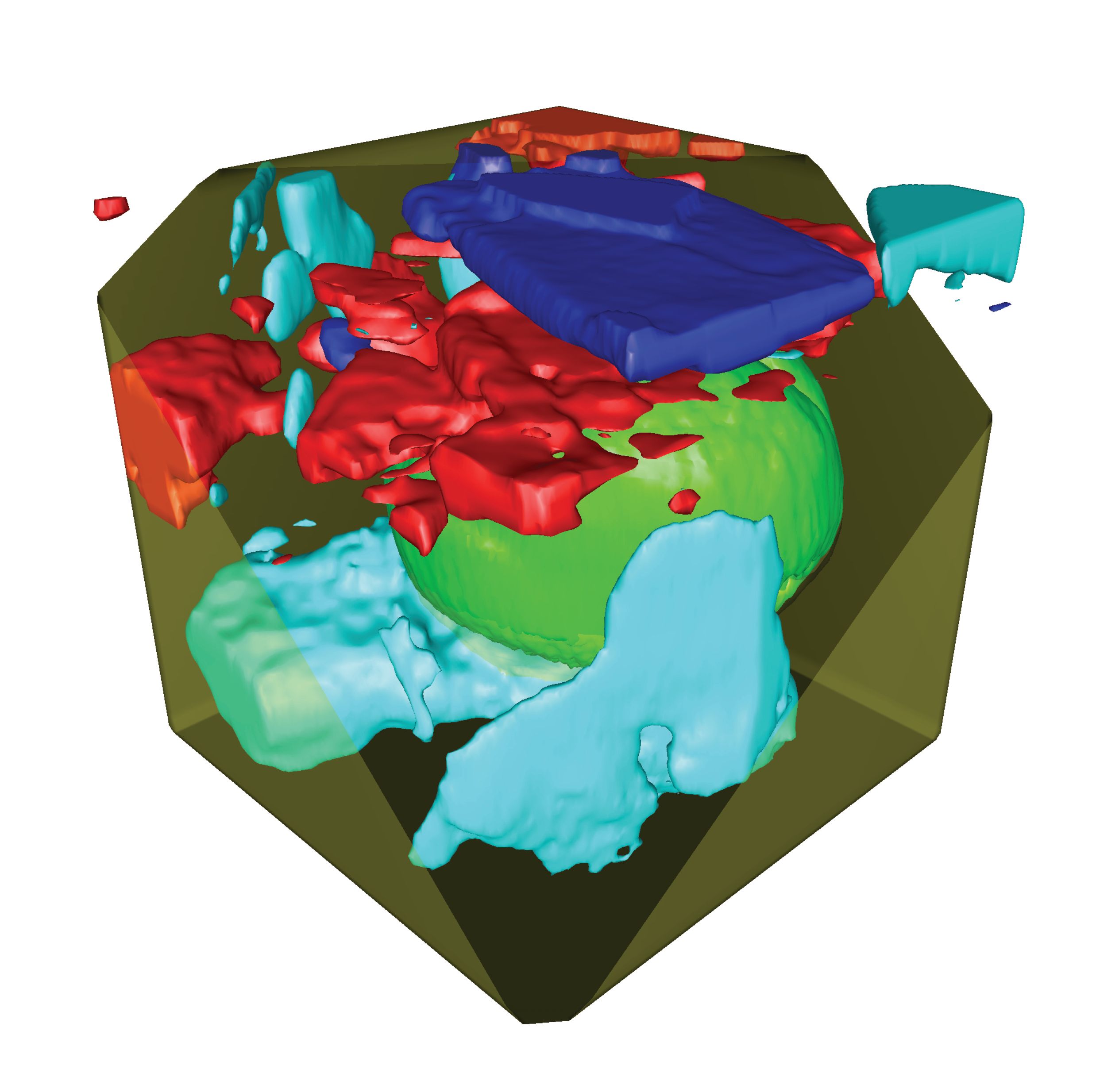
When Raman spectra are collected at each measurement point using a confocal microscope combined with a spectrometer, a Raman image can be generated that visualizes the distribution of the sample’s components. Due to the high confocality of WITec Raman systems, volume scans and 3D images can also be generated from a stack of 2D images acquired at different focal planes as shown in Figure 1.
Figure 1: A 3D Raman image of a honey droplet: This image was generated from 50 individual 2D confocal Raman images acquired along the Z-axis. It shows a pollen grain (green) and several crystalline phases of sugars (red, blue, cyan).

Emulsions and fat spreads
Fat spreads such as butter or margarine are water-in-oil emulsions. The microstructure of fat spreads determines properties such as suppleness, texture, and spreadability. These traits, as well as stability, depend on the fat crystal network at the interface and the emulsifier, and are strongly influenced by the production process. Therefore, manufacturers of emulsions and fat spreads analyze their products in detail to understand the composition-process-structure-function relationships.
In a study from the Dutch company Unilever (3), van Dalen et al. describe the development of the hyperspectral data analysis of Raman images of fat spreads including data pre-processing and multivariate curve resolution (MCR). With confocal Raman imaging, they not only localized the molecular compounds in fat spreads, but could also relate the microstructure of spreads to their production processes. The results (Figure 2) show that water forms droplets in a continuum of sunflower oil, stabilized at the interface by an emulsifier (monoglycerides) and lipids in the crystalline phase. The lipid crystalline phase (solid fat) is also found in the continuous phase of the emulsion, forming a network between the different water droplets. The image on the right shows the competition/co-crystallization between the solid fat and emulsifier at the droplet interface. The authors of the study conclude “This method can be applied to a wide range of different food emulsions such as butter, margarine, mayonnaise, and salad dressings.”
Figure 2: Raman images of a water-in-oil emulsion: From left to right, the concentration of sunflower oil, water, solid fat, and emulsifier is shown in single images, with the highest concentrations in red and the lowest in dark blue. In the rightmost image, solid fat and emulsifier Raman signals are overlaid. Image parameters: 20 x 20 μm2, 86 x 86 pixels. Images courtesy of Gerard van Dalen and colleagues, Unilever, Vlaardingen, NL

References
- G. P. Smith et al., J Raman Spec. 48, 374 (2016).
- E. M. Both et al., Food Res Int 109, 448 (2018).
- G. van Dalen et al., J Raman Spec 48, 1075 (2017).
WITec GmbH
Lise-Meitner-Str.6 89081 Ulm, Germany
Tel. +49 731 140 70 0, fax +49 731 140 70 200
Website: www.witec.de

Newsletter
Get essential updates on the latest spectroscopy technologies, regulatory standards, and best practices—subscribe today to Spectroscopy.


