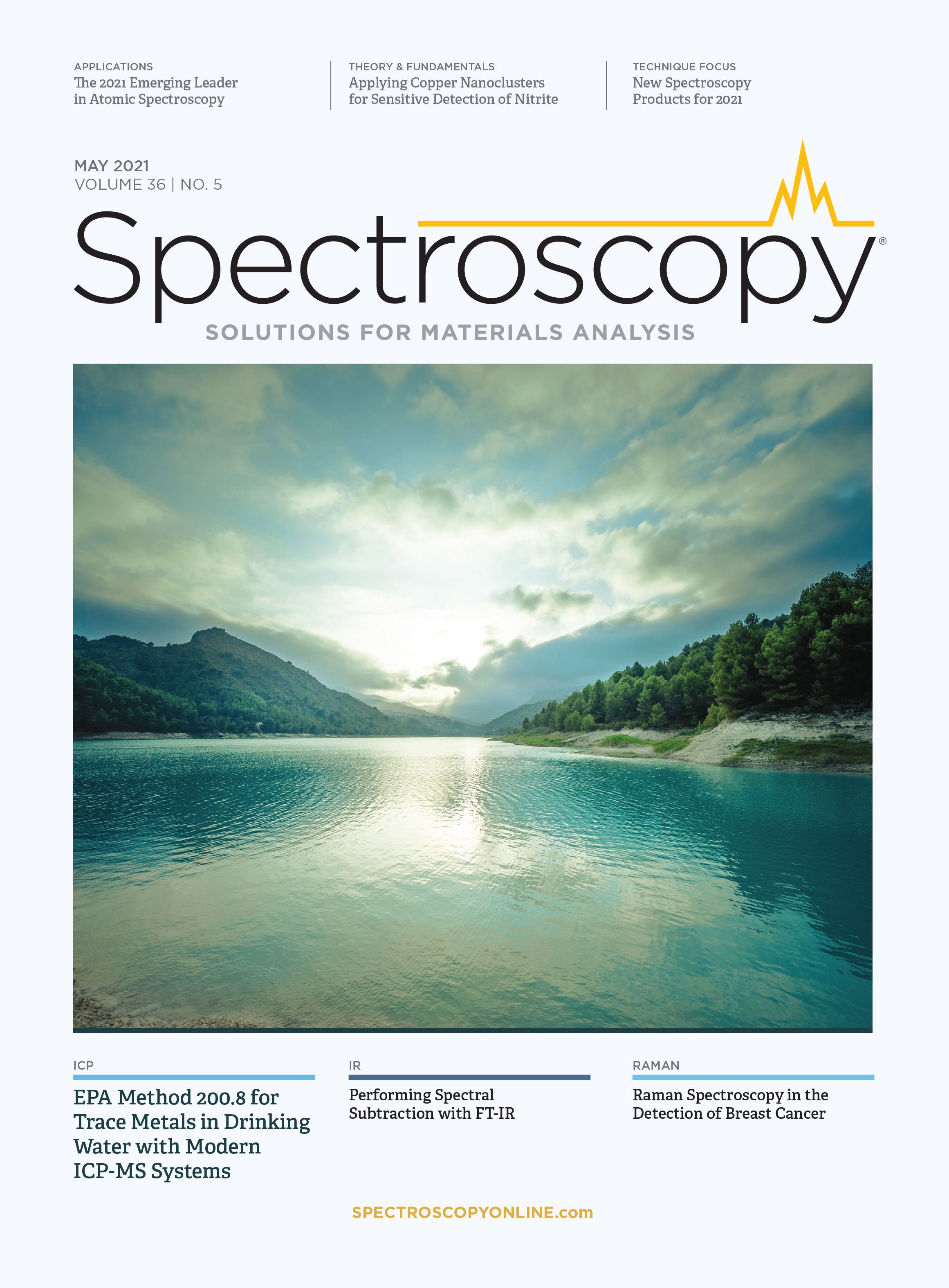
Raman Spectroscopy in the Detection of Breast Cancer
To explore the feasibility and reliability of using the Raman signature of aromatic amino acids as a marker in the detection of the presence of breast cancer, and perhaps even predicting cancer development in very early stages of onset, Rina Tannenbaum and her colleagues at Stony Brook University in Stony Brook, New York, collected the most recent and relevant literature where Raman spectroscopy was used as an analytical tool in the evaluation of breast cell lines and breast tissue. They then extracted the relevant Raman spectra from each paper, processed and normalized them, and then re-analyzed them. All spectral bands from each re-analyzed spectrum indicative of aromatic amino acids were plotted without bias to determine whether a pattern shedding light on a possible diagnostic classification could be detected. Tannenbaum discussed her findings, and the paper resulting from them (1), with Spectroscopy.
What is special or meaningful about aromatic amino acids as a marker for breast cancer, and why is Raman spectroscopy useful for detecting them? What motivated you to focus on this specific application?
Raman spectroscopy is a non-destructive and sensitive technique for exploring the structure and chemical composition of biological materials and could be a very effective tool for the delineation of cancer tissue, histological analysis of biopsies, in vivo detection of tumors, and intraoperative imaging. Raman spectroscopy is based on the inelastic scattering of monochromatic infrared light upon interaction with molecular vibrations, phonons, and other excitations, generating shifts in the energy of the incident light. The shift in energy gives information about the vibrational modes of the molecules in the system. Each molecule in a given system exhibits a precise set of vibrational modes, depending on their chemical composition, chemical environment, and spatial organization. These vibrational modes constitute the fingerprints of the given system and allow the identification of molecular species in the system and their interactions. Raman microscopy for biological and medical specimens generally uses near-infrared (NIR) lasers (785 nm), which reduces the risk of damaging the specimen by applying higher energy wavelengths and practically eliminates fluorescence.
Aromatic amino acids have a rigid, planar structure and possess added stability due to the π-electron cloud situated above and below the plane of the aromatic ring. The aromatic amino acids can undergo aromatic-aromatic interactions, consisting of hydrogen bonding coupled with attractive, noncovalent, dipole, and van der Walls interactions and pi-stacking of the benzene rings. These aromatic-aromatic interactions have been found to play an important role in the stabilization of the overall structure of protein molecules and protein-DNA/RNA complexes and constitute a crucial element in the proper functioning of protein molecules, which in turn impacts various cellular processes in which these proteins take part.
To be able to assess your hypothesis, you collected most recent and relevant literature in which Raman spectroscopy was used as an analytical tool in the evaluation of breast cell lines and breast tissue, re-analyzed all the Raman spectra, and extracted all spectral bands from each spectrum that were indicative of aromatic amino acids. Was there any specific “common ground” in that literature that stood out?
At first glance, there did not seem to exist a specific “common ground” among the various publications, other than the commonality of the presence of the characteristic Raman bands of the aromatic amino acids. However, once all the band intensities had been normalized and plotted together, various patterns started to emerge. We applied various statistical methods to analyze the large number of data points that we obtained, and observed that irrespective of the frequency of the incident infrared light or the specific Raman measurement techniques, the presence of aromatic amino acids was predominantly higher in samples originating from breast cancer cells or tissues.
What conclusions that you have come to so far in your research?
It has been observed in recent years that various types of cancers exhibit an overexpression of aromatic amino acids. Because aromatic amino acids play important roles in a variety of metabolic processes, as well as key functions in the structural integrity of cellular and membrane proteins, these anecdotal observations regarding their enhanced presence in cancerous tissues have prompted more dedicated and targeted inquiries. The applicability of Raman spectroscopy as a diagnostic tool in the prediction, detection, and evaluation of cancer tissues has been gaining considerable momentum in recent years. For us to be able to correlate Raman spectra with specific cellular features, it would be imperative to determine which molecular details would provide a unique signature that would allow a definitive discernment between healthy and cancerous tissues. In this context, we have set out to probe the role of aromatic amino acids in breast cancer and to ascertain the possibility of using their overexpression as a diagnostic tool. The emphasis of our analysis centered on the spectral region of 500–1200 cm-1 that has been shown as the most characteristic for aromatic amino acids, specifically tryptophan, phenylalanine, and tyrosine. We have been able to demonstrate that cancerous breast tissues and cells markedly exhibit overexpression of aromatic amino acid and that the difference between the extent of their presence in cancerous cells and healthy cells is decisive. On the basis of this analysis, we conclude that it is possible to use the signature Raman bands of aromatic amino acids as a biomarker for the detection, evaluation, and diagnosis of breast cancer.
In the paper, you state that you did not seek to establish Raman spectroscopy as a de-facto standard for the prediction or diagnosis of cancer, but rather as a tool with the ability to add to, as well as complement, the myriad of tools available to physicians and patients. That said, were there any benefits specific to using Raman spectroscopy that make this technique especially useful in this for clinical applications? What are the limitations to using this technique in a medical application?
There is no doubt that current diagnostic techniques such as mammography, ultrasound, MRI, and tissue biopsies are the cornerstone assessment tools for the establishment of tumor malignancy. This, however, does not preclude the development of new imaging modalities with potentially sub-cellular resolution that would initially complement current modalities, but as they evolve and improve, might eventually become powerful diagnostic tools in their own right. Raman spectroscopic imaging of biological samples has been generally plagued by weak spectral intensities, however new sampling and signal enhancement techniques developed in recent years have brought this method into focus and have generated great interest in its potential application in cancer detection and diagnosis.
Regarding the importance of the aromatic amino acids as breast cancer markers, the increasing data found in the recent literature demonstrate that cancerous breast tissues and cells exhibit a marked overexpression of these molecules and that the difference between the extent of their presence in cancerous cells and healthy cells is beyond statistical fluctuations. Based on this analysis, we concluded that it may indeed be possible to use the signature Raman bands of aromatic amino acids as a biomarker for the detection, evaluation, and diagnosis of breast cancer. We do not suggest that the Raman signature of aromatic amino acids could be used as a predictive biomarker and an early indication of disease onset. The limited data regarding the Raman signature of aromatic amino acids in breast cancer allows only a qualitative assessment of the possible clinical usefulness of this methodology.
What are the biggest challenges that you have encountered in developing this method? What options or alternatives are available to overcome these challenges?
The biggest challenge is access to breast tissue. While initial work can be done on cell lines, the analysis of actual breast tissue in imperative to determine the selectivity of the Raman method in the context of a larger and more diverse biological environment. Given that I am a materials scientist and chemical engineer, the extension of the work requires partnering with researchers from the relevant clinical disciplines and access to histological samples repository.
What are your next steps in this work?
As a first step, we just submitted a proposal to the National Cancer Institute of the National Institutes of Health. We have to better understand the relationship between the various types of breast cancer and the specific degree of the aromatic amino acid overexpression. We need to understand the structural effects of the changes in aromatic amino acids variations in various proteins that are characteristic to each type of breast cancer. There is no doubt that a greater amount of data is required to expand this work from the observational and perhaps anecdotal level to the verified phenomenological level. I need to establish collaborations with clinical researchers and recruit highly talented students to push the experimental work forward.
References
(1) S. Contorno, R.E. Darienzo, and R. Tannenbaum, Sci. Rep. 11, 1698 (2021) https:// www.nature.com/articles/s41598-021- 81296-3t
Rina Tannenbaum is a professor in the Program of Chemical and Molecular Engineering in the Department of Materials Science and Engineering at the State University of New York at Stony Brook. Direct correspondence to: Irena.Tannenbaum@stonybrook.edu

John Chasse is Managing Editor for Spectroscopy. Direct correspondence about this article to: jchasse@mjhlifesciences.com ●

AI-Powered SERS Spectroscopy Breakthrough Boosts Safety of Medicinal Food Products
April 16th 2025A new deep learning-enhanced spectroscopic platform—SERSome—developed by researchers in China and Finland, identifies medicinal and edible homologs (MEHs) with 98% accuracy. This innovation could revolutionize safety and quality control in the growing MEH market.
New Raman Spectroscopy Method Enhances Real-Time Monitoring Across Fermentation Processes
April 15th 2025Researchers at Delft University of Technology have developed a novel method using single compound spectra to enhance the transferability and accuracy of Raman spectroscopy models for real-time fermentation monitoring.
Nanometer-Scale Studies Using Tip Enhanced Raman Spectroscopy
February 8th 2013Volker Deckert, the winner of the 2013 Charles Mann Award, is advancing the use of tip enhanced Raman spectroscopy (TERS) to push the lateral resolution of vibrational spectroscopy well below the Abbe limit, to achieve single-molecule sensitivity. Because the tip can be moved with sub-nanometer precision, structural information with unmatched spatial resolution can be achieved without the need of specific labels.