Surface-Enhanced Raman Scattering
Columnist Fran Adar discusses surfaced-enhanced Raman scattering (SERS). The phenomenon is described and the enhancement factors that make it so attractive for analytical purposes are pointed out. In particular, she reviews the state-of-the-art from the point of view of the instrumentation and the robustness of the measurements.

In my last column, I anticipated writing about the state of the art of Raman imaging — spatial resolution and speed. However, in light of the recent buzz created by the Defense Advanced Research Projects Agency (DARPA) request for proposals regarding surface enhanced Raman scattering (SERS), I have decided to postpone the Raman imaging discussion to review SERS. I am less directly involved in this field than I am in Raman microscopy, but I have been interested in its potential. I hope that I get it right, and my apologies for anything that I get wrong.

Fran Adar
In 1974, Fleischmann and colleagues (1) reported enormous enhancements of Raman signals of pyridine adsorbed on a silver electrode roughened by successive oxidation–reduction cycles, using an argon ion excitation line. Calculations showed that the signals were 105 to 106 higher than expected, and they were originally explained as being due to the additional surface area provided by the roughening of the surface. However, others (2,3) showed that minimally roughened surfaces also showed enhancements, and these enhancements could not be explained by the surface area. Over the last 30-some years, SERS experiments and theory have been quite active in an attempt to understand the phenomenon and to make it reproducible and thus useful for analytical applications.
Making SERS measurements robust has been, in a sense, a holy grail for Raman spectroscopy. Those of us who do Raman will tell you how wonderful it is at extracting all kinds of information that is uniquely revealed by the technology. But the reality is that even with the advances of the last 20 years, it is still more work-intensive than other analytical measurements such as UV-vis or fluorescence spectroscopies. If the signal strengths could be enhanced by six or more orders of magnitude, Raman spectroscopy could become much more interesting from a practical standpoint. And there is even the possibility for single molecule detection (4).
Background
So 30 years is a long time. What is the problem? The problem has been the reproducibility of the technique – from day to day in a given laboratory, but this can sometimes be controlled; but also the inability to reliably reproduce results from laboratory to laboratory is even more important. It is easy to see how the technique would become competitive, controversial, and confusing. As an onlooker, a pivotal article in Science explained to me what was going on. In 1997, Nie and Emory (5) used atomic force microscopy (AFM) to isolate particular silver nanoparticles from a heterogeneous population and to measure the SERS signals from those particles. The enhancement factors for some of these particles were on the order of 1014 to 1015, which was orders of magnitude larger than the average enhancement factor measured. This means that a very small number of particles are supplying the overall enhancements that are measured in the bulk. In addition, because the "hot" particles were visualized, it was possible to document the effects of particle size, shape, orientation (relative to the laser polarization), and aggregation that already had been suggested as effecting the enhancement.
Historically, there have been two major fields of thought about the enhancement mechanism. Otto (6) proposed a "strong Raman enhancement for an adsorbate on a silver surface is only possible when the adsorbate is bound to an atomic scale roughness, e.g., an adatom." While many publications attempted to reconcile the observations that could not be explained by the electromagnetic theory (see below), this chemical effect currently receives less attention.

Figure 1
The electromagnetic effect, in particular surface plasmon-assisted resonance, is currently considered to hold the secrets of the enhancement mechanism of the SERS phenomenon. Reviewing the theories based on the interaction between the electromagnetic radiation and the metal particles is certainly beyond this column, so readers are pointed to the literature. A good start would be the recently released text by Professor Ricardo Aroca (7) at the University of Windsor in Ontario, Canada. It is interesting to note the comments at the beginning of the preface to this book: "Surface-enhanced Raman scattering (SERS) is a moving target. Every time you look at it, it mutates, and new speculations are suddenly on the horizon." Aroca also comments on the lack of determinism when working in the single molecule regime, which is "compounded by the fact that the enhanced signal is the result of several contributions, and their separation into well-defined components is virtually impossible."
So the last 10–20 years have seen efforts to determine the important parameters determining the enhancements. Our goal in this column is not to make final judgments on the field, but to assess what provides reasonably reproducible, enhanced signals that can be used for analytical purposes. In evaluating any technology, keep in mind that different molecules can have vastly different SERS activity, even under ideal circumstances. So how well SERS is going to work to solve a particular problem will depend on the chemical particulars of that system.
Commercialized SERS Products
During this period, numerous SERS preparations have appeared on the market. These products are usually in the form of sols of silver or gold particles or coated substrates that can be deposited by evaporation of metal or precipitation of particles from solution. Some of the products on the market of which we are aware will be described.
Real-Time Analyzers (Middletown, Connecticut) offers 2-mL vials whose inner surface is coated with a patented Ag sol gel that enhances signal up to six orders of magnitude. They claim detection limits of parts per billion, millimolar, or milligrams per liter, with less than 20% variability from vial to vial. The shelf life of the vial is greater than 1 year. They list applications in agriculture (pesticide residue), antiterrorism (chemical agents, hydrolysis products, bioagents), biochemistry (amino acids, DNA bases, and so forth), biomedical (physiological chemicals in blood and urine), chemistry (trace components), drug enforcement (trace drugs on surfaces), forensics (trace drugs in body fluids), pharmaceutical (drug discovery and development), and liquid chromatography detection.
Mesophotonics (Southhampton, UK) offers substrates of photonic crystals called Klarite. Note that this technology is a departure from creation of substrates by deposition. Using lithographic techniques developed for the integrated ciruit industry, they create two-dimensional arrays of holes, each a few hundred nanometers deep. Such an array forms a photonic band gap representing "a range of energies where no permitted states are allowed and the light of a wavelength residing within is forbidden to propagate" (8). The silicon surface is gold coated and the resulting surface plasmons determine the SERS amplification. The relative standard deviation achieved with 633-or 785-nm excitation is better than 10%. Using these substrates, Raman signals can be achieved from concentrations as low as parts per billion. Listed applications include analytical chemistry, pharmaceutical drug development, forensics, medical diagnosis, trace analysis, homeland security, and chemical–biological detection. Mesophotonics has recently transferred its SERS business to d3 Technologies (Glasgow, UK).
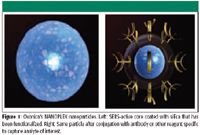
Oxonica (Mountain View, California) offers products in the NANOPLEX technology line. The nanoparticles in this line provide for a biomarker detection platform that enables simultaneous detection of multiple disease markers using SERS tags embedded in silica particles whose surfaces have been functionalized to tag specific biological targets. This concept solves several problems. The SERS-active molecule is preselected for its known SERS activity. Its signal strength is known and reproducible. And being trapped inside the silica coating, it will be chemically stable. Then, because of the selectivity of the surface functionalization, these tags can be exposed to untreated clinical fluids and selectively capture the analyte of interest, if it is present. And detection can be multiplexed because each analyte can be conjugated with its own SERS tag, which can be differentiated spectrally. This technology is specifically targeted to medical applications such as immunodiagnostics, molecular diagnostics, and proteomics. A wide range of formats are being offered for "disposable, rapid lateral flow tests to highly innovative assay formats for near patient and hospital laboratory testing, to high-throughput multi-well screens" (9). Of interest is the company's announcement in August 2006 of a licensing agreement for its NANOPLEX technology with Becton, Dickinson and Company (Franklin Lakes, New Jersey), a leading global medical technology company.
SERS Detectors
A number of companies are currently offering portable, sometimes handheld Raman detectors. Unless noted otherwise, these systems use 785-nm lasers, which are well-matched for the SERS products on the market. Here are a few that we located by scanning the Internet, listed in alphabetical order:
Ahura's (Wilmington, Massachusetts) TruScan handheld instrument is designed for "point and shoot" product identification, and it was designed for the homeland security and forensics markets.
B&Wtek (Newark, Delaware) offers Min Ram and Min RamII.
Delta Nu, now a part of Intevac (Santa Clara, California), provides benchtop (Advantage Series) and handheld (Inspector Raman) instruments for homeland security and forensics markets.
Horiba Jobin Yvon (Edison, New Jersey) offers OEM miniature Raman systems and components, in compact, desktop, and handheld versions (785 nm and 532 nm).
InPhotonics' (Norwood, Massachusetts) InPhotote instrument provides rapid chemical identification and is targeted for homeland defense and forensics.
Oxonica, in partnership with others, is developing novel instruments as part of its NANOPLEX biomarker detection platform.
Raman Systems Inc. (Austin, Texas) offers a SERS reader with 532- and 785-nm options.
Real Time Analyzers offers a portable Raman analyzer based on a Michelson interferometer and 1064-nm laser, as well as a battery-operated analyzer in a suitcase that uses a 785-nm laser (100 mW).
Professor Richard vanDuyne at Northwestern University (Chicago, Illinois), who has been active in SERS since its inception, has predicted that there will be SERS readers the size of credit cards for several thousand dollars in the near future.
State of the Art
So what can we say about the current situation? More than 30 years of publications, commercialized SERS products, and portable handheld Raman products certainly indicate that we are close to practical analysis. But an interesting development this year indicates the importance and credibility of this field. DARPA has put out a request for proposals (10) (posted September 12, with a due date of October 31) to determine definitively the physical underpinning of the phenomenon and to develop fabrication methods for analytical purposes. Here is what they say: "The goal of this program is to understand the physical origin of SERS enhancement factor and to prepare functionalized nanoparticles and other substrates optimized for future chemical and biochemical nanosensor technologies." What this means is that the potential of the technology as a detection scheme with detailed chemical information of minute amounts of material is important enough for targeted governmental support — a validation of what researchers in the field have been saying for quite a while.
Rather than trying to paraphrase DARPA's detailed goals, I will quote another paragraph from their announcement:
"DARPA is therefore interested in both theoretical and experimental approaches aimed at understanding SERS phenomena. In particular, research directed toward the fundamental understanding of both electromagnetic enhancement (EM) and chemical enhancement (CE) mechanisms commonly cited in the literature are sought through this BAA [broad agency announcement]. Understanding of the electromagnetic mechanism where illumination intensity is enhanced due to sharp metal surfaces, plasmonic effects, or near-field coupling between metal particles in nanoantenna-like structures is of interest. In particular, understanding the EM phenomena often described by plasmonic excitation leading to hot spots around nanometer-sized metal particles is desired. Similarly, research directed toward understanding the nature of chemical enhancement associated with the charge transfer excitation between molecules and metal particles is also of interest."
There will be three phases to the program scope:
Phase I: SERS modeling, design, and synthesis will combine the development of physical models, methods of materials synthesis, fabrication of nanostructures, and analytical characterization methods.
Phase II: SERS enhancement factor characterization and optimization will include reliability tests and delivery of 1-in. diameter samples to be tested in Department of Defense (DoD) laboratories.
Phase III: Performance optimization and scale-up will include adjustments to design and fabrication of SERS nanostructures with delivery of 6-in. diameter samples to DoD labs.
So expect important developments. One can guess that SERS will be used as a reliable method of analysis in a short period of time. Before I wrap up, I would like to include one more area of SERS activity somewhat afield of what has been discussed above, but still offering unique potential.
Tip-Enhanced Raman Spectroscopy
In studying microstructures, microscopists eventually will hit the spatial limit of this technology. Normally, the physical limit is determined by the wavelength of the probing particle. For optical microscopy, that is smaller than 0.5 μm. For electron microscopy, that is on the order of a few angstroms. So if you need nanometer resolution, you use electron microscopy, but if you need chemical (molecular) information, then you can't get what you need from electron microscopy. What are the alternatives? The atomic force microscope can provide resolution of 50–100 nm with topographical information. Replacing the metallic tip with a fiber optic pulled to a sub-wavelength diameter enables performing what has been called near field scanning optical microscopy (NSOM). In this scheme light is transmitted through the fiber through the tip to a sample that is in proximity to the tip, and then optical signals can be measured. While this has been tried for Raman, it has been found to be impractical because the transmission of the tip is too low, and increasing the power leads to melting of the fiber itself. [Having said this, there is, however, a recent report of UV-excited work in a 100-nm aperture probe fabricated from a Si3N4 AFM tip (11).]
The alternative that is currently being explored is tip-enhanced Raman spectroscopy (TERS) (12–16). In this scheme, an AFM tip is either coated with a SERS-active metal (silver or gold) or terminated with a Ag or Au metal particle. The hope is that when the tip is in proximity to the sample, there will be enhancement, and when it is raised there will not. The raised position will give the standard far-field signal, which can be subtracted from the signal acquired when the tip is close to the sample. There are a number of laboratories worldwide working on this technology. Providing the optical, mechanical, and software coupling between an atomic force microscope and a Raman microscope is fairly straightforward, even if it is not easy. The success of the technology, however, will clearly depend on the performance of the tips, which is currently the focus of the research in this field.
Summary
SERS is a field that is more than 30 years old. Although its robustness is quite improved from the early years, as evidenced by the availability of SERS-active products and small Raman analyzers, its ultimate analytic reliability should be improved further by the infusion of DoD funds. For chemical microscopy with nanometer resolution, an atomic force microscope with a SERS-active tip offers potential for the future of microspectroscopy.
Fran Adar is the Worldwide Raman Applications manager for Horiba Jobin Yvon (Edison, NJ). She can be reached by email at: fran.adar@jobinyvon.com
References
(1) M. Fleischmann, P.J. Hendra, and A.J. McQuillan, Chem. Phys. Lett. 26, 163 (1974).
(2) C.L. Jeanmaire and R.P. Van Duyne, J. Electroanal. Chem. 84, 1 (1977).
(3) M.G.Albrecht and J.A. Creighton, J. Am. Chem. Soc. 99, 5215 (1977).
(4) K. Kneipp and H. Kneipp, Appl. Spectrosc. 12, 322A–334A (2006).
(5) S. Nie and S.R. Emory, Science 275, 1102–1106 (1997).
(6) A. Otto, J. Timper, J. Billman, G. Kovacs, and I. Pockrand, Surf. Sci. 92, L55 (1980).
(7) R. Aroca, Surface Enhanced Vibrational Spectroscopy (John Wiley and Sons, Ltd., Chichester, UK, 2006).
(8) http:/www.mesophotonics.com/photonic_crystals.html
(9) www.oxonica.com/diagnostics/diagnostics_biodiangositcs.php
(10) www.darpa.mil/mto/solicitations/baa07-61/index.html
(11) M. Yoshikawa and M. Murakami, Appl. Spectrosc. 60, 479–482, (2006).
(12) M.S. Anderson, Appl. Phys. Lett. 76, 3130–3132 (2000).
(13) B. Pettinger, G. Picardi, R. Schuster, and G. Ertl, Single Molecules3, 285–294 (2002).
(14) R.M. Stockle, Y.D. Suh, V. Deckert, and R. Zenobi, Chem. Phys. Lett. 318, 131–136 (2000).
(15) D. Mehtani, N. Lee, R.D. Hartschuh, A. Kisliuk, M.D. Foster, A.P. Sokolov, and J.F. Maguire, J. Raman Spectrosc. 36, 1068–1075 (2005).
(16) A. Hartschuh, M.R. Beversluis, A. Bouhelier, and L. Novotny, Phil. Trans. R. Soc. Lond. A 362, 807–819 (2004).

A Proposal for the Origin of the Near-Ubiquitous Fluorescence in Raman Spectra
February 14th 2025In this column, I describe what I believe may be the origin of this fluorescence emission and support my conjecture with some measurements of polycyclic aromatic hydrocarbons (PAHs). Understanding the origin of these interfering backgrounds may enable you to design experiments with less interference, avoid the laser illuminations that make things worse, or both.
Raman Microscopy for Characterizing Defects in SiC
January 2nd 2025Because there is a different Raman signature for each of the polymorphs as well as the contaminants, Raman microscopy is an ideal tool for analyzing the structure of these materials as well as identifying possible contaminants that would also interfere with performance.